


César Augusto Zago Ferreira1; Vinícius de Souza1; Hélio Amante Miot2; Juliano Vilaverde Schmitt2
Submitted on: 12/01/2021
Approved on: 14/02/2021
Financial support: None
Conflict of interest: None
How to cite this article: Ferreira CAZ, Souza V, Miot HA, Schmitt JA. Development and validation of an artificial neural network to support the diagnosis of melanoma from dermoscopic images. Surg Cosmet Dermatol. 2021;13:e20210015.
INTRODUCTION: With the advancement of digital image analysis, predictive analysis, and machine learning methods, studies have emerged regarding the use of artificial intelligence in imaging tests such as dermoscopy.
OBJECTIVE: Construction, testing, and implementation of an artificial neural network based on characteristics of dermoscopic images.
METHODS: 1949 images of melanocytic nevi and melanomas were included, both from the authors’ files and from dermoscopic image banks available on the internet, and routines and plugins were developed to extract 58 features applied to a multilayered neural network construction algorithm. Also, 52 dermatologists assessed 40 random images and compared the results compared.
RESULTS: The training and testing of the neural network obtained a correct percentage of classification of 78.5% and 79.1%, respectively, with a ROC curve covering 86.5% of the area. The sensitivity and specificity of dermatologists were 71.8% and 52%. For the same images and a cutoff point of 0.4 (40%) of the output value, the application obtained 62% and 56% values, respectively.
CONCLUSIONS: Multilayer neural network models can assist in the dermoscopic evaluation of melanocytic nevi and melanomas regarding the differential diagnosis between them.
Keywords: Artificial intelligence; Diagnosis; Melanoma; Nevus
Melanoma, like most cancers, has a better prognosis and availability of less morbid treatments if diagnosed early. There are several tools for the early diagnosis of melanoma. Dermoscopy is the most prominent one given the accessibility of the skin to visual assessment and the practicality of the exam. Also, it can be performed on an outpatient basis at the time of dermatological consultation.1
Despite being widely used, its increased diagnostic accuracy for melanoma compared to examination with the naked eye has been more effectively evidenced in the last two decades. A meta-analysis published in 2008 showed a significant increase in sensitivity, from 71% to 90%, but no significant difference in specificity. Likewise, Hoorens et al. identified a 3.5% reduction in specificity despite a substantial increase in the sensitivity for diagnosing malignant skin neoplasms.2-4
With the advancement of digital image analysis, predictive analysis, and machine learning methods, studies regarding the use of artificial intelligence in imaging exams such as dermoscopy have emerged. Thus, the results obtained by convolutional neural networks with thousands of neurons stand out, and recent studies indicate diagnostic accuracy for melanoma superior to the examination by specialists. On the other hand, such mathematical models and algorithms usually require high computational power to obtain the results.5,6
Less complex models of predictive analysis or artificial intelligence through machine learning have a lower computational cost. They can be applied as collaborative tools in dermatological assessment, although they may present less accurate results.7,8
In the present study, we constructed, tested, and implemented an artificial neural network based on global dermoscopic imaging of melanocytic nevi and melanomas to predict the type of image analyzed.
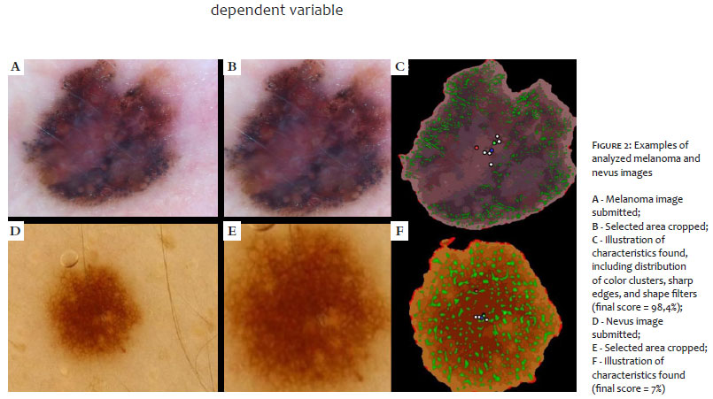
Images of melanocytic nevi and melanomas were included, both from the authors’ files and from dermoscopic image banks available on the internet (The International Skin Imaging Collaboration - ISIC - https://www.isic-archive.com/). We included only images of lesions with a histopathologically confirmed diagnosis, and non-pigmented lesions. We excluded non-pigmented lesions, with coarse hair, of the mucosa, in the nail or palmoplantar region, or those that extrapolated the image field of the dermoscopic photo for better performance of the model. Images that showed peripheral objects, such as dermatoscope edges, were cut in a rectangular shape to exclude them (Figure 2-A, D).9
The study was conducted between April and July 2018. The images were pre-processed using the imageJ 1.48 software, and routines and plugins were developed to extract 58 image characteristics. After segmentation between lesion and background, image features included the distribution, variability and color entropy (25 items), histogram (16 items), shape (five items), borders (four items) and size (two items), and the distribution of shape filters applied to the image (six items). Each image was assessed in two different dimensions to reduce the effects of cropping objects. However, overall, the lesions covered more than one-fifth of the pixels in the images submitted to feature extraction.
The 58 characteristics obtained from each of the 1,949 images (50.3% melanoma) were tabulated and analyzed using the IBM SPSS 20v software (Multilayer Perceptron Network) with the standardization of input data. The sample was divided into 80% training and 20% testing, with hyperbolic tangent activation function, softmax function output, and optimization conjugate gradient method, obtaining a network with a hidden layer of seven perceptrons.
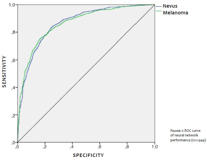
A percentage value (pseudoprobability) resulting from the softmax function in the output layer, ranging from 0-100%, characterizes the result of the neural network. So values higher than 50 were predicted as melanoma and values lower than 50, as nevus (Figure 1).
With an average of 9.3 years of dermatological practice and 7.6 years of dermoscopy use, 52 dermatologists randomly selected 40 images from the image bank excluded from the neural network training. The evaluators were informed that these were melanocytic lesions and answered whether each lesion was benign or malignant (including in situ).
The training and testing obtained a correct rating percentage of 78.5% and 79.1%, respectively, with a ROC curve covering 86.5% of the area (Figure 1). The weights and parameters obtained from the neural network were used to develop an application (Figure 2) hosted on a public web server, allowing the experimental online evaluation of dermoscopic images (http://200.145.131.197/mmview/index.php/).
Difficulty or uncertainty in analyzing lesions by dermatologists had a mean value of 3.3 on a scale of 0 to 5. The overall sensitivity and specificity of the 2,080 assessments by dermatologists were 71.8% and 52%, respectively. For the same images and a cutoff point of 0.4 (40%) of the output value, the application obtained 62% and 56% values, respectively.
The study results demonstrate that less complex predictive methods such as artificial neural networks can bring significant results despite their limitations. The online and open availability of the studied algorithm can add information in decision-making about melanocytic lesions, mainly when more extreme values are obtained. Nevertheless, it should be recognized that the tool has performance limitations. It was trained only with selected images of nevi and melanoma, not with coarse hair or pigmented lesions.
Other classification algorithms such as k-nearest neighbors algorithm (k-NN) and Support Vector Machine (SVM) may have different performances than the artificial neural network, and the group will explore them later. Also, extracting new variables from image analysis can lead to system performance gain.10
Computer vision methods have evolved significantly with cloud computing-based systems, spreading the use of convolutional neural networks with up to billions of neurons. Still, they depend on a high number of images for learning and significant maintenance costs. Nevertheless, machine learning will probably become more frequent in medical activities, especially in image analysis, as it is in other human activities.
We developed and implemented a neural network based on dermoscopic images, which can collaboratively assist in the differential diagnosis between melanocytic nevus and melanoma.
César Augusto Zago Ferreira 0000-0001-7299-1710
Approval of the final version of the manuscript; study design and planning; preparation and writing of the manuscript; data collection, analysis, and interpretation; critical revision of the manuscript.
Vinícius de Souza 0000-0001-8819-6906
Approval of the final version of the manuscript; data collection, analysis, and interpretation; critical revision of the manuscript.
Hélio Amante Miot 0000-0002-2596-9294
Statistical analysis; approval of the final version of the manuscript; study design and planning; preparation and writing of the manuscript; critical revision of the manuscript.
Juliano Vilaverde Schmitt 0000-0002-7975-2429
Statistical analysis; approval of the final version of the manuscript; study design and planning; preparation and writing of the manuscript; data collection, analysis, and interpretation; active participation in research orientation; intellectual participation in propaedeutic and/or therapeutic conduct of studied cases; critical literature review; critical revision of the manuscript.
1. Souza RJ, Mattedi AP, Rezende ML, Corrêa Mde P, Duarte EM. An estimate of the cost of treating melanoma disease in the state of Sao Paulo - Brazil. An Bras Dermatol. 2009;84(3):237-43.
2. Vestergaard ME, Macaskill P, Holt PE, Menzies SW. Dermoscopy compared with naked eye examination for the diagnosis of primary melanoma: a meta-analysis of studies performed in a clinical setting. Br J Dermatol. 2008;159(3):669-76.
3. van der Rhee JI, Bergman W, Kukutsch NA. The impact of dermoscopy on the management of pigmented lesions in everyday clinical practice of general dermatologists: a prospective study. Br J Dermatol. 2010;162(3):563-7.
4. Hoorens I, Vossaert K, Lanssens S, Dierckxsens L, Argenziano G, Brochez L. Value of dermoscopy in a population-based screening sample by dermatologists. Dermatol Pract Concept. 2019;9(3):200-6.
5. Cui X, Wei R, Gong L, Qi R, Zhao Z, Chen H, et al. Assessing the effectiveness of artificial intelligence methods for melanoma: a retrospective review. J Am Acad Dermatol. 2019;81(5):1176-80.
6. Brinker TJ, Hekler A, Enk AH, Berking C, Haferkamp S, Hauschild A, et al. Deep neural networks are superior to dermatologists in melanoma image classification. Eur J Cancer. 2019;119:11-7.
7. Basheer IA, Hajmeer M. Artificial neural networks: fundamentals, computing, design, and application. J Microbiol Methods. 2000;43(1):3-31.
8. Cullell-Dalmau M, Otero-Viñas M, Manzo C. Research techniques made simple: deep learning for the classification of dermatological images. J Invest Dermatol. 2020;140(3):507-14
9. ISIC. The International Skin Imaging Collaboration. Available from: https://www.isic-archive.com/#!/topWithHeader/wideContentTop/main. 2020. Acessed 21 Oct 2020.
10. Islam MM, Iqbal H, Haque MR, Hasan MK. Prediction of breast cancer using support vector machine and K-Nearest neighbors. IEEE Region 10 Humanitarian Technology Conference (R10-HTC), Dhaka. 2017;226-9.
 All content the journal, except where identified, under the Creative Commons Attribution 4.0 International licence - ISSN-e 1984-8773


 Read in Portuguese
 Portuguese PDF
 Print
Print
 Send this article by email
 How to cite this article
 Submit a comment
 Mendeley
 Pocket