


John Verrinder Veasey1; Bárbara Arruda Fraletti Miguel2; Roberta Buense Bedrikow2
The dermatologist's clinical practice is based on the analysis of cutaneous lesions that is carried out mainly by clinical observation, and currently supplemented with tests such as dermoscopy and confocal microscopy. Despite its low cost, the Wood's lamp has been decreasingly used as an auxiliary diagnostic method. The authors of the present study describe several cases of use of the Wood's lamp where it provided valuable assistance to the dermatologist, aiming at encouraging the use of this device in the daily practice.
Keywords: fluorescence; diagnosis; malassezia; propionibacterium acnes; porphyrias; vitiligo; melanosis; erythrasma; corynebacterium; tinea capitis
Dermatology is a medical specialty in which the observation of clinical lesions is crucial for diagnosis. New devices — such as the dermatoscope and the scanning confocal electron microscope — have been developed over time aimed at aiding the analysis of lesions during medical examination. With this, the use of centenarian apparatuses like the Wood’s lamp (WL) has come into disuse.
The WL was described in 1903 by physicist Robert W Wood and is based on the principle of fluorescence emitted by the skin when illuminated by a short wavelength source (340-400nm). The human eye receives the photons emitted by the skin, both those originating from the reflection of visible light (400-700nm wavelength) and those originated from fluorescence. However, the amount of photons originated from the reflection is much greater than that originated from fluorescence, which prevents naked eye observation of the latter.
Therefore, in order to identify the skin’s fluorescence, the patient should undergo irradiation with WL (320-400nm wavelength) in a dark environment, in the absence of visible light.1,2
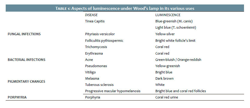
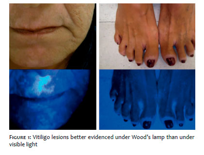
The use of the WL is comprehensive, and each dermatosis may show specific color under fluorescence (Table 1). It can be used in pigmentation disorders (hypo/hyperpigmentation) both for allowing the precise evaluation of the lesion’s limits and characteristics and for analyzing possible subclinical lesions not evidenced by the reflection phenomenon, but only by its fluorescence. For instance, this is the case of vitiligo 3 (Figure 1) and melasma.1 Its use has also been described in neoplastic diseases for the analysis of lesions and, more recently, for the surgical programming of lesions, determining margins more accurately.4
The diagnosis of infectious dermatoses also benefits from the use of WL. In such cases, the fluorescence is usually not emitted by the skin, but rather by the infectious agent and / or its metabolites.1,2,5
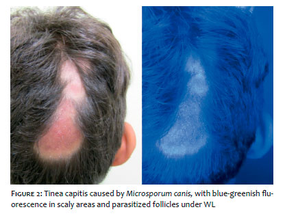
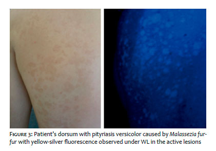
Tinea capitis, caused by some fungal species, can emit fluorescence, as in the case of parasitism by the genus Microsporum sp, emitting a blue-greenish coloration (Figure 2), and by Trychophy-ton schoenleinii, emitting a light blue coloration.1,2 In infections caused by malassezia, among them pityriasis versicolor (Figure 3), the lesions’ fluorescence can be evidenced. Nevertheless, this only happens in the lesions caused by the species Malassezia furfur, which has this characteristic due to the fact it produces fluorescent metabolites, such as pityrialactone.1,2
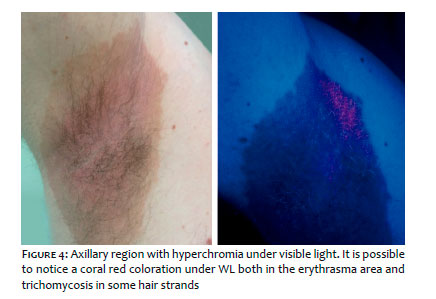
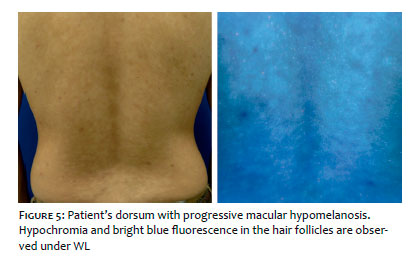
Erythrasma and trichomycosis (Figure 4), which are diseases respectively caused by the infestation of Corynebacterium minutissimum and C. tenuis, have a red-coral fluorescence.1,2 Dermatoses with parasitism of the bacterium Propionibacterium acnes, as is the case of acne and progressive macular hypomelanosis (Figure 5), may also emit fluorescence.5
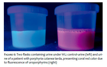
Just as WL can evidence infectious agents’ metabolites that parasitize human beings causing dermatoses, it also makes it possible to evaluate the metabolites produced by human beings. An example is the presence of porphyrin in the urine of patients bearing some porphyrias (Figure 6), of which the cutanea tarda variant is the most known.1,2
The WL is a small, durable, inexpensive, safe and very easy-to-use device. It provides rapid results, which can be very useful in the diagnosis and follow-up of the diseases, from pigmentation disorders to skin and cutaneous adnexa infections. We believe that the iconography presented in the present study may stimulate dermatologists to use the device, which will make their daily practice easier.
John Verrinder Veasey:
Study’s conception and planning, preparation and writing of the manuscript. Data collection, analysis and interpretation. Practical participation in the guidance of the research. Intellectual participation in the propaedeutic and / or therapeutic approach in the studied cases. Approval of the manuscript’s final version
Barbara Arruda Fraletti Miguel:
Data collection, analysis and interpretation. Practical participation in the guidance of the research. Intellectual participation in the propaedeutic and / or therapeutic approach in the studied cases
Roberta Buense Bedrikow:
Data collection, analysis and interpretation
1. Klatte JL, van der Beek N, Kemperman PM. 100 years of Wood's lamp revised. J Eur Acad Dermatol Venereol. 2015;29(5):842-7
2. Asawanonda P, Taylor CR. Wood's light in dermatology. Int J Dermatol. 1999;38(11):801-7.
3. Alghamdi KM, Kumar A, Taïeb A, Ezzedine K. Assessment methods for the evaluation of vitiligo. J Eur Acad Dermatol Venereol. 2012; 26(12):1463-71.
4. Walsh SB, Varma R, Raimer D, Keane JC, Cantor A, Theos A, et al. Utility of Wood's light in margin determination of melanoma in situ after excisio-nal biopsy. Dermatol Surg. 2015; 41(5):572-8
5. Relyveld GN, Menke HE, Westerhof W. Progressive macular hypomelanosis: an overview. Am J Clin Dermatol. 2007;8(1):13-9.
This study was performed at the Dermatology Department of the Hospital da Santa Casa de São Paulo - São Paulo (SP), Brazil.
 All content the journal, except where identified, under the Creative Commons Attribution 4.0 International licence - ISSN-e 1984-8773


 Read in Portuguese
 Portuguese PDF
 Print
Print
 Send this article by email
 How to cite this article
 Submit a comment
 Mendeley
 Pocket