


Maria Lopes Lamenha Lins Cavalcante1; Ana Cecília Duarte Pinto1; Fernanda Freitas de Brito1; Gardênia Viana da Silva2; Sadamitsu Nakandakari3; Cleverson Teixeira Soares4
Squamous cell carcinoma is related to exposure to UVB radiation and the presence of chronic cutaneous lesions. Folliculitis decalvans is a cicatricial alopecia that develops with the inflammation of the scalp. In the medical literature, the association between folliculitis decalvans and squamous cell carcinoma is rare, a factor motivating this report. A 66-year-old male patient, bearer of folliculitis decalvans since childhood, complained of progressive growth of lesions in the alopecia area. The biopsy confirmed squamous cell carcinoma. This case report covers the concept of "Marjolin ulcer" for including folliculitis decalvans as inflammatory lesion precursor of squamous cell carcinoma.
Keywords: ALOPECIA; CARCINOMA, SQUAMOUS CELL; FOLLICULITIS
Non-melanoma skin cancer is the most frequent form of cancer in humans - both in men and women. The estimated incidence of non-melanoma cutaneous neoplasias in Brazil in 2014 was 98,420 new cases in men and 83,710 in women. 1 Squamous cell carcinoma (SCC) is the second most common skin cancer and results from the malignant proliferation of keratinocytes. It has an incidence of 100 to 150 per 100,000 inhabitants, and is ten times more common in people over 75-years-old. It is a multifactorial disease and is mainly related to exposure to ultraviolet B radiation (UVB), sunburns in childhood, ionizing radiation, fair skin, genodermatoses, infection with oncogenic strains of human papilloma virus (HPV), immunosuppression, chemical agents, and chronic skin lesions.2 Folliculitis decalvans (FD) is a rare sub-group of cicatricial alopecias and presents as a chronic and recurring course. Although the precise cause is unknown, the association with Staphylococcus aureus and immune mechanisms have been postulated in order to justify the related follicular destruction. The classic clinical picture of FD, described by Quinquaud in 1888, is characterized by follicular pustules associated with central cicatricial areas devoid of hair, secondary to aggression by the etiologic agent and/or to pro-inflammatory processes.3 In the literature, there are few cases of patients with folliculitis decalvans who developed SCC. This rare association motivated the report of the present case.
Case report
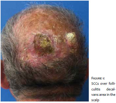
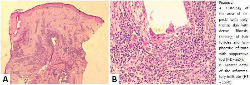
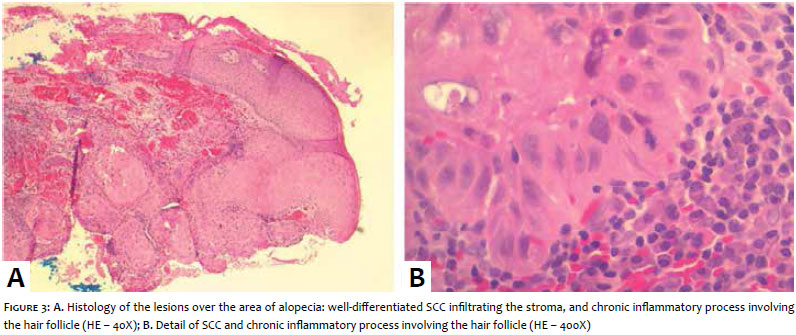
A 66-year-old white male patient bearing folliculitis decalvans with histological diagnosis carried out 26 years before at a reference center, described having these lesions since childhood. He had already made use of various treatments with antibiotics, including tetracycline and dapsone, with partial improvement. The patient had not been followed up with for the past 10 years, having returned in April 2014 complaining of lesions with progressive growth in the vertex, over the alopecia area. He could not assert the duration of the development. The dermatological examination revealed a plaque of alopecia in the described region of the scalp, with polytrichia in the periphery. At the center of this area, two lesions could be observed: a circumferential exulceration of about 4 cm with erythematous, scaly edge topped by a hematic crust and nodules of approximately 3 cm with their surfaces covered with a cutaneous horn (Figure 1). With these clinical findings, the diagnosis of SCC over the folliculitis decalvans area was hypothesized. Three biopsies were then performed: one in each of the lesions and another comprising the polytrichia area. The latter evidenced "scalp with dense fibrosis, thinning of hair follicles and lymphocytic infiltrate with suppurative focuses compatible with decalvans folliculitis" (Figure 2), while the others showed SCC (Figure 3). The patient was referred for surgical evaluation and is being followed up with by the service.
Malignant transformation in burn scars was first described by Jean-Nicholas Marjolin, in 1828. The term "Marjolin ulcer" is currently used when malignant neoplasms, especially SCC, occur over chronic ulcers, fistulas, and scars of various etiologies, including those of an infectious nature, such as leprosy, tuberculosis and lobomycosis.5, 6
The literature describes a case of a patient with a case of long course folliculitis decalvans who later developed SCC. The constant production of pro-inflammatory cytokines and tissue remodeling in chronic inflammatory disorders provide a favorable environment for malignant transformation. The relative contribution of ultraviolet radiation is difficult to assess. However, it is pertinent to advise the continuous use of photoprotection, as well as to maintain ambulatorial follow up with these patients. Therefore, the emergence of a nodule or ulcer within an area of folliculitis decalvans requires careful clinical evaluation and biopsy when there is a suspicion of neoplasia.7
1. BRASIL. Ministério da Saúde. Incidência de Câncer no Brasil: estimativa 2014. Rio de Janeiro: INCA; 2015.
2. Terzian LR, Festa Neto C, Pimenta ERA. Fatores preditivos do maior número de estádios na cirurgia micrográfica de Mohs para o tratamento do carcinoma espinocelular da cabeça. An Bras Dermatol. 2008;83(3):221-6.
3. Bunagan MJ, Banka N, Shapiro J. Retrospective Review of Folliculitis Decalvans in 23 Patients with Course and Treatment Analysis of Long-standing Cases. J Cutan Med Surg. 2015;19(1):45-9.
4. Bauk VOZ, Assunção AM, Domingues RF, Fernandes NC, Maya TC, Maceira JP. Úlcera de Marjolin: relato de 12 casos. An Bras Dermatol. 2006;81(4):355-8.
5. Gontijo GMA, Nogueira L, Mendes L, Rodrigues CAC, Santos M, Talhari S, Talhari C. Lobomicose e carcinoma espinocelular. An Bras Dermatol. 2013;88(2):297-9.
6. Maclean GM, Coleman DJ. Three fatal cases of squamous cell carcinoma arising in chronic perineal hidradenitis supurativa. Ann R Coll Surg Engl. 2007;8997):709-12.
7. Yip L, Ryan A, R. Sinclairs R. Squamous cell carcinoma arising within folliculitis decalvans. Br J Dermatol. 2008;159(2):481-2.
This study was carried out at the Instituto Lauro de Souza Lima (ILSL) - Bauru (SP), Brazil.
 All content the journal, except where identified, under the Creative Commons Attribution 4.0 International licence - ISSN-e 1984-8773


 Read in Portuguese
 Portuguese PDF
 Print
Print
 Send this article by email
 How to cite this article
 Submit a comment
 Mendeley
 Pocket