


Felipe Maurício Soeiro Sampaio1; Isabela Coelho Guimarães2; Bruno Lopes da Silva Ramos3
Financial support: None.
Conflicts of interest: None.
Submitted on: 01/15/2024
Approved on: 03/11/2024
How to cite this article: Sampaio FMS, Guimarães IC, Ramos BLS. Tip for achieving hemostasis in the paramedian interpolation flap pedicle. Surg Cosmet Dermatol. 2025;17:e20250342.
Postoperative bleeding from the vascular pedicle of the paramedian flap is a common complication. We describe the use of simple sutures on the raw lateral surface of the pedicle as an alternative method to reduce this risk.
Keywords: Surgical Flaps; Carcinoma Basal Cell; Mohs Surgery; Hemorrhage.
Postoperative bleeding from the raw area of the vascular pedicle of the paramedian flap is a common complication. Electrocoagulation of the raw portion of the pedicle, as well as the use of skin grafts and porcine xenografts, are described methods aimed at reducing postoperative bleeding.1 Wrapping the pedicle with oxidized regenerated cellulose-impregnated gauze,2 antibiotic ointment-impregnated gauze,3 or Vaseline-impregnated gauze4 at the raw surface of the flap has also been reported, with some positive results. We describe the use of simple interrupted sutures on the lateral surface of the pedicle as an alternative method to reduce this complication.
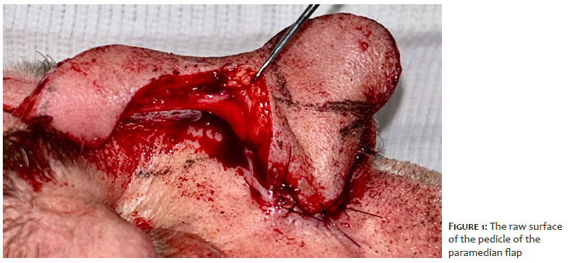
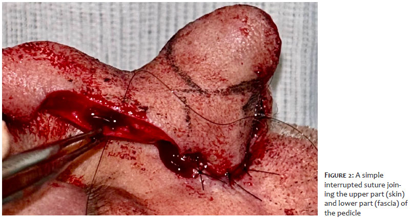
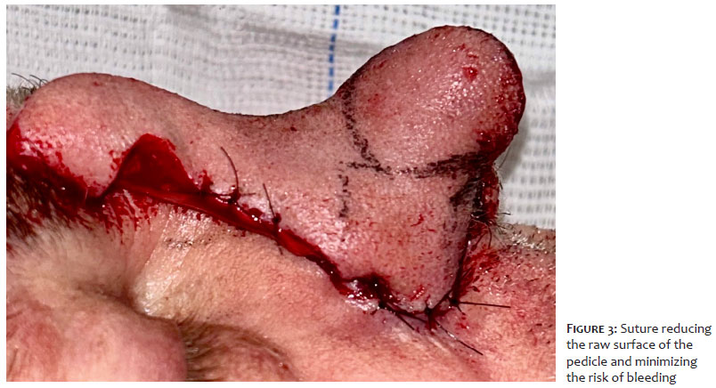
Immediately after completing nasal reconstruction with the paramedian flap, the surgeon should carefully observe the raw area of the pedicle to identify potential bleeding points (Figure 1). Once identified, a simple interrupted suture with 6-0 monofilament nylon is performed. The needle is inserted through the skin at the upper part of the pedicle and reinserted into the fascia at the lower part (Figure 2). This suture brings the skin closer to the fascia, locally reducing the raw surface of the pedicle and consequently minimizing the risk of bleeding (Figure 3). On average, four sutures are placed on each side of the pedicle, depending on the length of the flap and the presence of bleeding points. No vascular compromise or congestion has been observed in the distal part of the flap using this technique. The dressing is applied only after hemostasis has been reassessed.
The use of simple interrupted stitches on the lateral surface of the paramedian interpolation flap pedicle is a simple, safe, and easy-to-perform technique. It provides a comfortable postoperative period for both the patient and the surgical team, reduces the need for dressings, minimizes comorbidities, and is low cost.
Felipe Maurício Soeiro Sampaio
ORCID: 0000-0002-2235-5473
Statistical analysis; approval of the final version of the manuscript; study design and planning; manuscript preparation and writing; data collection, analysis, and interpretation; active participation in research supervision; intellectual contribution to the conduct of the preoperative and/or therapeutic management of the studied cases; critical review of the literature; critical review of the manuscript.
Isabela Coelho Guimarães
ORCID: 0009-0002-8142-6072
Manuscript preparation and writing; critical review of the literature.
Bruno Lopes da Silva Ramos
ORCID: 0009-0008-1148-211X
Approval of the final version of the manuscript; intellectual contribution to the conduct of the preoperative and/or therapeutic management of the studied cases; critical review of the literature.
1. Viscusi KS, Merritt B. Porcine xenografts for the optimization of pedicle care in interpolation flaps. Dermatol Surg. 2014;40(11):1262-5.
2. Christenson LJ, Otley CC, Roenigk RK. Oxidized regenerated cellulose gauze for hemostasis of a two-stage interpolation flap pedicle. Dermatol Surg. 2004;30(12 Pt 2):1593-4.
3. Brodland D. Paramedian forehead flap reconstruction for nasal defects. Dermatol Surg. 2005; 31:1046–52.
4. Goldman GD, Dzubow LM, Yelverton CB. Facial flap surgery. Burlington (VT): McGraw-Hill; 2012.
 All content the journal, except where identified, under the Creative Commons Attribution 4.0 International licence - ISSN-e 1984-8773


 Read in Portuguese
 Portuguese PDF
 Print
Print
 Send this article by email
 How to cite this article
 Submit a comment
 Mendeley
 Pocket