


Mariana Abdo de Almeida1; Michele Maria Reis-Feroldi2; Marcia Lanzoni Alvarenga Lira3
Data de submissão: 15/04/2021
Decisão final: 13/10/2021
Fonte de financiamento: Nenhuma
Conflito de interesse: Nenhuma
Como citar este artigo: Almeida MA, Reis-Feroldi MM, Lira MLA. Desenvolvimento de alopecia fibrosante frontal em duas pacientes usuárias de toxina botulínica: relação ou coincidência? Surg Cosmet Dermatol. 2021;13:20210036.
Frontal fibrous alopecia is a primary lymphocytic scarring alopecia that is difficult to control, with a perifollicular lichenoid infiltrate marking its histology. Since its description in 1994 by Kossard, there has been an increase in cases reported worldwide. This article reports two cases that underwent incisional biopsies with longitudinal and transverse sections in areas presenting higher disease activity and reviews some aspects of this dermatosis. It also addresses the suspicion of a possible relationship with the application of botulinum toxin – a question already raised by other authors but with no established confirmation.
Keywords: Alopecia; Botulinum toxins; Botulinum toxins type A
Frontal fibrosing alopecia (FFA) has a still unknown etiology and has evolved increasingly worldwide. Kossard first described it in 1994,1 in Australia, and it is more common in postmenopausal women, although cases in childbearing age women and men have also been reported.2
FFA is characterized by the loss of terminal hair and/or vellus follicles in the frontotemporal region, associated with perifollicular erythema and follicular hyperkeratosis. It may also present nonspecific symptoms such as itching and trichodynia. It presents fibrosing, scarring, progressive, and, most of the time, irreversible characteristics.3 It is currently classified in the spectrum of lichen planopilaris (LPP) – an entity that has been subdivided today into FFA, classic lichen planopilaris, and Graham-Little-Piccardi-Lasseur Syndrome (GLPLS).
It is common to observe terminal follicles and tufted hair loss in the front temporal region during the dermatological examination, associated with erythema and flaking in the alopecia plaque.1,2 The affected area may be slightly atrophic, with follicular ostia loss. It is also common to observe perifollicular erythema at the frontal hairline.2,3
Dermoscopy is a non-invasive method that assists in disease diagnosis and follow-up, and it provides a better site identification for a biopsy. The dermoscopy contributes to findings such as perifollicular erythema, follicular hyperkeratosis, follicular ostia reduction, and absence of vellus follicles, which are significant findings for better diagnostic clarification.3
Although it is an invasive procedure, scalp biopsy contributes significantly to the diagnosis through histopathological examination. For the biopsy, the material must be collected from a hair carrier area with clinical signs of active disease, which endorses the importance of using dermoscopy to identify the best site.4
Histopathological findings of FFA are similar or almost identical to those seen in the lichen planopilaris. Both diseases exhibit lymphocytic inflammatory infiltrate involving the isthmus and infundibulum hair follicles. The follicular epithelium shows exocytosis of lymphocytes, apoptotic and/or dyskeratotic cells, concentric fibroplasia around the inflamed follicles, and subsequent destruction of some follicles.5
A possible correlation between botulinum toxin application and the presence of FFA has been recently investigated.6,7
Two cases report
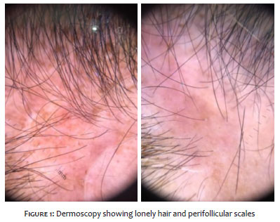
1. A 54-year-old-woman received botulinum toxin application (Botox® - Allergan, Irvine, CA, USA) on the upper third of the face and filling with hyaluronic acid in the nasolabial folds for more than five years before the dermatological examination, presenting no local complaints. The patient reported frontal hair loss and thinning hair presence in the frontal region. The dermatological examination evidenced high capillary implantation when raising the patient’s bangs and decreased vellus in the frontal hairline. Dermoscopy examination showed lonely hair, perifollicular scales, and black spots without eyebrows changes (Figure 1). An incisional biopsy was performed. In the optical microscopy, the cross-sections presented cicatricial tracts at the follicular isthmus levels, and a moderate perifollicular lymphomononuclear inflammatory infiltrate at the level of the infundibulum and follicular isthmus. The follicular epithelium showed lymphocyte exocytosis and vacuolar alteration. The treatment consisted of topical therapy with clobetasol propionate and 5% minoxidil solution on the scalp and eyebrows. After eight months of clinical follow-up, the patient remained without signs of FFA activity.
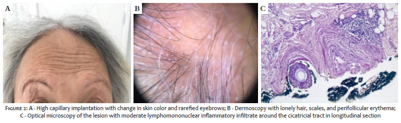
2. A 79-year-old woman, hypertensive, received botulinum toxin (Botox® - Allergan, Irvine, CA, USA) applications on the upper third of the face one year before the dermatological examination; filling with hyaluronic acid five years before examination on the nasolabial folds; and a complete facial lifting 15 years before examination. She presented a surgical scar on the front hairline implantation. The patient sought dermatological care for a new botulinum toxin application in the upper third of the face. She did not report any hair or scalp complaints, and she used a haircut with bangs that completely covered her frontal region to hide her frontal hairline loss. The dermatological examination showed high capillary implantation noticed when raising her bangs; alteration of the skin color in the frontal hairline capillary implantation; contraction of the frontal muscle more evident with enlargement of the hairline capillary implantation; vellus hair reduction on the frontal hairline; and eyebrows thinning (Figure 2A). Dermoscopy showed lonely hair; scales; perifollicular erythema; capillary rarefaction in the frontal, midline, and vertex areas; and rarefaction in the eyebrows (Figure 2B). An incisional biopsy was performed. The microscopy examination revealed a peripheral lymphomononuclear inflammatory infiltrate at the level of the infundibulum, perifollicular fibrosis, and a moderate lymphomononuclear inflammatory infiltrate around the scar tract (Figure 2C). The patient underwent oral therapy with finasteride, topical therapy with clobetasol propionate and 5% minoxidil solution on the scalp and eyebrows. After seven months of clinical follow-up, she remained free of FFA activity signs.
In the two cases reported, the patients were women aged between 54 and 79 years. Also, the patients received botulinum toxin (Botox® - Allergan, Irvine, CA, USA) applications and hyaluronic acid. The second patient also had a complete facial lifting 15 years before the dermatological examination, presenting a surgical scar on the frontal hairline. We found a report in the literature about a 55-year-old patient presenting scleroatrophy on the frontal hairline of the scalp insertion ten days after local injection of botulinum toxin type A for aesthetic purposes. However, the authors did not discard an inappropriate toxin application or an overdose of the drug.6 Another article reports five cases of patients who were submitted to botulinum toxin periodically. These cases reported regression of the frontal hairline, which led the authors to question a correlation known as “botulin-induced frontal alopecia (BIFA)”. However, it was impossible to confirm this likely disease’s aspects because the patients did not accept to undergo biopsy.7
A recent case-control study showed that the relationship between FFA and environmental factors is still controversial. According to this study, FFA was associated with the use of facial moisturizers, non-dermatological facial soaps, and hair straightening with formalin. Literature has suggested the use of capillary anti-residue solutions and tobacco as protective factors. There was no association with the use of sunscreens. The application of botulinum toxin or hyaluronic acid was not mentioned in this study.8 The cases reported in the present study showed an association of FFA with the application of botulinum toxin. Further studies are needed to ascertain this possible relationship, a fact that corroborates with the Piraccini6 and Persechino7 studies.
Regarding the patient who also underwent a facelift, the possible correlation between the procedure and FFA was described in a case series where three patients developed FFA after a facelift. A possible explanation for this correlation is the induction of the Koebner phenomenon in which a trauma or non-specific inflammatory process in healthy skin occurs in susceptible individuals.9
We report two cases of alopecia in the present study, in women aged between 54 and 79 years, with clinical, dermoscopic, and histological findings compatible with FFA. They were treated and presented no progression of the lesions in clinical follow-up. Both had a previous history of a single application of botulinum toxin (Botox® - Allergan, Irvine, CA, USA). The present study addresses a possible relationship between FFA and botulinum toxin’s application and presents studies that suspected this relationship.
Mariana Abdo de Almeida 0000-0002-7080-689X
Statistical analysis; approval of the final version of the manuscript; study design and planning; preparation and writing of the manuscript; data collection, analysis and interpretation; active participation in research orientation; intellectual participation in propaedeutic and/or therapeutic conduct of studied cases; critical literature review; critical revision of the manuscript.
Michele Maria Reis-Feroldi 0000-0003-4900-3598
Approval of the final version of the manuscript; study design and planning; preparation and writing of the manuscript; data collection, analysis and interpretation; active participation in research orientation; intellectual participation in propaedeutic and/or therapeutic conduct of studied cases; critical literature review.
Marcia Lanzoni Alvarenga Lira 0000-0002-1208-7911
Approval of the final version of the manuscript; study design and planning; preparation and writing of the manuscript; data collection, analysis and interpretation; active participation in research orientation; intellectual participation in propaedeutic and/or therapeutic conduct of studied cases; critical literature review; critical revision of the manuscript.
1. Kossard S. Postmenopausal frontal fibrosing alopecia. Scarring alopecia in a pattern distribution. Arch Dermatol. 1994;130(6):770-4.
2. Dawn G, Holmes SC, Moffat D, Munro CS. Post-menopausal frontal fibrosing alopecia. Clin Exp Dermatol. 2003;28(1):43-5.
3. Crisóstomo MR, Crisóstomo MCC, Crisóstomo MGR, Gondim VJT, CrisóstomoMR, Benevides NA. Perda pilosa por líquen plano pilar após transplante capilar: relato de dois casos e revisão da literatura. Na Bras Dermatol. 2011;86(2):359-62.
4. Donati A, Molina L, Doche I, Valente NS, Romiti R. Facial papules in frontal fibrosing alopecia: evidence of vellus follicle involvement. Arch Dermatol. 2011;147(12):1424-7.
5. Sperling LC, Cowper SE, Knopp EA. An Atlas of Hair Pathology with Clinical Correlations. 2nd ed. Florida: CRC Press; 2012.
6. Di Pietro A, Piraccini BM. Frontal alopecia after repeated botulinum toxin type A injections for forehead wrinkles: an underestimated entity? Skin Appendage Disord. 2016;2(1-2):67-9.
7. Persechino S, Lupi F, Di Vito E, Romano I, Persechino F, et al. Sclero- Atrophic Reaction after Botulinum Toxin Injection. Clin Res Dermatol Open Access. 2016;3(2):1-2.
8. Ramos PM, Anzai A, Duque-Estrada B, Farias DC, Melo DF, Mulinari-Brenner F, et al. Risk factors for frontal fibrosing alopecia: a case-control study in a multiracial population. J Am Acad Dermatol. 2021;84(3):712-8.
9. Chiang YZ, Tosti A, Chaudhry IH, Lyne L, Farjo B, Farjo N, et al. Lichen planopilaris following hair transplantation and face-lift surgery. Br J Dermatol. 2012;166(3):666-370.
 All content the journal, except where identified, under the Creative Commons Attribution 4.0 International licence - ISSN-e 1984-8773


 Read in Portuguese
 Portuguese PDF
 Print
Print
 Send this article by email
 How to cite this article
 Submit a comment
 Mendeley
 Pocket