


Cristina Diniz Borges Figueira de Mello1; Milena da Rocha e Souza2,3; Nilton Gioia Di Chiacchio3,4; Nilton Di Chiacchio3
Received on: 28/11/2019
Approved on: 14/12/2019
Financial support: None
Conflict of interests: None
Research performed at Hospital do Servidor Público Municipal de São Paulo, São Paulo (SP), Brazil
The “harpoon nail” is a variant of the ingrown toenail. In this condition, the nail spicule pierces the distally growing periungual tissue and may emerge through the hyponychium. Its diagnosis can be confirmed by high-frequency ultrasound, facilitating the therapeutic programming.
Keywords: Dermoscopy; Nail diseases; Onychomycosis
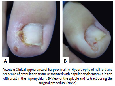
"Harpoon nail" is a variant of ingrown toenail1 (Figure 1A). The etiology is similar to that of onychocriptosis and can be secondary to over-curvature of the nail plate or hypertrophy of nail folds, resulting in distal ingrowth.1,2 Inadequate trimming of the nails to relieve the pain results in the formation of a lateral spicule that grows distally, covered by the skin, perforating it at the tip of the toe and emerging through the hyponychium (Figure 1B).
The clinical picture is similar to that of onychocryptosis, associated with an erythematous-edematous papule and hematic crust in the hyponychium (Figure 1A). In the absence of treatment, the canal that contains the spicule may epithelize and the inflammation may disappear, leading to the chronic form.2 High-frequency ultrasound is a noninvasive imaging method that is useful in the diagnosis of harpoon nail. The following ultrasound findings are possible in the diagnosis of harpoon nail:
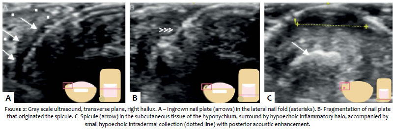
Characterization of ingrowing nail plate, in a comparative study with the normal toe, highlighting the hypoechoic soft tissues adjacent to the nail plate (Figure 2A) with or without vascularization on Echo Doppler.
Identification of the hyperechoic spicule in communication with the nail plate (Figure 2B), located in the subcutaneous tissue of the nail fold and the hyponychium (Figure 2C), surrounded by a hypoechoic inflammatory halo with or without increased vascularization on Echo Doppler (depending on the inflammatory activity).
Characterization of periungual or subcutaneous hypoechoic or anechoic collections with hemorrhagic and/or purulent content.
High-frequency ultrasound in experienced professional hands proves to be a valuable tool for ruling out possible differential diagnoses of harpoon nail (e.g. onychoclavus, inclusion cyst, etc.), confirming the diagnosis and facilitating surgical programming.
Cristina Diniz Borges Figueira de Mello | 0000-0001-7199-8451
Case submission; writing of manuscript; final revision; overall revision.
Milena da Rocha e Souza | 0000-0002-0732-0432
Case submission; writing of manuscript; final revision; overall revision.
Nilton Gioia Di Chiacchio | 0000-0001-5944-7737
Case submission; writing of manuscript; final revision; overall revision.
Nilton Di Chiacchio | 0000-0001-9536-2263
Case submission; writing of manuscript; final revision; overall revision.
1. Richert B, Caucanas M, Di Chiacchio N. Surgical Approach to Harpoon Nail: A New Variant of Ingrowing Toenail. Dermatol Surg. 2014;40(6):700-1.
2. Richert B, Di Chiacchio N, Caucanas M, Di Chiacchio NG. Definition Pathogenesis Risk Factors – Classification – Scoring. In: Management of Ingrowing Nails. 1.ed. Switzerland: Springer International Publishing; 2016. p.51-53.
 All content the journal, except where identified, under the Creative Commons Attribution 4.0 International licence - ISSN-e 1984-8773


 Read in Portuguese
 Portuguese PDF
 Print
Print
 Send this article by email
 How to cite this article
 Submit a comment
 Mendeley
 Pocket