


Rachel de Avila Coelho; Luiz Fernando de Oliveira Santana; Juliana Cristina Silva Fraga
Received on: 07/07/2018
Approved on: 11/04/2019
This study was performed at the Minas Gerais Military Police Hospital - Belo Horizonte (MG), Brazil.
Financial support: None
Conflict of interests: None
Skin biopsy is an important introductory tool and also essential for the dermatologist. The conventional biopsy method, with the use of local anesthetics, sterile fields, surgical instruments (punch, scalpel, forceps, scissors) and suture material demand staff, time and considerable financial investment. Criobiopsy is a simple method, where by using the cryocautery an extraction of good quality skin fragments is made for histopathologic analysis, making it an excellent alternative to the conventional biopsies.
Keywords: Ambulatory Surgical Procedures; Biopsy; Cryosurgery; Dermatology; Histology
The histological study of skin fragments is an indispensable propaedeutic method in the daily routine of dermatologists. Conventional skin biopsies require sterilized surgical instruments, operating room, nursing team and longer surgical time, as well as local anesthesia, which can cause pain and distress in patients who have phobia to needles.1 In Brazil, there is a complicating factor since the issuance of Resolution CFM (Brazilian Federal Council of Medicine) No. 2,056/2013, requiring that premises where procedures using local anesthesia are carried out be furnished with minimal equipment and medication to deal with intercurrences, such as cardiorespiratory arrest and anaphylaxis – which makes mandatory that items such as emergency oxygen kits and aspirators, defibrillators, and vasoactive drugs be made available in the dermatological practice, a space in which small surgeries are performed. This requirement discourages the performance of conventional biopsies in dermatological practices. As a result, cryobiopsy becomes a good alternative to harvest cutaneous specimens to be studied.
Technique description
The technique consists of:
1) Topical antisepsis of the site where the cryobiopsy will be performed. 2
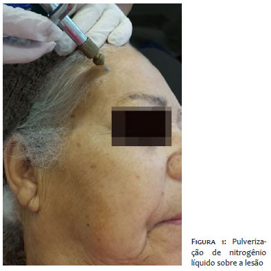
2) Spraying of liquid nitrogen over the lesion from a distance of 2.5 to 3.8cm, for 4 to 5 seconds, up until the complete freezing of the lesion, visually verified based on the whitening of the region (Figure 1).2
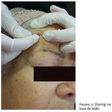
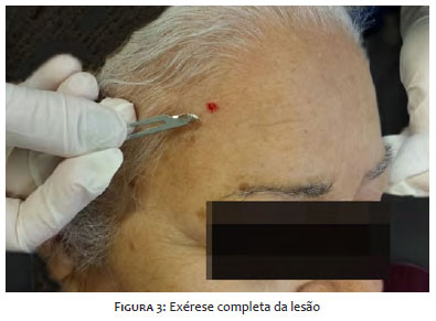
3) Shaving at the base of the lesion, using a scalpel with n.15 blade, as soon as the thawing of the skin begins, while the tissue still offers some resistance (Figures 2 and 3).2
4) Once removed, the specimen is immediately transferred to a flask containing formaldehyde solution.2
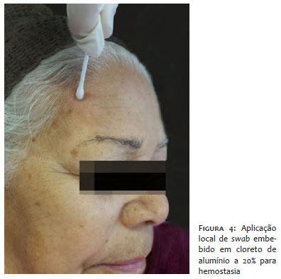
5) Prior to the complete thawing of the skin, a swab soaked in 20% aluminum chloride is gently pressed onto the site for hemostasis (Figure 4).2
Cryobiopsy is a method used to obtain cutaneous specimens for histological analysis that uses equipment that is usually employed in dermatological practices and dismisses the use of local anesthetics and complex infrastructure to be performed.
The procedure is indicated for benign, pre-malignant lesions and non-melanoma skin cancers. Thus, the technique can be performed in most cases where routine biopsy is indicated (e.g. basal and squamous cell carcinomas, actinic keratoses, nevi and inflammatory or infectious lesions with dubious diagnosis – such as facial granuloma, annular granuloma, molluscum contagiosum, angiomas, soft fibromas, seborrheic keratoses, viral warts etc).2,3
Few conditions limit the use of cryobiopsy, which should, nevertheless, not be performed in patients with pathologies that can be induced or exacerbated by exposure to cold – such as cryoglobulinemia, Raynaud's disease, cold-induced urticaria, and previous history of cold-induced injury – or in sites with poor blood circulation.4
Complications arising from the procedure are usually mild and dependent on the physician's familiarity with the technique. The most common is dyschromia, in special hypopigmentation, which is the most common type due to the destruction of melanocytes caused by the low temperature.4 In individuals with high skin phototypes, postinflammatory hyperpigmentation may also occur. The onset of depressed scars is a result of deep cryobiopsy, however it usually resolves spontaneously.4 In hairy areas, permanent cicatricial alopecia may occur, resulting from the destruction of the hair follicle by freezing. Finally, if cryobiopsy is performed on the nail matrix or cartilage (nasal or auricular), tissue distortion may occur secondarily to the formation of indentations or retractions.4
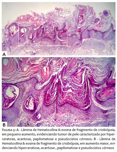
Regarding the quality of the obtained skin specimen via cryobiopsy, the histological analysis of the fragments does not show any tissue damage formation or artifacts in the evaluated pieces after they have been subjected to low temperatures (Figure 5). To avoid cell autolysis, the histological specimen is placed in formalin aiming at preserving the morphology and chemical composition of the tissue's components, which contributes to a reliable histological examination.5-7.
The authors of the present study describe a new technique with a view to raising the awareness regarding a safe, almost painless, cost effective and efficacious procedure that can be of great value in the daily dermatological practice.
In face of the new enforced legislation, the described technique is a viable alternative to most dermatologists and provides more agility in performing skin biopsies.
The promising experience of cryobiopsies in diverse fields of medicine, such as pneumology, which has been conducting tracheobronchial biopsies since the 1980s, demonstrates how dermatological surgery can still develop and benefit from the more frequent use of this technique.5,6,8-10
Rachel de Avila Coelho | ORCID 0000-0002-7947-7754
Approval of the final version of the manuscript, study design and planning, preparation and drafting of the manuscript, critical review.
Luiz Fernando de Oliveira Santana | ORCID 0000-0002-7793-5360
Approval of the final version of the manuscript, study design and planning, preparation and drafting of the manuscript, critical review.
Juliana Cristina Silva Fraga | ORCID 0000-0002-1593-8742
Approval of the final version of the manuscript, study design and planning, effective participation in the research guidance, intellectual participation in the propaedeutic and / or therapeutic approach of the cases studied, critical review of the literature, review of the manuscript.
1. Pasquali P, Freites-Martinez A, Fortuño-Mar A. Cryobiopsy: Analternative technique to conventional shavebiopsy. J Am Acad Dermatol. 2015;73(5):867-8.
2. Pasquali P, Freites-Martinez A, Fortuño-Mar A. Use of cryobiopsy in dermatological practice. J Am Acad Dermatol. 2015;72(2):e63-4.
3. Pasquali P. The cryosurgery alternative. Int J Dermatol. 2007;46(5):511-3.
4. Prohaska J, Badri T. Cryotherapy. [Updated 2019 Apr 4]. In: StatPearls [Internet]. Treasure Island (FL): StatPearls Publishing; 2019 Jan-. Available from: https://www.ncbi.nlm.nih.gov/books/NBK482319/.
5. Casoni GL, Tomassetti S, Cavazza A, Colby TV, Dubini A, Ryu JH, et al. Transbronchial lung cryobiopsy in the diagnosis of fibrotic interstitial lung diseases. PLOS ONE. 2014;9(2):e86716.
6. Ohno N, Terada N, Saitoh S, ZhouH,Fujii Y, Ohno S. Recent development of in vivo cryotechnique to cryobiopsy for living animals.. Histol Histopathol. 2007;22(11):1281-90.
7. Junqueira LCU, Carneiro J. Histologia Básica: texto e atlas. 4 ed. Rio de Janeiro: Editora Guanabara Koogan; 1979. p. 1.
8. Ohno N, Terada N, Bai Y, Saitoh S, Nakazawa T, Nakamura N, et al. Application of cryobiopsy to morphological and immune histochemical analyses of xenografted human lung can cert issues and functional blood vessels. Cancer. 2008;113(5):1068-79.
9. Olariu B. Endoscopic cryobiopsy in tracheobronchial pathology. Rev Chir Oncol Radiol O R L Oftalmol Stomatol Otorinolaringol. 1983;28(3):225-7.
10. Hetzel J, Hetzel M, Hasel C, Moeller P, Babiak A. Oldmeetsmodern: the use of traditional cryoprobes in the age of molecular biology. Respiration. 2008;76(2):193-7.
 All content the journal, except where identified, under the Creative Commons Attribution 4.0 International licence - ISSN-e 1984-8773


 Read in Portuguese
 Portuguese PDF
 Print
Print
 Send this article by email
 How to cite this article
 Submit a comment
 Mendeley
 Pocket