


Elisete Isabel Crocco1; Vanessa Alice Amorim2; Renata Oliveira Alves3
Angioma serpiginosum is a rare and benign vascular ectasia that clinically arises as small red or purple dots, with serpiginous configuration. Controlled light pulses or intense pulsed light is the light source that emits high energy light within the continuous waves spectrum from 500nm to 1,200nm. Among its characteristics is the indication for the treatment of vascular lesions, which is the oxyhemoglobin chromophore’s pigment. This paper describes a case of a 12-year-old patient with angioma serpiginosum on the right arm that receded after 6 intense pulsed light sessions.
Keywords: intense pulsed light therapy; hemangioma; capillary hemangioma; vascular diseases
Angioma serpiginosum is a rare and benign vascular nevoid disorder that arises with punctate red and purplish lesions arranged in a gyroscopic or serpiginous configuration that do not disappear under pressure. On histological examination, it presents dilation and ectasia of capillary vessels in the papillary dermis. Cases are sporadic, however an autosomal dominant inheritance pattern has also been described.1
Intense pulsed light (IPL) is a source of pulsed light that emits non-coherent high-energy light ranging from 500nm to 1,200nm. The pulses’ duration ranges from 0.5ms to 20ms and can be applied in single, double or triple pulse sequences. Among its characteristics are indications for the treatment of vascular lesions, reaching the oxyhemoglobin chromophore.2
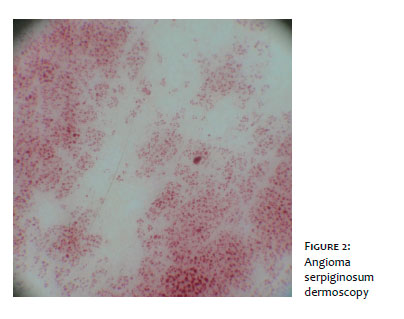
Numerous relatively well-demarcated small red lagoons can be observed on dermoscopy, ranging from round to oval in shape.3 Dermoscopy is indicated for the differentiation of angioma serpiginosum vis a vis purpuric dermatoses.
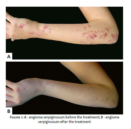
A 12-year old female patient attended a consultation visit bearing an angioma serpiginosum in the right arm that receded after six IPL sessions.
A 12-year old girl presented red spots on the right forearm and dorsum of the right hand. Her mother described having noticed the eruption from birth. The physical examination revealed multiple and confluent punctiform lesions, varying from red to violet in color, and arranged in a serpiginous pattern. The lesion did not disappear under diascopy, with the observed der-moscopic features revealing numerous relatively well demarcated small red lagoons, varying from round to oval in shape.
The condition was diagnosed as angioma serpiginosum. The treatment chosen for this case was IPL (Starlux®, Palomar, Burlington, MA, USA), Max G tip, 500 - 670nm and 870 -1,200nm, 22J / cm2 fluence, 10ms). The lesion was treated in 6 sessions with an interval of 1 month between procedures.
The patient responded very well to the treatment, without developing scars or adverse effects.
Angioma serpiginosum was first described by Hutchinson in 1889, however this nomenclature was proposed by Rad-cliffe-Crocker in 1893. Clinically, the lesion consists of multiple tiny macules ranging from red to purple in color, grouped in serpiginous patterns (Figure 1). Although the purpuric point does not disappear completely after application of pressure, there is no evidence of bleeding. The distinction between purpuric dermatoses and angioma serpiginosum is important. Predominantly seen in eruptive hemangiomas and angiokeratomas, the well-demarcated red lagoons represent one of the vascular patterns seen through dermoscopy (Figure 2). This appearance is due to enlarged and dilated vascular spaces within the papillary dermis. When these spaces are located deeper in the skin, a variable reddish-blue color to blackish pattern may occur.4
Histological examination shows dilatation with capillary ectasia in the papillary dermis.1
The main complaint linked to angioma serpiginosum concerns the aesthetic appearance.5 Although asymptomatic it can be severely disfiguring and cause significant psychological stress to the patient.6
The IPL system emits polychromatic light from a high-intensity source. Its effect is based on the principle of selective photothermolysis. The heat generated by the application of light causes thermal damage to the target tissue without causing damage to the epidermis or surrounding healthy tissue. The use of optical filters generates a well-defined wavelength range, having been established between 560nm and 1,000nm for the patient in question. The duration and sequence of pulses can also be varied. In this way, the system allows to modify the parameters considerably aiming at adjusting them to the specific treatment.5
Intense pulsed light has been used in the treatment of venous malformations, infantile hemangioma, angioma serpig-inosum, tufted angioma, pyogenic granuloma, lymphangioma, Fabry angiokeratoma and multinucleated cell angiohistiocyto-ma. Intense pulsed light may be considered a treatment option that yields reasonable results for venous malformations (with 3c evidence level). In fact, the study designs did not receive a high index of evidence; nevertheless the results were uniformly favorable, with all the studied patients having their lesions improved from 70% to 100%.7 These outcomes are comparable to the case reported by the authors of the present paper.
In the present case, IPL was a viable technique not only for the results, but also due to the low cost of the treatment.2
Elisete Isabel Crocco:
Responsible for the clinical case, participated in the preparation of the manuscript and in the English version copy-editing
Vanessa Alice Amorim:
Preparation of the manuscript, application of IPL sessions
Renata Oliveira Alves:
Participation in the preparation of the manuscript, application of pulsed light
1. Madan V, August PJ, Ferguson JE. Pulsed-dye laser treatment of angio- 5. ma serpiginosum. Clin Exp Dermatol. 2009;34(5):e186-8.
2. Crocco E, Abramavicus A, Russo C, Zaitz C, Nunes K. Treatment of port wine stain with pulsed light of the square pulse type. J Am Acad Der- 6. matol. 2010;62(3 Suppl 1):AB147.
3. Ghanadan A, Kamyab-Hesari K, Moslehi H, Abasi A. Dermoscopy of angioma serpiginosum: a case report. Int J Dermatol. 2014;53(12):1505-7.
4. Ilknur T, Fetil E, Akarsu S, Altiner DD, Ulukus C, Günes AT. Angioma serpiginosum: dermoscopy for diagnosis, pulse dye laser for treatment. J Dermatol. 2006;33(4):252-5.
5. Poenitz N, Koenen W, Utikal J, Goerdt S. Angioma serpiginosum following the lines of Blaschko - an effective treatment with the IPL technology. J Dtsch Dermatol Ges. 2006;4(8):650-3.
6. Rho NK, Kim H, Kim HS. Successful treatment of angioma serpiginosum using a novel 532 nm potassium titanyl phosphate (KTP) laser. J Dermatol. 2014;41(11): 996-8.
7. Wat H, Wu DC, Rao J, Goldman MP. Application of intense pulsed light in the treatment of dermatologic disease: a systematic review. Dermatol Surg. 2014;40(4):359-77.
This study was performed at Dermatology Department of the Hospital e Faculdade de Medicina da Santa Casa de São Paulo, São Paulo (SP), Brazil.
 All content the journal, except where identified, under the Creative Commons Attribution 4.0 International licence - ISSN-e 1984-8773


 Read in Portuguese
 Portuguese PDF
 Print
Print
 Send this article by email
 How to cite this article
 Submit a comment
 Mendeley
 Pocket