


Tábata Natasha Almeida Rodrigues1; Luiz Eduardo Garcia Galvão1; Heitor de Sá Golçalves1; Maria Araci de Andrade Pontes2
The sebaceous nevus of Jadassohn is a congenital hamartoma that may develop into a malignant cutaneous neoplasia. Photodynamic therapy is used to treat actinic keratoses and superficial or nodular basal cell carcinomas, and the cutaneous field cancerization can be observed using the Wood's lamp during the performance of the technique. This article describes a case of photodynamic therapy used in the treatment of a basal cell carcinoma, which developed on a sebaceous nevus, where the field cancerization was demonstrated through the use of Wood's lamp. The procedure is a non-surgical alternative for the treatment of the basal cell carcinoma, with excellent aesthetic outcome. The patient is on clinical follow-up, with absence of recurrence of the neoplasia 18 months after the treatment.
Keywords: CARCINOMA, BASAL CELL; NEVUS, SEBACEOUS OF JADASSOHN; PHOTOCHEMOTHERAPY
The sebaceous nevus of Jadassohn is a congenital hamartoma 1 commonly located on the scalp and face, being found in 0.5 to 1% of the population. Its etiology is unknown, however recent studies suggest a possible link with the human papilloma virus or mutations in the patched gene (PTCH).2 The development of malignant neoplasms in this lesion is rare, and typically involves basal and squamous cell carcinomas.3
Photodynamic therapy (PDT) is currently used for the treatment of actinic keratoses on the face and scalp and as an alternative to surgical treatment of superficial or nodular basal cell carcinomas (BCCs).4 The visualization of the cancerization field in PDT is possible with the assistance of a Wood’s lamp three hours after the use of methyl aminolevulinate (MAL) photosensitizing cream, followed by occlusion with clear plastic film and aluminum foil.
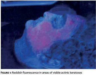
On examination, a reddish fluorescence is observed in the areas corresponding to the actinic keratoses visible on dermatological examination or in subclinical lesions 5 (Figure 1). In BCC lesions, it is possible to delimit the margins of poorly defined lesions, which helps in future surgical procedures.6 The authors describe a clinical case of a patient who underwent PDT for the visualization of the cancerization field and treatment of nodular BCC that developed over a sebaceous nevus.
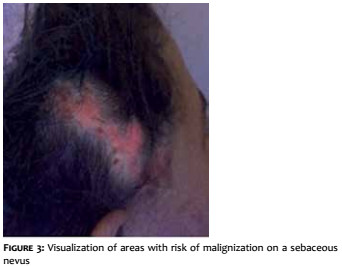
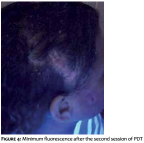
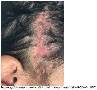
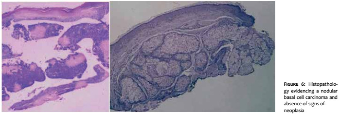
A 28-year-old female patient (Fitzpatrick phototype IV) with history of congenital lesion in the right temporal region sought medical advice. Irritation and pruritus had emerged at the site six months before, with biopsies having been performed in two points: one at the lesion’s upper border – whose anatomopathological study confirmed the presence of a sebaceous nevus – and the other at the lesion’s lower third – which indicated the presence of a nodular BCC, with compromised margins (Figure 2). The proposed treatment comprised two sessions of PDT using MAL cream, with an interval of one week. Curettage of crusty and rough areas of the nevus was performed on the day of the first procedure, with the subsequent application of MAL cream and occlusion with plastic film and aluminum foil for three hours. The dressing and product were removed after that period, when the field cancerization could be visualized through fluorescence with the assistance of a Wood’s lamp (Figure 3). Irradiation with red light was then applied for eight minutes with a diode light emitting equipment whose wavelength ranged between 631 and 637nm, at a fluence of 37 J/cm2. After a one-week interval, the procedure was repeated. Re-examination with Wood’s lamp did not show fluorescence, evidencing the therapeutic effect of the photosensitizing agent in the neoplastic areas or at risk of malignization (Figure 4). A new biopsy was then performed in two points in the lower third of the sebaceous nevus, where a BCC lesion had been identified. Both specimens showed absence of signs of neoplasia (Figure 5). The cutaneous lesion histopathological images obtained before and after the therapy are shown in Figure 6.
The patient remains under clinical follow-up, with no signs of neoplasia recurrence 18 months after the treatment.
Sebaceous nevi have the clinical appearance of yellow or orange plaques with a well-circumscribed verrucous surface.7 Dermoscopic observation reveals isolated or grouped yellow-whitish rounded structures, which correspond to mature superficialized sebaceous glands.8 From the histological perspective, there are a series of abnormalities in the sebaceous and sweat glands, and in hair follicles, which are not well differentiated.2 The lesion’s size increases proportionally to the patient’s age, presenting velvety appearance, small protrusions and verrucous surface at puberty.7 Some neoplasms can arise over sebaceous nevi, especially after puberty: 1 benign tumors (trichoblastoma and syringocystadenoma papilliferum) and malignant tumors, among which BCC is the most common,7 arising in approximately 0.8% of patients with this type of lesion.1 There is no consensus regarding an ideal therapy. Some authors recommend early surgical excision in order to prevent malignant and aesthetically disfiguring transformations. Nevertheless, others advocate a more conservative approach. Future research should identify molecular markers or genetic alterations that might indicate a greater risk of neoplastic transformation, in this manner avoiding unnecessary surgical interventions.1 Lasers and PDT are currently being explored for the treatment of sebaceous nevi, with different degrees of response.2
Photodynamic therapy with MAL has been used in the treatment of superficial or nodular BCC with varying rates of efficacy and recurrence depending on the lesion’s characteristics, namely dimensions, periorificial location and recurrent nature. It is also effective for the mapping of actinic keratoses in the field cancerization.9,10
In this way, in the case in question, it was possible to show: i) the efficacy of PDT-MAL as an alternative to surgery in patients with BCC growth over sebaceous nevi, and ii) the usefulness of the Wood’s lamp as a tool for observing subclinical malignant lesions on hamartomas.
Photodynamic therapy is an effective treatment option for BCC as a non-surgical alternative and also useful for visualizing areas of risk of cancerization in a sebaceous nevus, as observed in the present case.
1. Pereira FB, Cuzzi T. Carcinoma basocelular, estruturas crisálides, oncogenes e nevo sebáceo: algumas considerações. Surg Cosmet Dermatol. 2012;4(1):97-9.
2. Moody MN, Landau JM, Goldberg LH. Nevus sebaceous revisited. Pediatric Dermatology. 2012;29(1): 15-23.
3. Enei ML, Paschoal FM, Valdés G, Valdés R. Carcinoma basocelular aparecendo em um nevo sebáceo de Jadassohn: características dermatoscópicas. An Bras Dermatol. 2012;87(4):640-2.
4. Morton CA, McKenna KE, Rhodes LE. Guidelines for topical photodynamic therapy: update. Br J Dermatol. 2008;159(6):1245-66.
5. Torezan LAR, Festa-Neto C. Cutaneous field cancerization: clinical, histopathological and therapeutic aspects. An Bras Dermatol. 2013;88(5):775-86.
6. Foley P, Freeman M, Menter A, Siller G, El-Azhary RA, Gebauer K, et al. Photodynamic therapy with methyl aminolevulinate for primary nodular basal cell carcinoma: results of two randomized studies. Int J Dermatol. 2009;48(11):1236-45.
7. Kamyab-Hesari K, Seirafi H, Jahan S, Aghazadeh N, Hejazi P, Azizpour A, et al. Nevus sebaceus: a clinicopathological study of 168 cases and review of the literature. Int J Dermatol. 2016;55(2): 193-200.
8. Bruno CB, Cordeiro FN, Soares FES, Takano GHS, Mendes LST. Aspectos dermatoscópicos do siringocistoadenoma papilífero associado a nevo sebáceo. An Bras Dermatol. 2011;86(6):1213-6.
9. Telfer NR, Colver GB, Morton CA; British Association of Dermatologists. Guidelines for the management of basal cell carcinoma. Br J Dermatol. 2008;159(1):35-48.
10. Braathen LR, Morton CA, Basset-Seguin N, Bissonnette R, Gerritsen MJ, Gilaberte Y, et al. Photodynamic therapy for skin field cancerization: an international consensus. International Society for Photodynamic Therapy in Dermatology. J Eur Acad Dermatol Venereol. 2012;26(9): 1063-6.
This study was carried out at the Centro de Referência Nacional em Dermatologia Sanitária Dona Libânia, Fortaleza (CE), Brazil.
 All content the journal, except where identified, under the Creative Commons Attribution 4.0 International licence - ISSN-e 1984-8773


 Read in Portuguese
 Portuguese PDF
 Print
Print
 Send this article by email
 How to cite this article
 Submit a comment
 Mendeley
 Pocket