


Laís de Abreu Mutti1; Marta Regina Machado Mascarenhas1; João Marcos Goes de Paiva1; Solange Pistori Teixeira2; Samira Yarak2
Introduction: Introduction: Not focused ultrasound is a noninvasive alternative to improve body contour.
Methods: It's reported five cases treated with eight weekly sessions of not focused ultrasound on the abdomen.
Results: The treatment did not affect the routine of patients; it was comfortable and safe for most. There was a reduction in the thickness of the fat layer evaluated by ultrasound and in the circumferential measures in all patients, with clinical improvement evidenced in the photographic documentation especially in patients with lesser thickness of subcutaneous.
Conclusion: The device was safe, and was shown clear reduction in abdominal subcutaneous tissue.
Keywords: ADIPOSE TISSUE; ULTRASONIC THERAPY; ULTRASONOGRAPHY; ABDOMINAL SUBCUTANEOUS FAT
Although highly effective, surgical treatment of body remodeling requires anesthesia and a long recovery time.1 Stimulated by an increasing demand for procedures with minimal recovery time and few side effects, several modalities of non-invasive treatment have arisen with the promise to improve the body’s contour.2,3
A significant number of such devices are based on ultrasonic energy – including with focused and non-focused ultrasound devices, depending on how the energy is delivered to the tissues.4 Focused ultrasound devices cause necrosis to fat cells in the treated area. Non-focused ultrasound devices act by altering the permeability of adipocytes, reducing their volume, with absence of cellular necrosis, leading to minimal discomfort.4,6
The MedContour® (General Project, Montespertoli, Italy) is a non-focused ultrasound device that has a handpiece equipped with two non-focused ultrasound angled transducers, aimed at treating adipose tissue (AT) between 1 and 5cm below the skin surface. Non-focused beams can create a weakly focused ultrasound field at the point where the beams overlap. The vacuum’s action pulls the AT into the handpiece, allowing the ultrasonic beams to be directed exclusively to the treated area’s AT, without exposing adjacent structures to risk. This mechanism alters the adipocyte’s plasma membrane’s permeability, releasing intracellular lipids into the interstitial fluid without evidence of cellular apoptosis.1,4,6 The device also has a separate vacuum handpiece for lymph node stimulation and lymph drainage.
According to the scientific literature, non-invasive imaging, carried out with soft tissue ultrasound,7 can evidence the reduction in the subcutaneous
The present study was aimed at describing the effect of transcutaneous, non-focused ultrasound on subcutaneous abdominal tissue of five patients.
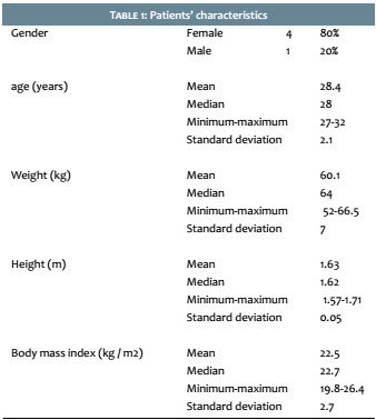
The present paper describes five cases of patients (Table 1) treated with eight 1-hour weekly sessions in the abdominal region (power = 2-3 watts, vacuum = 25mmHg, wave frequency = 1Mhz modulated between 20-50Khz), using the MedContour® (General Project, Montespertoli, Italy) device, from November to December 2014.
The objective and subjective parameters evaluated before and one week after the last session were: a) circumferential measurements b) ultrasound assisted measurements of the abdominal subcutaneous tissue thickness and c) digital photographs taken with a Sony Cyber-shot DSC-W380® digital camera (Sony, Tokyo, Japan). The patients were instructed not to change their eating habits and physical exercise routine.
The treatment was described by patients as comfortable, with only burning sensation being reported when the handpiece was not well coupled to the treatment area. Only one patient reported burning sensation in the abdomen’s lateral regions, which required several pauses for cooling.
There was erythema and heat sensation immediately after the session, which resolved within hours without intervention. Ecchymosis occurred in one patient, with complete remission after two weeks. The treatment did not interfere in the patients’ routine, and there were no reports of other adverse events.
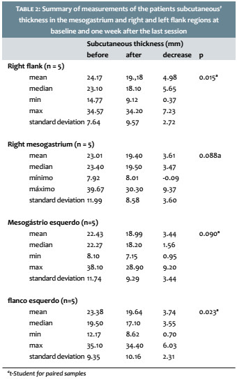
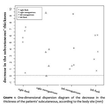
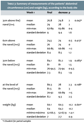
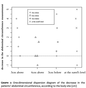
There was a significant reduction in the subcutaneous tissue’s thickness, measured by ultrasonography (Table 2, Graph 1), with a maximum decrease of 9.4 mm. In the circumferential measurements (Table 3, Graph 2), a patient had an increase of the circumference of up to 1cm in two of the measurements (Graph 2), which, however, was not confirmed on ultrasonography (Graph 1).
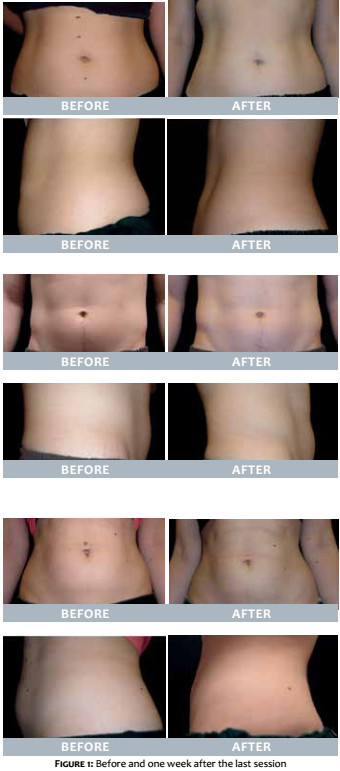
The majority of the patients had a slight clinical improvement (Figures 1).
Although most modalities of noninvasive treatments for the improvement of the body’s contour are safe, there is little scientific proof of the efficacy of the various modalities. In addition, most of the published studies use subjective parameters or circumferential measurements with little standardization.1,8
Ultrasound has been used in studies on cryolipolysis due to the fact that it is capable to objectively evidence the thickness of the subcutaneous tissue.3,9 Using ultrasonography, Coleman et al.9 verified a mean reduction of 20.4% in the subcutaneous tissue that was not correlated with body weight, after two months of treatment. The authors of the present study also observed a reduction of the subcutaneous weight that was not associated with body weight.
All individuals presented circumferential reduction in at least one measurement. Two patients presented a reduction greater than 4cm (Graph 2), a decrease similar to that of the focused ultrasound study.10 The authors of the present study emphasize that, although objective, circumferential measurement is subject to many sources of possible imprecision, such as adequate positioning, greater or lesser compression during measurement, and even interference from breathing.
In a controlled study by Jewell et al.8 with high intensity focused ultrasound, there was a significant reduction of the abdominal circumferential measures. However, 7.6% (9/118) reported severe pain during the procedure, and 22.2% required analgesia before, during and after the procedure. Alterations in the sensitivity for up to six weeks are described in cold-based therapies.9 In contrast, in the present study, tolerability was excellent and there was no need for any analgesia, and absence of alterations in sensitivity. It is worth noting that the mechanism of action of the device in question is the alteration of the adipocyte’s permeability, with absence of apoptosis (as is the case with cryolipolysis) or fat cell necrosis (as is the case with focused ultrasound). In this way, the authors of the present study believe that, in face of the fact that no long term follow up has been carried out aimed at verifying whether the decrease in measurements would be sustained, results may be less durable.
Of all presented measurements, the most reliable was the thickness of the subcutaneous abdominal region taken via ultrasonography. In this evaluation, the five patients experienced reduction, which although millimetric, is significant, especially in lean individuals, since it corresponds to the AT’s thickness and not to the circumferential measurement. As for the clinical photographs, standardization was flawed, which may have interfered with the evaluation of clinical improvement.
The fact that reductions of up to 9mm in the thickness of the subcutaneous tissue were found is promising. Nevertheless, in individuals with great subcutaneous’ thickness, this reduction leads to limited benefits. In these cases, even if there is a 9mm reduction in thickness, most of the AT remains in place. This might justify the limited improvement seen in the photographic records of patients with voluminous abdomens.
The major complication of all these noninvasive technologies used for improving the body’s contour is the patient’s dissatisfaction due to unreal expectation with the outcomes of the procedures.2 In line with this, most patients in the present study were dissatisfied with the final results because they expected better outcomes. Many patients believe they will experience outcomes similar to those obtained with liposuction, entailing that and it is crucial to educate them about what to expect from the treatment.
Patients who desire non-invasive body contouring need to be carefully selected, and the best candidates are those who are likely to accept modest results and those who do not want to undergo surgery.
Cryolipolysis presents robust results, for instance a 30-50% reduction in the thickness of the fat layer.3,9 Although safe, most often there is discomfort during the procedure, ecchymosis, and temporary dysesthesia in up to 20% of patients 9 with risk of paradoxical hypertrophy of the subcutaneous. In addition, outcomes can only be observed after several months.3
A lower cost (there is absence of consumables) and probably faster initial results are some of the advantages of non-focused ultrasound for improving the body’s contour in patients with small localized increases in the AT, as compared to cryolipolysis.
The present study shows that the non-focused ultrasound is able to offer localized reductions of the AT. Studies with larger, randomized and controlled samples are however necessary to better evaluate the percentage of the reduction in the AT. The authors also suggest that other studies should be performed with longer follow-up periods and that ultrasound based subcutaneous measurement be used, in this manner allowing uniformity of methods and better scientific evidence.
1. Atluri P, Barone F, Cervone J, Chavez L, Davis G, DiLaurao M, et al. Clinical effects of noninvasive ultrasound therapy for circumferential reduction. Am J Cosmet Surg. 2012;29(2):114-20.
2. Mulholland RS, Paul MD, Chalfoun C. Noninvasive body contouring with radiofrequency, ultrasound, cryolipolysis, and low-level laser therapy. Clin Plast Surg. 2011;38(3):503-20, vii-iii.
3. Avram MM, Harry RS. Cryolipolysis for subcutaneous fat layer reduction. Lasers Surg Med. 2009;41(10):703-8.
4. Garcia O Jr, Schafer M. The effects of nonfocused external ultrasound on tissue temperature and adipocyte morphology. Aesthet Surg J. 2013;33(1):117-27.
5. Kennedy JE, Ter Haar GR, Cranston D. High intensity focused ultrasound: surgery of the future? Br J Radiol. 2003;76(909):590-9.
6. Bani D, Quattrini Li A, Freschi G, Russo GL. Histological and ultrastructural effects of ultrasound-induced cavitation on human skin adipose tissue. Plastic Reconstr Surg Glob Open. 2013;1(6):e41.
7. Kleinerman R, Whang TB, Bard RL, Marmur ES. Ultrasound in dermatology: principles and applications. J Am Acad Dermatol. 2012;67(3):478-87.
8. Jewell ML, Baxter RA, Cox SE, Donofrio LM, Dover JS, Glogau RG, et al. Randomized sham-controlled trial to evaluate the safety and effectiveness of a high-intensity focused ultrasound device for noninvasive body sculpting. Plastic Reconstr Surg. 2011;128(1):253-62.
9. Coleman SR, Sachdeva K, Egbert BM, Preciado J, Allison J. Clinical efficacy of noninvasive cryolipolysis and its effects on peripheral nerves. Aesthetic Plastic Surg. 2009;33(4):482-8.
10. Ascher B. safety and efficacy of ultrashape contour i treatments to improve the appearance of body contours: multiple treatments in shorter intervals. Aesthet Surg Journal. 2010;30(2):217-24.
The graphs show the distribution of the decrease in the patients’ abdominal circumference measurements between Week 1 and the final experimental timepoint. The points above the dashed line correspond to patients who experienced a decrease in the measurements (positive decrease), and the points below that line correspond to patients who had an increase in the measurements (negative decrease). Dashed line = zero decrease.
This study was carried out at the Escola Paulista de Medicina of the Universidade Federal de São Paulo (EPM / Unifesp) - São Paulo (SP), Brazil.
 All content the journal, except where identified, under the Creative Commons Attribution 4.0 International licence - ISSN-e 1984-8773


 Read in Portuguese
 Portuguese PDF
 Print
Print
 Send this article by email
 How to cite this article
 Submit a comment
 Mendeley
 Pocket