


Aline Soares de Sousa1; Osório Alves Corrêa de Castro Lara2; Ed Wilson Tsuneo Rossoe3
The reconstruction of the eyelid is a considerable challenge for surgeons due to the fact that it is a delicate organ of the utmost importance. The procedure is indicated in defects of congenital origin, post-trauma, or neoplasia. There are many techniques described in the literature. The authors report a case of a female patient bearer of xeroderma pigmentosum with extensive basal cell carcinoma in the lower right eyelid, who underwent tumor resection and reconstruction of the lower eyelid using the modified Fricker technique with excellent functional and aesthetic result.
Keywords: CARCINOMA, BASAL CELL; EYELIDS; SURGICAL FLAPS
The eyelid is a complex structure with an important role in protecting the eyeball against injury and other damage, in addition to being responsible for the secretion, distribution, and drainage of the lacrimal film.1,2 Eyelids can be affected by birth defects, trauma and tumors, and basal cell carcinoma (BCC) - the latter being the most frequent, especially in fair-skinned people and in those exposed to ultraviolet radiation. The incidence of this tumor in the eyelid area has the following distribution: 70% in the lower eyelid, 20% in the medial canthus, 7% in the upper eyelid, and 3% in the lateral canthus. The treatment depends on the size and location of the lesion, and the surgeon's experience.3,4
The present paper is aimed at demonstrating the reconstruction of the lower eyelid using the modified Fricker method, with good aesthetic and functional outcomes in a patient with BCC that affected around 50% of the lower eyelid.
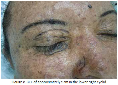
A 48-year-old female patient with a history of xeroderma pigmentosum and multiple resections of BCCs and squamous cell carcinomas. The dermatological examination evidenced a pigmented nodular lesion with elevated and pearly borders, measuring roughly two centimeters in diameter and affecting about 50% of the right lower eyelid. (Figure 1) Once the diagnosis of pigmented BCC was confirmed through an incisional biopsy, a decision was made to resect the tumor and reconstruct the lower eyelid using the modified Fricker method.
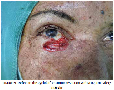
After the surgical marking, antisepsis with polyvinylpyrrolidone-iodine (PVPI) and local anesthesia using the tumescent technique with 0.5% lidocaine, the tumor was resected with 0.5 cm safety margins. (Figure 2)
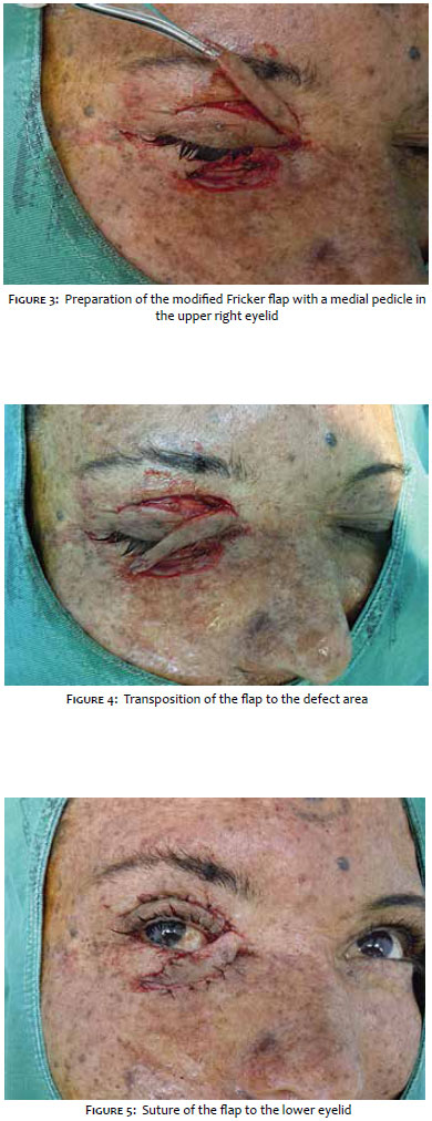
Due to the extent of the defect, the reconstruction of the lower eyelid was performed using the modified Fricker method with a medial pedicle. (Figures 3 to 5)
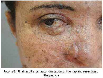
After the autonomization of the flap, the pedicle resection was performed in a second surgical procedure. (Figure 6)
Skin flaps are indicated when it is not possible to carry out a primary closure of the lesion. In the lower eyelid area, direct closure is only possible when the defect affects up to 30% of the eyelid.1 In the present case, the resulting defect covered around 50% of the right lower eyelid, requiring the performance of a skin flap. Several flaps are described for correcting this deformity, and the authors chose the modified Fricker technique.
The Fricker technique was first described in 1829 by Johann Karl Fricke as a temporal flap in the supraorbital area of the frontal region and is used to reconstruct the lower and upper eyelids, and the lateral canthus.4-6 The authors used a myocutaneous transposition flap with a medial pedicle, composed of skin and the pre-septal portion of the orbicularis muscle of the upper eyelid. The pedicle can be medial or lateral to the palpebral aperture, depending on the location of the defect. This type of flap offers a good level of thickness for the reconstructed eyelid and may require a second surgical event for the resection of the pedicle following its autonomization (usually 21 days after the first surgery).
This flap is easy to perform and provides excellent aesthetic results and low complication rates.1
1. Subramanian N. Reconstructions of eyelid defects. Indian J Plast Surg. 2011;44(1):5-13.
2. Peruzzo A, Garcia A, Cutait V, Mendes A, Farah A, Nahas FX. Reconstrução total de pálpebra inferior com retalho frontal. Arquivos Catarinenses de Medicina. 2007;36(Supl 1):175-7.
3. Bekir Atik, MD; Onder Tan, MD; Mehmet Bekerecioglu, MD; Adnan Cinal, MD; Lutfi Tekes, MD. Reconstruction of Lower Eyelid Defects Using a Cross Upper Eyelid Flap Composited with Ear Cartilage. Dermatol Surg. 2007;33(6):709-12.
4. Neto GH, Sebastiá R, Viana GAP, Machado F. Reconstrução palpebral com retalho de Fricke: relato de dois casos. Arq Bras Oftalmol. 2006;69(1):123-6.
5. Gómez JB, Mendoza OZ, Briseño II,Tadeo MTS, Gómez JFB, Frechero NM et al. Total lower-eyelid reconstruction: Modified Fricke's cheek flap. J Plast Reconstr Aesthet Surg. 2011;64(11):1430-5.
6. Martin I. Newman and Henry M. Spinelli. Reconstruction of the eyelids, correction of ptosis, and canthoplasty. In: Thorne CH, Barlett SP, Beasley RW, Aston SJ, Gurtner GC, Spear SL, editors. 6th edition. Grabb and Smith's Plastic Surgery. Philadelphia: Lippincott Williams & Wilkins; 2007. p.397-415.
This study was carried out at Complexo Hospitalar Padre Bento de Guarulhos - Guarulhos (SP), Brazil.
 All content the journal, except where identified, under the Creative Commons Attribution 4.0 International licence - ISSN-e 1984-8773


 Read in Portuguese
 Portuguese PDF
 Print
Print
 Send this article by email
 How to cite this article
 Submit a comment
 Mendeley
 Pocket