


Ana Paula Weber; Giulia Maria dos Santos Goedert; Barbara Brunca; Hamilton Ometto Stolf; Paula Tavares Colpas
Funding: None
Conflict of interest: None
Submitted on: 04/02/2026
Final decision: 05/23/2026
How to cite this article: Weber AP, Goedert GMS, Brunca B, Stolf HO, Colpas PT. Nodular hidradenoma: diagnostic innovation in a rare case. Surg Cosmet Dermatol. 2026;18(2):e20260571.
Nodular hidradenoma is a rare benign adnexal tumor originating from the sweat glands, which clinically presents as a tumor mimicking primary or metastatic malignant lesions. Dermoscopy, as well as clinical characteristics, reveals structural patterns that allow for broad differential diagnoses. Dermatological ultrasonography has become a scientifically recognized diagnostic tool to enable early and accurate diagnosis of hidradenoma, supporting surgical treatment and histopathological analysis. This study reports a rare case of nodular hidradenoma, highlighting the diagnostic innovation achieved through low-frequency ultrasonography, which includes clinical, radiological, and histopathological characteristics.
Keywords: Skin neoplasms; Ultrasonography; Diagnostic imaging; Clinical diagnosis
Nodular hidradenoma is a rare benign adnexal tumor originating from the sweat glands and represents a diagnostic challenge because it may clinically mimic malignant lesions, including basal cell carcinoma and melanoma. It affects predominantly women, with a mean age at presentation of 37 years. It is more commonly found on the scalp, face, and upper limbs and is rare on the lower limbs. It has a low potential for malignant transformation; however, diagnosis may be delayed because of its variety in clinical and morphological patterns, including diverse histopathological and clinical presentations.1,2 Currently, apocrine hidradenomas are the most common subtype and are defined by histopathological examination, since no confirmatory radiologic criteria have been described, contributing to inappropriate therapeutic approaches and a higher likelihood of local recurrence.2 In this context, dermatological ultrasonography, described in a limited number of prospective studies, has emerged as an essential tool to assist in early and accurate diagnosis, enabling appropriate surgical management.3 The present case demonstrates a rare clinical presentation of nodular hidradenoma on the lower limb (thigh), associated with diagnostic investigation using high-frequency cutaneous ultrasonography, identifying the two characteristic ultrasonographic signs first described in 2018, thereby facilitating appropriate surgical treatment and corresponding histopathological evaluation. It is important to report diagnostic innovations confirmed in a rare clinical and surgical dermatologic case.
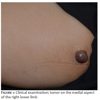
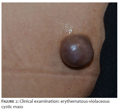
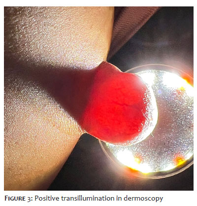
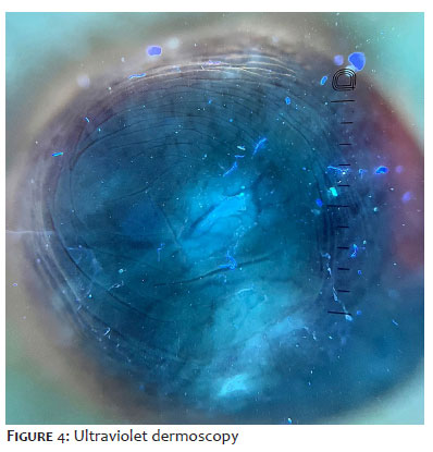
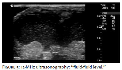
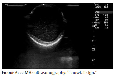
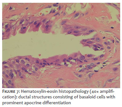
A 50-year-old female patient presented with a violaceous mass located on the medial aspect of the right thigh, first noticed 7 months earlier, with progressive enlargement and impairment of quality of life. The patient denied any previous cutaneous disorders. Dermatological examination revealed a cystic tumor (Figures 1 and 2), slightly infiltrated, with positive transillumination (Figure 3). Ultraviolet dermoscopy showed a central homogeneous pink area and arborizing vessels (Figure 4). Diagnostic investigation was performed using dermatological ultrasonography with a 22-MHz high-frequency linear transducer, revealing a hypoechoic nodular lesion in the dermis and subcutaneous tissue, with the presence of the snow-globe sign, also known as the “snowfall sign” (Figure 5), and a fluid-fluid level (Figure 6), measuring 2.23 × 2.34 cm. The ultrasonographic findings were suggestive of nodular hidradenoma. After combined assessment of the clinical, dermoscopic, and ultrasonographic features, complete excision of the lesion was performed, given its presumed benignancy and low malignant potential. Histopathological examination described a dermal epithelial lesion composed of a densely cellular basaloid proliferation surrounded by fibrosis, with apocrine ductal differentiation, cystic spaces, and pinkish hyaline fluid content (Figure 7), consistent with nodular hidradenoma. The patient remains under follow-up with the clinical and surgical dermatology team, with no signs of local recurrence and satisfied with the treatment provided.
Nodular hidradenoma belongs to the group of benign skin adnexal tumors with a broad range of morphologic differential diagnoses, clinically simulating various primary and metastatic tumors. Clinically, it presents as a solitary nodule with cystic and serous areas, showing a predilection for the scalp and upper limbs. It is exceedingly rare on the lower limbs, as in the present case. Growth is slow and generally asymptomatic. Macroscopically, positive transillumination is explained by serous areas containing fluid illuminated by the polarized light of the dermoscope.1 Dermoscopic findings described in the literature include a homogeneous pinkish area, whitish structures, and a variable vascular pattern, including arborizing and atypical polymorphous vessels, most often mimicking basal cell carcinoma and melanoma.4
Given the clinical diagnostic challenge, high-frequency dermatological ultrasonography, considered an advantageous noninvasive method, was important in establishing the diagnosis through the characteristic findings of nodular hidradenoma, including the “snowfall sign” and the “fluid-fluid level,” both identified in the present case. These findings were first described in 2018 in a case series, highlighting the importance of this imaging method. It is also important to emphasize that there is no consensus regarding diagnostic and therapeutic criteria, further underscoring the importance of ultrasonography.3 Accordingly, diagnostic investigation was essential in determining the surgical approach, with juxtalesional excision representing one of the available options given the low probability of progression to hidradenocarcinoma. In this case, surgery is considered curative, although recurrence following incomplete excision has been reported. Histopathological examination remains the gold standard for diagnostic confirmation, described by a well-circumscribed lesion composed of solid and cystic spaces, allowing identification of ductal structures with basaloid cells.2 Therefore, an accurate diagnosis in this rare case is essential for follow-up and surgical management
We report a rare benign neoplasm in an unusual location, for which ultrasonography was fundamental to diagnosis and surgical planning. However, further scientific studies are still needed to establish ultrasonographic diagnostic criteria.
1. Arthi M, Joseph LD, Arun Kumar K. Clinicopathological profile of nodular hidradenoma: A ten year study in a tertiary care center. J Cutan Aesth Surg. 2023;16(1):34.
2. Nandeesh BN, Rajalakshmi T. A study of histopathologic spectrum of nodular hidradenoma. Am J Dermatopathol. 2012;34(5):461-70.
3. Wortsman X, Reyes C, Ferreira-Wortsman C, Uribe A, Misad C, Gonzalez S. Sonographic characteristics of apocrine nodular hidradenoma of the skin. J Ultra Med. 2017;37(3):793-801.
4. Serrano P, Lallas A, Del Pozo LJ, Karaarslan I, Medina C, Thomas L, et al. Dermoscopy of Nodular Hidradenoma, a Great Masquerader: a morphological study of 28 cases. Dermatol. 2016;232(1):78-82.
Ana Paula Weber
ORCID: 0000-0002-8841-7817
Statistical analysis, Approval of the final version of the manuscript, Conception and design of the study, Preparation and writing of the manuscript, Acquisition, analysis and interpretation of data, Effective participation in the conduct of the study, Intellectual participation in the propaedeutic and/or therapeutic approach to the cases studied, Critical review of the literature, Critical revision of the manuscript.
Giulia Maria dos Santos Goedert
ORCID: 000.0002-6672-0276
Statistical analysis, Approval of the final version of the manuscript, Conception and design of the study, Preparation and writing of the manuscript, Acquisition, analysis and interpretation of data, Effective participation in the conduct of the study, Intellectual participation in the propaedeutic and/or therapeutic approach to the cases studied, Critical review of the literature, Critical revision of the manuscript.
Barbara Brunca
ORCID: 0000-0002-2492-8474
Statistical analysis, Approval of the final version of the manuscript, Conception and design of the study, Preparation and writing of the manuscript, Acquisition, analysis and interpretation of data, Effective participation in the conduct of the study, Intellectual participation in the propaedeutic and/or therapeutic approach to the cases studied, Critical review of the literature, Critical revision of the manuscript.
Hamilton Ometto Stolf
ORCID: 0000-0003-4867-0276
Statistical analysis, Approval of the final version of the manuscript, Conception and design of the study, Preparation and writing of the manuscript, Acquisition, analysis and interpretation of data, Effective participation in the conduct of the study, Intellectual participation in the propaedeutic and/or therapeutic approach to the cases studied, Critical review of the literature, Critical revision of the manuscript.
Paula Tavares Colpas
ORCID: 0000-0002-1389-0749
Statistical analysis, Approval of the final version of the manuscript, Conception and design of the study, Preparation and writing of the manuscript, Acquisition, analysis and interpretation of data, Effective participation in the conduct of the study, Intellectual participation in the propaedeutic and/or therapeutic approach to the cases studied, Critical review of the literature, Critical revision of the manuscript.
 All content the journal, except where identified, under the Creative Commons Attribution 4.0 International licence - ISSN-e 1984-8773




 Read in Portuguese
 Portuguese PDF
 Print
Print
 Send this article by email
 How to cite this article
 Submit a comment
 Mendeley
 Pocket
 Share on Facebook
 Share on Twitter