


Luiz Roberto Dal Bem Pires Júnior1,2; Maira Mitsue Mukai1
Funding: None
Conflict of interest: None
Submitted on: 11/19/2025
Final decision: 4/9/2026
How to cite this article:
Pires Júnior LRDB, Mukai MM. Complications in Dermatologic Surgery: A Case Series Study. Surg Cosmet Dermatol. 2026;18(2):e20260525.
INTRODUCTION: Dermatologic surgery is widely used in the treatment of cutaneous neoplasms, but early and late complications remain underreported despite their functional and aesthetic impact.
OBJECTIVE: To analyze complications in dermatologic surgery and their correlation with the repair technique, anatomical location, and tumor subtype.
METHODS: A retrospective study of 107 lesions from 86 patients who underwent resection of skin tumors under local anesthesia between May 2023 and March 2024. Clinical, histological, and technical variables were assessed. Early complications (≤ 4 weeks) and late complications (1-6 months) were analyzed using SPSS v.29 (p < 0.05).
RESULTS: Mean patient age was 73.3 years, and 57% were women. Most lesions were located on the face (69.2%), and basal cell carcinoma was the most frequent subtype (64.5%). Primary closure was performed in 49.5% of cases, and flaps in 44.9%. Early complications occurred in 42.1%, mainly dehiscence; late complications occurred in 13.1%, predominantly unaesthetic scars. Flaps had higher rates of early (53.7% vs. 30.2%) and late complications (25.9% vs. 1.9%). Lower-limb lesions showed a high complication rate. Early complications strongly correlated with late events (33.3% vs. 2.1%)..
CONCLUSIONS: Complications are frequent and influenced by technique and location. Complex techniques and early events increase the risk of late outcomes.
Keywords: Dermatology; Skin Neoplasms; Surgical Flaps; Medical Oncology; Postoperative Complications
Skin cancer is the most common malignant neoplasm worldwide, arising primarily from cumulative exposure to ultraviolet (UV) radiation. 1-3
Skin cancers are classified into melanoma and non-melanoma skin cancer (NMSC), the latter consisting primarily of basal cell carcinoma (BCC) and cutaneous squamous cell carcinoma (SCC), which together account for the majority of cases.4
Outpatient dermatologic surgery performed under local anesthesia is widely adopted and recognized for its safety and efficacy. 5-8
Multicenter studies have reported complication rates ranging from 1.6% to 12.7%, depending on procedure complexity, anatomical location, and patient risk factors.5-6,9
The most frequent complications include wound dehiscence (3.3%), surgical site infection (2.9%–8.7%), hematoma, and bleeding.5-6,10
Wound dehiscence represents an important complication resulting from the interaction between mechanical factors (such as excessive tension, inadequate closure technique, and inappropriate undermining) and physiological factors, including local vascularization, comorbidities, and characteristics of the excised tumor.6,11,12
Anatomical location plays a key role in determining the risk of complications. The risk of infection and overall complications in the lower limbs is up to three times higher due to reduced tissue perfusion, increased skin tension, and impaired lymphatic drainage.13-15 In contrast, the trunk exhibits the lowest complication rates, benefiting from better vascularization and reduced mobility.5,14 The face, although frequently subjected to complex reconstructions, has intermediate outcomes.10,16
The size of the surgical defect constitutes an independent risk factor for complications. Defects larger than 10 mm are associated with a 1.52-fold higher risk of complications, whereas defects exceeding 10 cm² increase the risk of infection by 3.64-fold.5,13,15
Tumor type also influences the risk of complications, with invasive and in situ SCC carrying a 2.96-fold greater risk of infection compared with other skin neoplasms.13,17,18
Appropriate surgical techniques are essential for minimizing complications. Proper undermining can reduce closure tension by as much as 83%–92%, making it essential in closure under tension.19-22 Deep-layer suturing is responsible for most of the approximation and eversion of wound edges, significantly reducing tension on the epidermal suture and the risk of wound dehiscence.23-26
Patient-related factors also influence surgical outcomes. Advanced age is associated with impaired wound healing due to changes in skin composition and thickness, reduced microcirculation, and compromised immune response.27-32 Immunosuppression and male sex, in turn, are risk factors for surgical site infection.13,33,34
Prolonged operative time (≥ 38 minutes) is also associated with an increased risk of complications, possibly reflecting procedural complexity.5
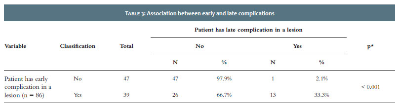
There is a strong correlation between early and late complications. Patients who develop complications in the immediate postoperative period are significantly more likely to develop long-term sequelae, such as hypertrophic scars, retractions, and functional impairment, highlighting the importance of rigorous early management of these complications to prevent their progression into chronic conditions.10,35
Given this context, the present study aimed to analyze postoperative complications, tumor control, functional preservation, and aesthetic outcomes in patients undergoing skin cancer resection, considering different reconstructive techniques and anatomical regions, based on data obtained from the outpatient Dermatology Department of a university hospital.
A retrospective observational study was conducted using medical record data and image analyses from the database of the Dermatology Department of a university hospital. Patients who underwent skin cancer resection followed by reconstruction in a single surgical time, on an outpatient basis and under local anesthesia, between May 10, 2023, and March 27, 2024, were selected.
Patients of all age groups and both sexes were included, provided they had complete surgical records, including details regarding the surgical techniques used and the body areas affected, and attended follow-up evaluations during the 6 months following surgery.
Early complications, such as wound dehiscence, hematoma, infection, necrosis, and bleeding, were monitored during the first 4 weeks after the procedure. Late complications, assessed during the 6-month follow-up period, included abnormal wound healing (hypertrophic, incomplete, or keloid scars), aesthetic alterations (facial or body asymmetry, changes in skin texture, or loss of function), and altered sensitivity.
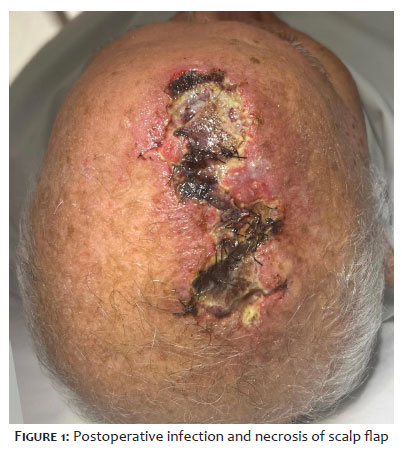
The data were organized in an Excel® spreadsheet and analyzed using IBM SPSS Statistics v.29.0 (Armonk, NY: IBM Corp.). Age data were described using means, standard deviations, medians, minimums, and maximums. Frequencies and percentages were presented for categorical variables. Associations between two categorical variables were assessed using Fisher’s exact test or the chi-square test. A p-value < 0.05 was considered statistically significant (Figure 1).
A total of 107 lesions from 86 patients who underwent outpatient dermatologic surgery for resection of skin neoplasms were analyzed. Most participants (81.4%) had only one lesion, while a single patient had four lesions treated during the study period.
Mean age at the time of surgery was 73.3 years (range: 40–94 years), with a predominance of individuals older than 60 years (91.9%). There was a slight predominance of female patients, accounting for 57% of participants.
Regarding the reconstructive techniques employed, primary closure was the most commonly used, being performed in 49.5% of lesions (53 cases). Skin flaps were used in 44.9% of cases (48 lesions), whereas skin grafts were employed in 3.7% (4 lesions). Two reconstructions involved a combination of techniques, including a flap associated with a graft or healing by secondary intention.
The anatomical distribution of lesions showed a predominance of facial involvement, with 74 cases (69.2%), followed by the trunk (10.3%), hands (5.6%), and lower limbs (4.7%). Other locations included the scalp, upper limbs, and cervical region, each accounting for less than 4% of cases.
From a histopathological perspective, BCC was the most prevalent tumor type, diagnosed in 69 cases (64.5%), followed by SCC in 26 cases (24.3%). Melanoma was identified in 6 cases (5.6%). Collision tumors (BCC + SCC) accounted for 2.8% of cases, while basosquamous carcinoma, sebaceous carcinoma, and eccrine spiradenoma accounted for one case each.
Positive surgical margins were observed in 7.5% of lesions. Among these, two lesions showed simultaneous involvement of both deep and lateral margins, whereas three had isolated lateral margin involvement and three had isolated deep margin involvement.
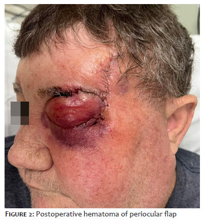
Early complications occurred in 42.1% of lesions (45 cases). Wound dehiscence was the most frequent complication, occurring in 25.2% of cases, followed by hematomas (16.8%) and infections (9.3%). Bleeding was the least frequent complication, occurring in only 3.7% of lesions (Figure 2).
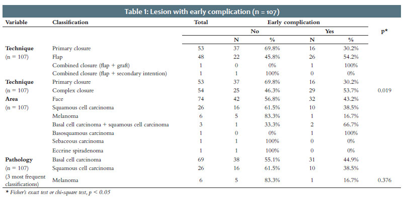
Early complications showed a variable distribution, depending on the surgical technique employed. Flaps presented the highest rate of complications (54.2%), followed by grafts (50.0%), whereas primary closure had the lowest rate (30.2%). When techniques were grouped into primary closures and complex closures (flaps, grafts, and combined techniques), the rate of early complications was 30.2% for primary closures compared with 53.7% for complex closures. This difference was statistically significant (p = 0.019) (Table 1).
Regarding anatomical location, lower limbs exhibited complications in 100% of treated lesions, whereas the trunk showed the lowest rate of early complications (18.2%) (Table 1).
Among tumor subtypes, BCC was most frequently associated with early complications (44.9%), followed by SCC (38.5%) and melanoma (16.7%). However, comparison among these variables did not find a statistically significant association (p = 0.376) (Table 1).
Late complications were less frequent, occurring in 14.0% of lesions (15 cases). Among these, abnormal scarring was the most common outcome (46.7%), followed by aesthetic complications (40.0%) and altered sensitivity (6.7%).
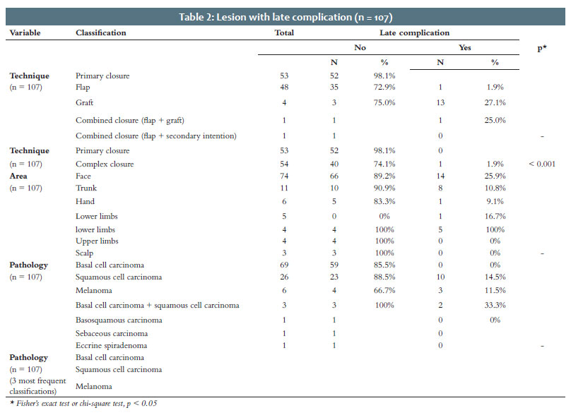
Late complications also showed statistically significant differences among the techniques used (p < 0.001), with rates of 25.9% for complex closures compared with 1.9% for primary closures. Among reconstructive techniques, flaps continued to exhibit the highest prevalence of late complications (27.1%), followed by grafts (25.0%) (Table 2).
Regarding anatomical location, the lower limbs again stood out, with a 100% rate of late complications. In contrast, the scalp, upper limbs, and cervical region showed no late complications (Table 2).
Among tumor subtypes, although the difference was not statistically significant (p = 0.396), melanoma had the highest rate of late complications (33.3%), followed by BCC (14.5%) and SCC (11.5%) (Table 2).
Furthermore, patients who experienced early complications were more likely to develop late complications (33.3%) than those without early complications (2.1%), and this difference was statistically significant (p < 0.001) (Table 3)
This study reinforces the safety and effectiveness of outpatient dermatological procedures for the treatment of skin neoplasms while identifying critical factors associated with the development of postoperative complications.
The demographic profile observed, with 91.9% of patients older than 60 years and a slight female predominance (57%), is consistent with the global epidemiological pattern of skin cancers, which predominantly affect older individuals due to the cumulative effects of sun exposure and the physiological changes associated with aging.1-6
The incidence of immediate postoperative complications was 42.1%, a value considerably higher than the 1.6%–12.7% range reported in large multicenter series.6-8 Wound dehiscence (25.2%) was the most common complication and substantially exceeded the previously reported rates of 0.5%–3.3%.5-7 This discrepancy may be attributed to the greater complexity of the reconstructions performed, the unfavorable anatomical distribution of the lesions, and the specific clinical characteristics of the study population, including advanced age, multiple comorbidities, and limited access to health care.
This complication occurs when there is an imbalance between the mechanical forces applied to the tissue—including excessive tension, suboptimal undermining technique, and inadequate suture method—and the physiological capacity for wound healing, which is influenced by tissue perfusion, associated clinical conditions, and the nature of the excised lesion.4,7,9,10 Layered closure transfers most of the mechanical load to the subdermal layers, significantly relieving tension on the superficial suture line and reducing the risk of wound edge separation.8,10,13,14
Other early complications included hematomas (16.8%), infections (9.3%), and bleeding (3.7%), all occurring at frequencies higher than those usually reported in the literature.5-7,12,14,15
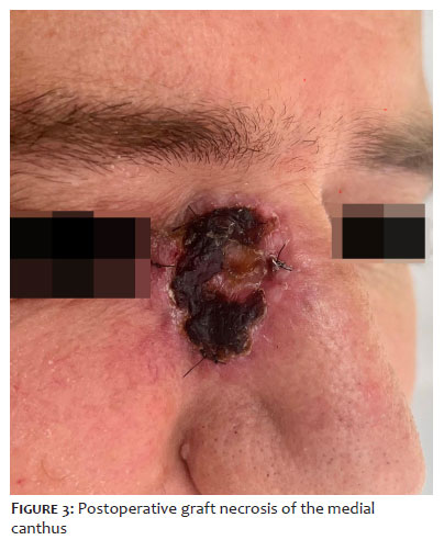
Comparative analysis revealed that complex reconstructions (flaps, grafts, and combined techniques) resulted in a 53.7% rate of early complications, compared with 30.2% for primary closures (p = 0.019).5-7 This disparity reflects the greater tissue trauma, longer operative times, and more demanding technical requirements needed to ensure adequate perfusion and satisfactory functional outcomes (Figure 3).5,6
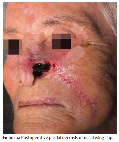
Lesion topography had a substantial influence on outcomes. The lower limbs exhibited complications in 100% of cases, confirming previous reports indicating the risk of infection and other complications is three times higher in this region, attributed to limited blood supply, increased skin tension, and impaired lymphatic drainage.14-16 In contrast, the trunk had the lowest rate of early complications (18.2%).6,15 The facial region exhibited intermediate outcomes, likely benefiting from its rich vascular network (Figure 4).5,17
Late complications occurred in 14% of postoperative cases, predominantly abnormal scarring (46.7%) and aesthetic impairment (40%). Analysis by reconstructive technique revealed a statistically significant difference, with late complication rates of 25.9% in complex reconstructions versus 1.9% in primary closures.5
The robust association between early and late complications represents a finding of particular clinical relevance. Patients who developed early postoperative complications were markedly more likely to experience persistent sequelae (33.3% vs. 2.1%; p < 0.001), underscoring the importance of prompt and rigorous intervention to prevent their progression into chronic conditions.4,5 Events such as wound dehiscence, blood pooling, and infection during the early postoperative period may compromise the healing process, ultimately resulting in permanent sequelae such as hypertrophic scars, tissue contractures, and functional deficits.4,9,10
Once again, the lower limbs ha a 100% rate of late complications, reinforcing the particular vulnerability of this anatomical region.14-16
Although no statistically significant association was identified between tumor subtype and complications, melanoma presents unique challenges. Its wide resections, often performed across skin tension lines, increase the risk of late complications, present in 33.3% of melanoma cases, compared with BCC (14.5% late complications; 44.9% early complications) and SCC (11.5% late complications; 38.5% early complications).
This study has limitations inherent to its retrospective design, single-center setting, and potential variability in technical expertise among surgeons. The limited number of cases involving rare neoplasms or severe complications restricted statistical analyses in specific subgroups. Future prospective multicenter studies with larger sample sizes will be necessary to validate and expand upon these findings
The outcomes of dermatologic surgery for skin cancer result from the interaction of anatomical, technical, and oncological factors. Differences between anatomical sites and the use of complex reconstructions were associated with increased complication rates, whereas early postoperative events were predictive of late complications. Differences among tumor subtypes imposed distinct technical demands and risk profiles. Therefore, the choice of reconstructive technique should consider not only tumor removal but also anatomical factors, tumor subtype, and functional and aesthetic impact.
Luiz Roberto Dal Bem Pires Júnior
ORCID: 0009-0001-6728-420X
Statistical analysis, Approval of the final version of the manuscript, Conception and design of the study, Preparation and writing of the manuscript, Acquisition, analysis and interpretation of data, Effective participation in the conduct of the study, Intellectual participation in the propaedeutic and/or therapeutic approach to the cases studied, Critical review of the literature, Critical revision of the manuscript
Maira Mitsue Mukai
ORCID:0000-0002-4591-8451
Conception and design of the study, Critical review of the literature
1. Zhou L, Zhong Y, Han L, Xie Y, Wan M. Global, regional, and national trends in the burden of melanoma and non-melanoma skin cancer: insights from the Global Burden of Disease Study 1990–2021. Sci Rep. 2025;15.
2. Wang R, Chen Y, Shao X, et al. Burden of skin cancer in older adults from 1990 to 2021 and modelled projection to 2050. JAMA Dermatol. 2025.
3. Arnold M, Singh D, Laversanne M, et al. Global burden of cutaneous melanoma in 2020 and projections to 2040. JAMA Dermatol. 2022;158(5):495–503.
4. Long GV, Swetter SM, Menzies AM, Gershenwald JE, Scolyer RA. Cutaneous melanoma. Lancet. 2023;402(10399):485–502.
5. Kato K, Kato H, Magara T, Morita A. Postoperative complications and risk factors in skin tumor excision with simple suturing under local anesthesia: a multicenter retrospective study in Japan. J Dermatol. 2025;52.
6. Amici JM, Rogues AM, Lasheras A, et al. A prospective study of the incidence of complications associated with dermatological surgery. Br J Dermatol. 2005;153(5):967–71.
7. Alam M, Schaeffer MR, Geisler A, et al. Safety of local intracutaneous lidocaine anesthesia used by dermatologic surgeons for skin cancer excision and postcancer reconstruction: quantification of standard injection volumes and adverse event rates. Dermatol Surg. 2016;42(12):1320–4.
8. Locke MC, Davis JC, Brothers RJ, Love WE. Assessing the outcomes, risks, and costs of local versus general anesthesia: a review with implications for cutaneous surgery. J Am Acad Dermatol. 2018;78(5):983–8.
9. O’Neill JL, Lee YS, Solomon JA, et al. Quantifying and characterizing adverse events in dermatologic surgery. Dermatol Surg. 2013;39(6):872–8.
10. Bordeaux JS, Martires KJ, Goldberg D, et al. Prospective evaluation of dermatologic surgery complications including patients on multiple antiplatelet and anticoagulant medications. J Am Acad Dermatol. 2011;65(3):576–83.
11. Sandy-Hodgetts K, Carville K, Santamaria N, Parsons R, Leslie GD. The Perth Surgical Wound Dehiscence Risk Assessment Tool (PSWDRAT): development and prospective validation in the clinical setting. J Wound Care. 2019;28(6):374–84.
12. Sandy-Hodgetts K, Carville K, Leslie GD. Surgical wound dehiscence: a conceptual framework for patient assessment. J Wound Care. 2018;27(3):119–26.
13. Schlager JG, Patzer K, Wallmichrath J, et al. Surgical site infection in skin surgery: an observational study. Int Wound J. 2023;20(1):85–94.
14. Schlager JG, Ruiz San Jose V, Patzer K, et al. Are specific body sites prone for wound infection after skin surgery? A systematic review and meta-analysis. Dermatol Surg. 2022;48(6):680–9.
15. Liu X, Sprengers M, Nelemans PJ, Mosterd K, Kelleners-Smeets NWJ. Risk factors for surgical site infections in dermatological surgery. Acta Derm Venereol. 2018;98(2):246–50.
16. Mori WS, Demer AM, Mattox AR, Maher IA. Mohs micrographic surgery at challenging anatomical sites. Dermatol Surg. 2019;45(3):401–12.
17. Nakamura Y, Sasaki K, Ishizuki S, et al. Invasive and in situ lesions of squamous cell carcinoma are independent factors for postoperative surgical-site infection after outpatient skin tumor surgery: a retrospective study of 512 patients. J Dermatol. 2021;48(8):1162–9.
18. Heal CF, Buettner PG, Drobetz H. Risk factors for surgical site infection after dermatological surgery. Int J Dermatol. 2012;51(7):796–803.
19. Boyer JD, Zitelli JA, Brodland DG. Undermining in cutaneous surgery. Dermatol Surg. 2000;26(4):327–31.
20. Chen DL, Carlson EO, Fathi R, Brown MR. Undermining and hemostasis. Dermatol Surg. 2015;41(3):e1–e7.
21. Melis P, Noorlander ML, Bos KE. Tension decrease during skin stretching in undermined versus not undermined skin: an experimental study in piglets. Plast Reconstr Surg. 2001;107(5):1201–5.
22. Huang L, Wenzhi L. The role of the undermining during circular excision of secondary intention healing. Am Surg. 2014;80(9):E246–8.
23. Joo JS, Zhuang AR, Tchanque-Fossuo C, et al. Dermal suture only versus layered closure: a randomized, split wound comparative effectiveness trial. J Am Acad Dermatol. 2019;81(3):721–7.
24. Gurusamy KS, Toon CD, Allen VB, Davidson BR. Continuous versus interrupted skin sutures for non-obstetric surgery. Cochrane Database Syst Rev. 2014;(2):CD010365.
25. Luo W, Tao Y, Wang Y, et al. Comparing running vs interrupted sutures for skin closure: a systematic review and meta-analysis. Int Wound J. 2022;19(5):1198–1208.
26. Yang CS, Yeh CH, Chen MY, et al. Mechanical evaluation of the influence of different suture methods on temporal skin healing. Dermatol Surg. 2009;35(2):227–33.
27. Li J, Zhu D, Zhang M, et al. Deciphering age-related differences in wound healing: insights from the interaction between endothelial cells and fibroblasts. Mol Med Rep. 2025.
28. Bentov I, Reed MJ. Anesthesia, microcirculation, and wound repair in aging. Anesthesiology. 2014;120(3):760–72.
29. Brubaker AL, Rendon JL, Ramirez L, Choudhry MA, Kovacs EJ. Reduced neutrophil chemotaxis and infiltration contributes to delayed resolution of cutaneous wound infection with advanced age. J Immunol. 2013;190(4):1746–57.
30. Sgonc R, Gruber J. Age-related aspects of cutaneous wound healing: a mini-review. Gerontology. 2012;59(2):159–64.
31. Dube CT, Ong YHB, Wemyss K, et al. Age-related alterations in macrophage distribution and function are associated with delayed cutaneous wound healing. Front Immunol. 2022;13:821143.
32. Greenhalgh DG. Management of the skin and soft tissue in the geriatric surgical patient. Surg Clin North Am. 2015;95(1):103–14.
33. Schlager JG, Hartmann D, Wallmichrath J, et al. Patient-dependent risk factors for wound infection after skin surgery: a systematic review and meta-analysis. Int Wound J. 2022;19(4):899–909.
34. Schwartzman G, Khachemoune A. Surgical site infection after dermatologic procedures: critical reassessment of risk factors and reappraisal of rates and causes. Am J Clin Dermatol. 2021;22(4):503–13.
35. Sandy-Hodgetts K, Carville K, Leslie GD. Determining risk factors for surgical wound dehiscence: a literature review. Int Wound J. 2015;12(3):265–75.
 All content the journal, except where identified, under the Creative Commons Attribution 4.0 International licence - ISSN-e 1984-8773




 Read in Portuguese
 Portuguese PDF
 Print
Print
 Send this article by email
 How to cite this article
 Submit a comment
 Mendeley
 Pocket
 Share on Facebook
 Share on Twitter