


Tânia Aparecida Meneghel1; Daniella De Grande Curi2
Financial support: None
Conflict of interest: None
How to cite this article: Meneghel TA, Curi DG. Photodynamic therapy with Curcuma longa for the treatment of Kerion celsi. Surg Cosmet Dermatol. 2023;15:e20230184.
Kerion Celsi is an inflammatory variant of tinea capitis and is usually caused by Microsporum canis. Griseofulvin is the gold standard treatment, but FDA approves its use only for children up to two years. Even though it is rare in children younger than three years, tinea capitis can still occur, as in the case of the one-year-old child who was successfully treated with photodynamic therapy combined with curcumin, resulting in total remission.
Keywords: Infant; Curcumin; Photochemotherapy; Tinea capitis.
Dermatophytosis affects the skin, nails, and hair. Its etiological agents are the three main genera of fungi: Microsporum, Trichophyton, and Epidermophyton. Microsporum canis has been reported as the primary agent of tinea capitis in South and Southeast regions of Brazil. The infection is acquired through direct contact with animals or a contaminated person, or through indirect contact with objects or places where fungal spores have been deposited.1
The characteristics of the lesion and the intensity of involvement depend on the interaction between the affected host and the etiologic agent. Exuberant, more inflamed lesions are commonly related to zoophilic species. Kerion celsi is a severe manifestation of tinea capitis, resulting from an intense immune response to the infection caused by the fungus.2
Clinically, tinea capitis presents as an erythematous-scaly plaque with tonsured (fractured) hair. At dermoscopy, there is the presence of comma-shaped and corkscrew hairs. In the most severe cases of Kerion celsi, a plaque of alopecia, with pustules and peripheral adenopathy, may occur.3
The treatment of choice for tinea capitis is systemic therapy, and griseofulvin is the drug of choice, especially when the etiologic agent is Microsporum canis.2
Curcumin, the main active compound in turmeric, is a curcuminoid extracted from the rhizomes of C. longa. Several data in the literature indicate several pharmacological activities for C. longa, proving its anti-inflammatory, antiviral, bactericidal, antioxidant, fungicidal, and anticarcinogenic activities, among others.4 Phenolic compounds, such as curcuminoids, inhibit the production of reactive oxygen species (ROS), protecting the body from damage caused by oxidative stress. C. longa compounds can also interfere with other cellular processes, such as apoptosis activation, platelet aggregation inhibition, inflammatory cytokine production, and cyclooxygenase (COX).5
Curcumin can be used in oil or ointment form and has been described in the treatment of onychomycosis and infected ulcers.
This report aims to demonstrate an innovative method to treat tinea capitis in its most severe form – Kerion celsi – with no oral antimycotics or systemic complications.
A patient of eight-month-old girl, presented an erythematous-scaly plaque on her scalp for six months. Her parents reported that the child had contact with a cat.
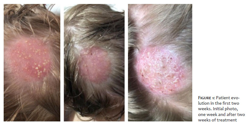
Clinical examination showed an erythematous-infiltrated alopecia plaque with pustules (Figure 1). There was the growth of Microsporum canis in the fungal culture.
The patient was initially treated at another service with ketoconazole shampoo, isoconazole nitrate cream (Icaden®), and isoconazole nitrate combined with diflucortolone valerate (Icacort®) cream. However, her condition worsened. The parents decided to seek a second opinion after the suggestion of starting oral antifungal therapy, as the child weighed only 8 kg, and they preferred to try a topical treatment.
Then, the patient received an application of curcumin 20% in tea tree and clove essential oils applied in equal proportions.
The solution was applied to the site, and occlusion was performed with aluminum foil for 10 minutes with subsequent exposure to blue LED for 10 minutes.
The photodynamic therapy used multiwaves equipment (Empresa Indústria/São Carlos) with blue LED, diode light intensity of 9.0mW/cm2, and energy of 5.40J/cm2.
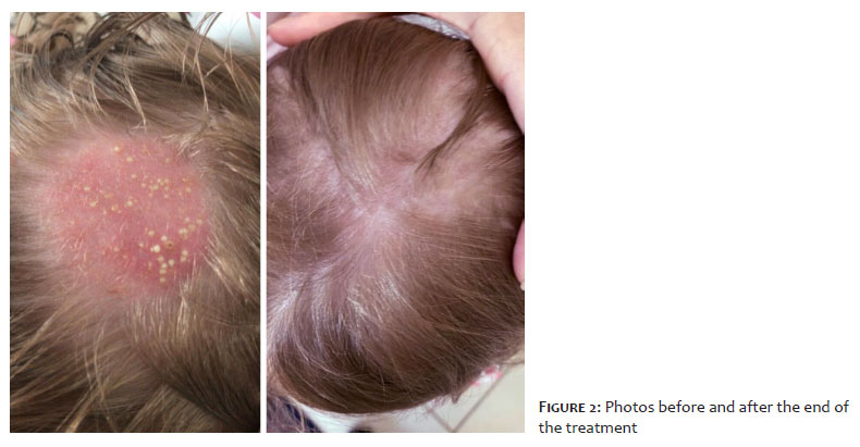
A total of 36 sessions were performed over four months according to the following schedule: first week, three sessions; the second week, four sessions; and, from the third week onwards, five sessions. After five weeks, there was an evident improvement. Thus, therapy was performed once or twice a week until completing 30 sessions (three months of treatment). After the cure, six more sessions were conducted for another month to avoid recurrence. In the last two months, topical amphotericin B once a day was associated with complete resolution of the condition (Figure 2).
Tinea capitis commonly affects children between three and seven years, and it’s rare after puberty or in children younger than one year of age.6ß
The treatment of choice for infections caused by Microsporum spp is griseofulvin, administered for six to 12 weeks, approved by the Food and Drug Administration (FDA) for children over two years of age. Topical treatment should be performed concurrently with oral treatment, such as ketoconazole 2% and ciclopirox olamine shampoo, until mycological cure.7 Terbinafine can be used but seems to be less effective in cases of infection by M. canis.3
Although Gilaberte et al.6 describe the uneventful use of griseofulvin to treat tinea capitis in six children under one year of age, there is an apprehension about using systemic medications in children in this age group. Thus, it is necessary to further research alternative treatments, since non-treatment can lead to definitive alopecia.
Some studies have reported photodynamic therapy (PDT) as a therapeutic option. The mechanism of action is due to the activation of a photosensitizing agent using light, generating reactive oxygen species (ROS) with consequent damage to cellular components.7,8
Brasch et al. demonstrated in 2018 that the association of curcumin with exposure to visible light could inhibit the growth of dermatophytes in vitro.9 Moghadamtousi et al. also reported the antifungal effect of curcumin on dermatophytes.10
There are no in vivo studies on the application of curcumin to treat ß, making our report significant for the development of research on the use of this substance.
The result of photodynamic therapy using curcumin 20% in tea tree and clove essential oils is very promising in cases of tinea capitis, notably the most severe form of Kerion celsi, a disease that until now has oral antimycotics as the gold-standard therapy. However, further studies on its application are necessary, in addition to standardizing concentrations and protocols.
Tânia Aparecida Meneghel 000-003-4670-9800
Data collection, analysis, and interpretation; active participation in research orientation; intellectual participation in propaedeutic and/or therapeutic conduct of studied cases.
Daniella de Grande Curi 000-002-3179-0485
Approval of the final version of the manuscript; preparation and writing of the manuscript; critical literature review; critical revision of the manuscript.
1. Silva CS, Neufeld PM, Gouvêa H, Abreu PA. Etiologia e epidemiologia da tinea capitis: relato de série de casos e revisão da literatura. Rev Bras Anal Clin. 2019;51(1):9-16.
2. John AM, Schwartz RA, Janniger CK. The kerion: an angry tinea capitis. Int J Dermatol. 2018;57(1):3-9.
3. Hay RJ. Tinea capitis: current status. Mycopathologia. 2017;182(1-2):87-93.
4. Araújo CC, Leon LL. Biological activities of Curcuma longa L. Me. Inst Oswaldo Cruz. 2001;96(5):723-8.
5. Balasubramanyam M, Koteswari AA, Kumar RS, Monickaraj SF, Maheswari JU, Mohan V. Curcumin-induced inhibition of cellular reactive oxygen species generation: novel therapeutic implications. J Biosci. 2003;28(6):715-21.
6. Gilaberte Y, Rezusta A, Gil J, Sáenz-Santamaría MC, Coscojuela C, Navarro M, et al. Tinea capitis in infants in their first year of life. Br J Dermatol. 2004;151(4):886-90.
7. Lee Y, Baron ED. Photodynamic therapy: current evidence and applications in dermatology. Semin Cutan Med Surg. 2011;30(4):199-209.
8. Wu MF, Lv T, Wang HW. Successful photodynamic therapy of tinea capitis child with liver dysfunction caused by oral antifungal drugs: a case report. Photodiagnosis Photodyn Ther. 2020;30:101745.
9. Brasch J, Beck-Jendroschek V, Mahn V. Photochemical inhibition of Trichophyton rubrum by different compoundings of curcumin. Mycoses. 2018;61(6):393-9.
10. Moghadamtousi SZ, Kadir HA, Hassandarvish P, Tajik H, Abubakar S, Zandi K. A review on antibacterial, antiviral, and antifungal activity of curcumin. Biomed Res Int. 2014;2014:186864.
 All content the journal, except where identified, under the Creative Commons Attribution 4.0 International licence - ISSN-e 1984-8773


 Read in Portuguese
 Portuguese PDF
 Print
Print
 Send this article by email
 How to cite this article
 Submit a comment
 Mendeley
 Pocket