


Luana Amaral de Moura1; Lucia Martins Diniz1; Robson Dettmann Jarske2; Emilly Neves Souza1; Marcela Scárdua Sabbagh de Azevedo1
Financial support: None
Conflict of interest: None
How to cite this article: Moura LA, Diniz LM, Jarske RD, Souza EN, Azevedo MSS. Chondroid syringoma: unusual presentation of a rare tumor. Surg Cosmet Dermatol. 2022;14:e20220171.
Chondroid syringoma, also known as a cutaneous mixed tumor, is a rare benign neoplasm originating from the sweat glands, composed of epithelial structures immersed in a myxochondroid stroma. It usually presents as a solid, single tumor located on the face or neck with a chronic and asymptomatic course. We report the case of a 75-year-old woman with a slightly elevated lesion on the forehead, whose diagnosis was defined by histopathological analysis.
Keywords: Adenoma, pleomorphic; Eccrine glands; Neoplasms.
Chondroid syringoma is a rare benign adnexal tumor of the skin, composed of epithelial structures immersed in myxochondroid stroma.1 It usually presents as a solid, single, asymptomatic tumor located on the face or neck, with chronic evolution, whose diagnosis is determined by histopathological analysis.2 We report a case of a 75-year-old woman with a slightly elevated lesion on the forehead.
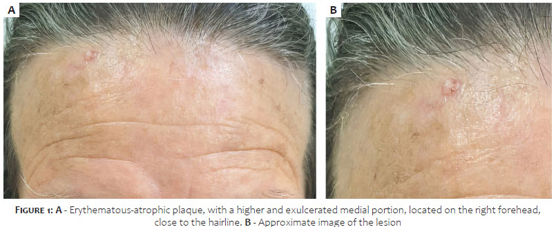
A 75-year-old woman with a history of photodamage and previous excision of three basal cell carcinomas (BCC) reported the appearance of an asymptomatic lesion on her forehead one year ago. On examination, she had an erythematous-atrophic plaque, with a higher and exulcerated medial portion located on the right forehead, close to the hairline (Figure 1).
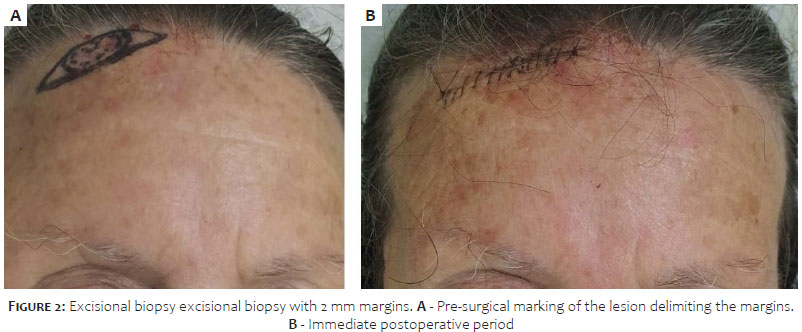
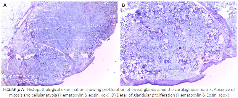
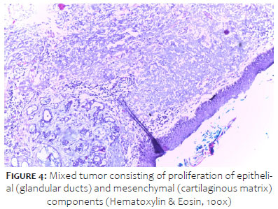
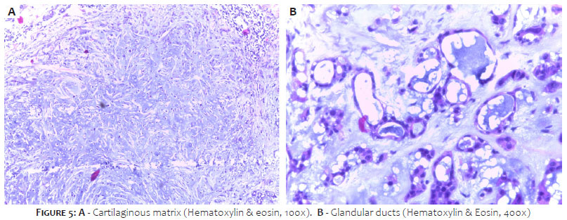
We considered BCC and performed an excisional biopsy with margins of 2 mm (Figure 2). Histopathological analysis showed a tumor characterized by the coexistence of eccrine glands (epithelial structures) and chondroid tissue (mesenchymal), without atypia, with infiltration up to the deep dermis and free resection limits (Figures 3, 4, and 5). Thus, we concluded the diagnosis of chondroid syringoma. The patient is being followed up and presents no clinical signs of recurrence six months after the excision.
Chondroid syringoma (CS), also known as a cutaneous mixed tumor, is a rare benign neoplasm originating from the sweat glands, which is part of the group of adnexal tumors.1,2 It corresponds to about 0.098% of all primary skin neoplasms, and it’s more frequent in men (2:1) between 20 and 60 years, which differs from the case reported.3
Typically, it manifests as a well-defined, firm, mobile, asymptomatic, slow-growing subcutaneous nodule measuring 0.5cm to 3cm.1,3,4 The most common locations are the nose, upper lip, scalp, forehead, chin, and malar region.2 A specific dermoscopic pattern has not yet been described.3,5 The patient reported a slightly elevated plaque on the forehead, differing from the classic picture, although in the usual topography.
Differential diagnoses include epidermoid cyst, dermatofibroma, cylindroma, eccrine poroma, and basal cell carcinoma.1,3
The diagnosis is essentially histopathological due to the non-specificity of the skin lesion. Anatomopathology shows a well-defined tumor in the dermis and/or subcutaneous tissue with overlapping epithelial and mesenchymal components. The epithelial portion includes gland-like structures and cell nests that form ducts and tubules. The mesenchymal element is generally composed of chondromyxoid stroma but focal lipomatous and bone metaplasias have been described.1-3,5
CS is a benign tumor with a good prognosis. However, a de novo malignant variety has been described after incomplete resection and in tumors larger than 3 cm. Histological analysis reveals anomalous features, such as asymmetry, cytological atypia, necrosis, involvement of deep structures, and satellite tumor nodules in these cases.1-3
Surgical excision is the treatment of choice. The literature also describes the use of electrocoagulation, dermabrasion, and CO2 laser.1,3 Clinical follow-up is indicated due to the possibility of local recurrence and malignancy. Wide excision is recommended in case of evidence of malignant transformation. Adjuvant radiotherapy may also be considered.2
The present report emphasizes the importance of considering chondroid syringoma as a differential diagnosis of nodular or flat lesions on the face and cervical region of adult and elderly patients, demonstrating a case of a rare tumor with a presentation that differs from the usual one.
Luana Amaral de Moura 0000-0002-3697-0186
Statistical analysis; approval of the final version of the manuscript; study design and planning; preparation and writing of the manuscript; data collection, analysis, and interpretation; active participation in research orientation; intellectual participation in propaedeutic and/or therapeutic conduct of studied cases; critical literature review; critical revision of the manuscript.
Lucia Martins Diniz 0000-0001-8107-8878
Approval of the final version of the manuscript; study design and planning; preparation and writing of the manuscript; active participation in research orientation; intellectual participation in propaedeutic and/or therapeutic conduct of studied cases; critical revision of the manuscript.
Robson Dettmann Jarske 0000-0003-0519-2032
Approval of the final version of the manuscript; study design and planning; active participation in research orientation; intellectual participation in propaedeutic and/or therapeutic conduct of studied cases.
Emilly Neves Souza 0000-0003-1151-8537
Approval of the final version of the manuscript; study design and planning; preparation and writing of the manuscript; critical literature review; critical revision of the manuscript.
Marcela Scárdua Sabbagh de Azevedo 0000-0002-1664-3217
Approval of the final version of the manuscript; preparation and writing of the manuscript; critical literature review; critical revision of the manuscript.
1. Agarwal R, Kulhria A, Singh K, Agarwal D. Cytodiagnosis of chondroid syringoma -Series of three cases. Diagn Cytopathol. 2021;49(9):E374-E7.
2. Linares González L, Aguayo Carreras P, Rueda Villafranca B, Navarro-Triviño FJ. Chondroid syringoma mimicking basal cell carcinoma. Actas Dermosifiliogr. 2020;111(4):341-3.
3. Vázquez Hernández A, Pérez Campos AE, Gamboa Jiménez TI, Fenton Navarro BF. Giant chondroid syringoma on the upper lip: a case report. Dermatol Online J. 2021;27(5).
4. Purkayastha P, Thomson R, Wilson Jones N, Ng S. Chondroid syringoma: an unusual presentation in a 7-year-old boy. BMJ Case Rep. 2021;14(7):e232943.
5. Palit A, Sethy M, Nayak AK, Ayyanar P, Behera B. Dermoscopic features in a case of chondroid syringoma. Indian J Dermatol Venereol Leprol. 2021;87(1):89-92.
 All content the journal, except where identified, under the Creative Commons Attribution 4.0 International licence - ISSN-e 1984-8773


 Read in Portuguese
 Portuguese PDF
 Print
Print
 Send this article by email
 How to cite this article
 Submit a comment
 Mendeley
 Pocket