


Rossana Cantanhede Farias de Vasconcelos; Leonardo Navroski Durski; Artur Antonio Duarte
Financial support: None
Conflict of interest: None
Submitted on: 19/09/2021
Approved on: 06/05/2022
How to cite this article: Vasconcelos RCF, Durski LN, Duarte AA. Reconstrução de hemiatrofia facial com hidroxiapatita de cálcio: relato de uma técnica segura e minimamente invasiva. Surg Cosmet Dermatol. 2022;14:e20220093
INTRODUCTION: Progressive Facial Hemiatrophy, also known as Parry Romberg Syndrome, is a rare form of linear scleroderma. The management of facial atrophy sequelae is challenging.
OBJECTIVE: This study aims to evaluate for the first time in literature the effectiveness of the calcium hydroxylapatite (CaHa) filler in the jawline reshaping of a patient with Parry Romberg Syndrome.
CASE REPORT: A 15-year-old woman with progressive atrophy of the right side of the face due to Parry Romberg Syndrome. After disease control, the patient’s main complaint was facial asymmetry, mainly in the jawline region.
DISCUSSION: CaHa is a biocompatible injectable filler that is gradually resorbed and replaced by fibrovascular stroma, mainly formed for new collagen, in a process that occurs without any immunological reaction. This safety profile makes CaHa a good choice for correcting stable scleroderma defects.
CONCLUSION: This report concludes that CaHa filler biocompatibility and suitability for the jawline correction are also applicable in reconstructive procedures for stable scleroderma, safely and minimally invasively, with optimal aesthetic results. The method must be programmed case-by-case, and a regular follow-up is also recommended.
Keywords: Scleroderma localized; Facial hemiatrophy; Dermal fillers
Localized scleroderma belongs to the systemic sclerosis complex, a rare autoimmune disease. The extent of cutaneous involvement and extracutaneous manifestations characterize the specific subgroup. Two categories of scleroderma are known: systemic sclerosis, characterized by cutaneous sclerosis and visceral involvement, and localized scleroderma, classically confined to the skin and/or underlying tissues. A worldwide classification for localized scleroderma still does not exist; however, the most commonly used is the Mayo Clinic classification, which includes five groups: 1) plaque morphea, 2) generalized morphea, 3) bullous morphea, 4) linear scleroderma (including linear scleroderma “en coup de sabre” and progressive facial hemiatrophy or Parry Romberg syndrome), and 5) deep morphea.
Progressive facial hemiatrophy, also known as Parry Romberg syndrome, is a rare form of linear scleroderma that usually develops between the first and second decades of life with a slow and self-limiting progression. It is characterized by unilateral atrophy of the skin, subcutaneous tissue, muscles, and bone structures in the area of one or more branches of the trigeminal nerve. Treatment should be started before complications occur. Options are corticosteroids, methotrexate, cyclosporine, and interferon. However, the management of facial atrophy is challenging and must be decided on a case-by-case basis. Most patients benefit from fat grafting and reconstruction with flaps, but new techniques must be incorporated to improve the quality of life of these patients.1
The jaw contour plays an essential role in facial aesthetics. Many studies and techniques seek to improve this area, and calcium hydroxyapatite (CaHa), an injectable filler, appears as an ideal agent to restore the mandible due to its ability to provide immediate and prolonged improvement. In addition to its effectiveness, this agent draws attention to aesthetic and reconstructive procedures for its safety, proven in electron microscopy studies, which demonstrated collagen deposition around the CaHa microspheres with a minimal inflammatory response.2
This study aims to evaluate, for the first time in the literature, the effectiveness of CaHa filling in the remodeling of the mandible of a patient with Parry Romberg syndrome, considering the challenge of treating facial hemiatrophy and the availability of an effective and safe product.
A 15-year-old woman sought medical care due to progressive atrophy of the right side of the face. She had no history of febrile illness or trauma. Her medical history and family history were not significant. On examination, there was facial asymmetry, with atrophy of the skin and underlying tissues of the forehead and jawline, on the right side of the face. No neurological signs were present, and there were no other alterations in the systemic and skin examination.
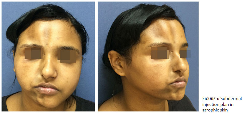
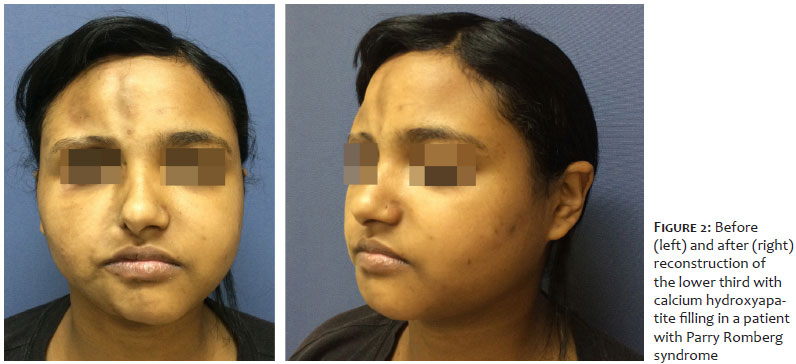
The clinical diagnosis was Parry Romberg syndrome, with prompt initiation of corticosteroids and methotrexate. After less than two years, the patient had all medications discontinued and no sign of disease activity. After two more years of follow-up, at 19 years of age, the patient’s main complaint was facial asymmetry, especially in the mandible and chin region (Figure 1).
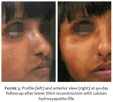
A CaHa filler (CaHA; Radiesse, Merz Pharmaceuticals GmbH, Frankfurt, Germany) was chosen for the reconstruction procedure, considering the patient’s biocompatibility and safety, and the author’s experience. This filler comes in a 1.5 mL syringe, and a correction factor of 1:1 is used, with isotonic saline and 2% lidocaine as diluents. For areas where more projection was intended, such as the chin line, pre-jowl, and mandible, the amount of diluent was only 0.4 ml. A white marker delimited the treated area, and we injected it with a 22-gauge cannula positioned in the subdermal plane to correct the mandible. In the chin area, we addressed the submuscular and anteromuscular planes. The insertion points to treat the jawline were the pre-jowl sulcus and the mandibular; and for chin augmentation, the anterior submental area served as an insertion point. The total volume of filler used was 3.0 cc or two syringes. The distribution of the filler was performed according to the needs of each area, using a fan technique, with injections of 0.2 ml. The aesthetic result was immediate, and no adverse events occurred during or after the procedure, with a 90-day follow-up (Figure 2 and 3).
Parry Romberg syndrome is a rare disease, with an estimated prevalence of 1 in 700,000 people, affecting three times more women than men. The etiology is still unknown, but it is believed that there is autoimmune pathogenesis, possibly triggered by events such as trauma or viral infection, which activate lymphocytes and endothelial cells, followed by excessive collagen synthesis by fibroblasts. The initial treatment is based on immunosuppressants, in line with the autoimmune etiology. After stabilization, the remaining damage caused by cutaneous, fatty, and bone atrophy has a psychosocial impact on the patient and represents a therapeutic challenge for physicians.
In the last analysis of the literature, the most frequent procedure for correction of scleroderma defects was surgery (59%), with autologous fat grafting being the most used (50% of procedures), followed by flaps (24%). Looking for less invasive and painful alternatives, the literature provides reports of correction of facial defects in localized scleroderma with hyaluronic acid, polymethylmethacrylate, and poly-L-lactic acid.
A perfect agent for injection must be non-immunogenic, biocompatible, and stable at the implantation site. Thus, this case report aimed to describe for the first time in the literature the successful use of CaHa in the correction of sequelae of Parry Romberg syndrome.
CaHa is a biocompatible injectable filler composed of microspheres of 25-45 micrometers of CaHa suspended in a carboxymethylcellulose gel. CaHa microspheres have a smooth appearance and are identical in composition to the mineral substance of human bones and teeth. After implantation, the carrier gel is reabsorbed and gradually replaced by fibrovascular stroma, formed mainly by new and organized collagen, generating lasting volumization and improved quality, roughness, and skin pigmentation.
An experimental study using hematoxylin and eosin staining, special staining with picrosirius red (PSR), and immunohistochemistry (IHC) assessed the nature of the newly deposited collagen fibers. PSR and IHC staining confirmed the presence of type I collagen. On the other hand, type III collagen was found in much smaller amounts in the biopsy samples, consistent with its gradual replacement by type I collagen in the remodeling process.3 In normal skin tissues, types I and III collagen are maintained in a relatively fixed ratio to each other, although there is an age-dependent increase in the ratio from type I to type III. During CaHa-induced neocollagenesis, mature type I collagen replaces gradually newly formed type III for optimal structural support and tensile strength.
Morphological evidence from a study using electron microscopy in tissues treated with CaHa filler suggests specific mechanisms involved in structural modifications, both in the filler microspheres and in the connective tissue cells. They demonstrate the absence of any immunological reaction and show that the filler used is very slowly modified over time by the action of connective tissue cells, without any phagocytosis activity.4 This safety profile makes CaHa a good choice for the correction of stable scleroderma sequelae. Our report seeks to support its use and contribute to improving treatment options for these patients.
In 2006, US Food and Drug Administration (FDA) approved injectable CaHa to correct signs of HIV-associated lipodystrophy and volumetric correction of soft tissue. In Europe, CaHa has approval for tissue augmentation, including treatment of nasolabial folds, marionette lines, and jaw contouring. The amount of filler injected varies depending on the location and extent of the restoration, but consensus recommendations guide both esthetic and reconstructive procedures. The consensus recommended a needle or cannula to correct the lower jawline. For cannulas the insertion points are at the mandibular angle or pre-jowl groove. The filler should be placed at the dermohypodermal junction or deep dermis using a fan technique.
In our experience, cannulas are safer, cause less trauma to atrophic skin, and allow a larger volume injection. The aesthetic result is immediate, and the adverse events are mild and transient, such as swelling and occasional bruising.
We conclude that the biocompatibility and technical adequacy of the CaHa filler, already known for the aesthetic correction of the mandible and chin, are also applicable in reconstructive procedures of stable scleroderma sequelae, in a safe and minimally invasive way, with excellent aesthetic results. The procedure should be scheduled on a case-by-case basis, and regular follow-up is also recommended.
Rossana Cantanhede Farias de Vasconcelos 0000-0002-6185-1840
Approval of the final version of the manuscript; study design and planning; preparation and writing of the manuscript; active participation in research orientation; intellectual participation in propaedeutic and/or therapeutic conduct of studied cases; critical literature review; critical revision of the manuscript.
Leonardo Navroski Durski 0000-0002-1566-7372
Statistical analysis; study design and planning; preparation and writing of the manuscript; data collection, analysis, and interpretation; critical literature review; critical revision of the manuscript.
Artur Antonio Duarte 0000-0003-0361-9776
Approval of the final version of the manuscript; study design and planning; active participation in research orientation; intellectual participation in propaedeutic and/or therapeutic conduct of studied cases; critical revision of the manuscript.
1. Marmur ES, Phelps R, Goldberg DJ. Clinical, histologic and electron microscopic findings after injection of a calcium hydroxylapatite filler. J Cosmet Laser Ther. 2004;6(4):223-6.
2. Ayoub, R, Saba, SC. Treatment of linear scleroderma “En coup de Sabre” with single‐ stage autologous fat grafting: a case report and review of the literature. J Cosmet Dermatol. 2021;20(1):285-9.
3. Zerbinati N, Calligaro A. Calcium hydroxylapatite treatment of human skin: evidence of collagen turnover through picrosirius red staining and circularly polarized microscopy. Clin Cosmet Investig Dermatol. 2018;11:29-35.
4. Zerbinati N, D'Este E, Parodi PC, Calligaro A. Microscopic and ultrastructural evidences in human skin following calcium hydroxylapatite filler treatment. Arch Dermatol Res. 2017;309(5):389-96.
 All content the journal, except where identified, under the Creative Commons Attribution 4.0 International licence - ISSN-e 1984-8773


 Read in Portuguese
 Portuguese PDF
 Print
Print
 Send this article by email
 How to cite this article
 Submit a comment
 Mendeley
 Pocket