


Flávia Machado Alves Basilio1; Fabiane Mulinari Brenner1; Betina Werner2
Submitted on: 15/06/2021
Approved on: 05/08/2021
Financial support: None
Conflict of interest: None
How to cite this article: Basilio FMA, Brenner FM, Werner B. Microneedling for female pattern hair loss: case report and histopathological changes. Surg Cosmet Dermatol. 2022;14:e20220074.
Microneedling has traditionally been used to induce collagen formation. Scalp microneedling has been seen to stimulate the capillary cycle and anagen phase, but studies demonstrating histopathological changes after this procedure are lacking. Here we present the case of a 37-year-old woman with a 15-year history of female pattern alopecia, with diffuse hair thinning prominent in the frontoparietal region and hair miniaturization seen in dermoscopy. The patient’s condition remained stable for seven years with use of spironolactone and topical minoxidil. The patient underwent three scalp microneedling sessions at monthly intervals. Histopathological analysis was conducted before the sessions and one month after the last session. Despite slight clinical and dermoscopic improvement, the alopecia pattern remained the same, without significant changes in follicle count after the interventions. Neither inflammation nor fibrous tracts were observed after the procedure. The histopathological analysis is essential to assess the safety of scalp microneedling in the short and long term, investigate signs such as inflammation and fibrosis, and determine the effectiveness of this procedure in treating alopecia. Studies with a more significant number of cases are necessary.
Keywords: Alopecia; Pathology; Treatment outcome
Microneedling has traditionally been used for collagen induction in facial scars and skin rejuvenation. Microwounds induce the healing process and create transdermal microchannels across the stratum corneum, increasing permeability for small molecule drugs. Recently, regeneration of hair follicles and resumption of the anagen phase has also been observed after this therapy.1,2
Hair regeneration is essential for alopecia treatment and in wound healing and skin regeneration. Epidermal stem cells from hair follicles are a significant supplier of keratinocyte proliferation in wounded skin.3 Wound regeneration is delayed when hair follicles are absent and is faster in the skin with anagen than with telogen hairs.3
These findings have led to the use of microneedling to stimulate hair follicle growth in androgenetic alopecia (AGA) and possibly modulate the hair cycle. But so far, no study has documented histopathological changes after scalp microneedling for AGA.
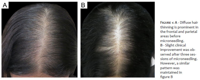
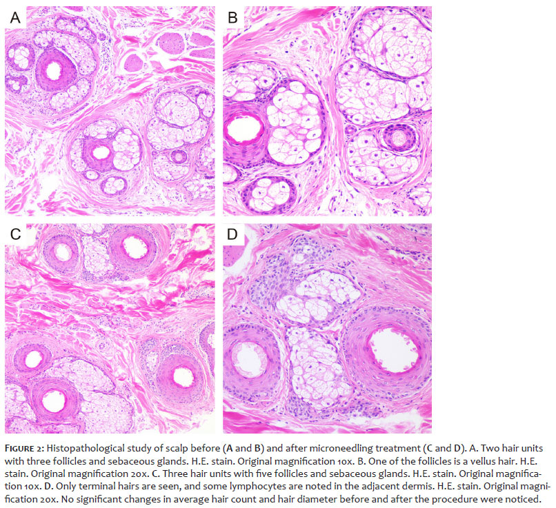
Our 37-year-old female patient had a 15-year history of progressive hair thinning. She also had pernicious anemia and hypothyroidism and received vitamin B12 and levothyroxine. Diffuse hair thinning was prominent in the frontal and parietal areas. Trichoscopy demonstrated miniaturization, an increase in single hair follicular units, and yellow dots in the frontal area (compared to the occipital area). Pull test was negative. Hair thinning was stable after treatment with spironolactone 150 mg/day and topical minoxidil 5% for seven years. However, the patient was unhappy with her hair density. Scalp microneedling with a 2.0 mm roller device was performed at monthly intervals. Slight clinical and dermoscopic improvement was observed after three sessions, as shown in Figures 1A and 1B. We conducted two 4 mm punch biopsies in the central and parietal scalp before the treatment, and another near the first site one month after the third session. Histopathological examination of all samples using transverse sections did not demonstrate significant changes in the follicular count before and after the intervention, and the standard female androgenetic alopecia pattern persisted (low terminal:vellus hair ratio). We observed a slight inflammatory lymphocytic infiltrate in the superficial dermis around the follicles after the microneedling procedures but no significant fibrosis (Figures 2A, 2B, 2C, 2D).
Microneedling is believed to increase hair regrowth in alopecia by increasing platelet-derived growth factor, epidermal growth factors, and bulge activation.1 Overexpression of Wnt proteins has also been seen after microneedling.4 Activation of the Wnt⁄b-catenin signaling pathway is essential to initiate and maintain hair morphogenesis.5 Wnt10b is responsible for proliferation and maintenance of trichogenesis-promoting ability. At the same time, Wnt3a is involved in hair follicle growth and melanocyte homeostasis. In AGA, androgens downregulate secreted factors involved in normal hair follicle stem cell differentiation by inhibiting the Wnt signaling pathway.5 Microneedling has been considered to increase long-term clinical results in AGA. Weekly microneedling associated with daily application of minoxidil 5% topical solution was proven to increase hair growth after 12 weeks. When the procedure was associated with 1 mg finasteride and 5% minoxidil, it demonstrated persistent results after 18 months of follow-up.4,6 However, it is still unknown whether microneedling alone can elicit the desired therapeutic response in AGA.
One concerning adverse event on the scalp is perifollicular fibrosis, which could hinder hair growth.2 In the case reported here, microneedling did not lead to unequivocal clinical or dermoscopic improvement. Slight clinical improvement was not correlated with average hair diameter and hair count on histopathological evaluation. Nevertheless, neither significant inflammation nor scarring tracts were detected after the procedure, in this case disproving the hypothesis that microneedling could induce fibrosis.
Further studies with a more significant number of cases are needed to demonstrate the therapeutic effect of microneedling in AGA. Histopathological assessments as part of these studies can help determine the safety and efficacy of this procedure on the scalp during the short and long terms.
Flávia Machado Alves Basilio 0000-0001-7426-9879
Approval of the final version of the manuscript; study design and planning; preparation and writing of the manuscript; data collection, analysis and interpretation; active participation in research orientation; intellectual participation in propaedeutic and/or therapeutic conduct of studied cases; critical literature review; critical revision of the manuscript.
Fabiane Mulinari Brenner 0000-0001-7970-522X
Approval of the final version of the manuscript; study design and planning; preparation and writing of the manuscript; data collection, analysis and interpretation; active participation in research orientation; intellectual participation in propaedeutic and/or therapeutic conduct of studied cases; critical literature review; critical revision of the manuscript.
Betina Werner 0000-0002-9671-5603
Approval of the final version of the manuscript; study design and planning; preparation and writing of the manuscript; active participation in research orientation; intellectual participation in propaedeutic and/or therapeutic conduct of studied cases; critical revision of the manuscript.
1. Fertig RM, Gamret AC, Cervantes J, Tosti A. Microneedling for the treatment of hair loss? J Eur Acad Dermatol Venereol. 2018;32(4):564-9.
2. Kim YS, Jeong KH, Kim JE, Woo YJ, Kim BJ, Kang H. Repeated microneedle stimulation induces enhanced hair growth in a murine model. Ann Dermatol. 2016;28(5):586-92.
3. Ansell DM, Kloepper JE, Thomason HA, Paus R, Hardman MJ. Exploring the "hair growth-wound healing connection": anagen phase promotes wound re-epithelialization. J Invest Dermatol. 2011;131(2):518-28.
4. Dhurat R, Sukesh M, Avhad G, Dandale A, Pal A, Pund P. A randomized evaluator blinded study of effect of microneedling in androgenetic alopecia: a pilot study. Int J Trichology. 2013;5(1):6-11.
5. Leirós GJ, Attorresi AI, Balañá ME. Hair follicle stem cell differentiation is inhibited through cross-talk between Wnt/β-catenin and androgen signalling in dermal papilla cells from patients with androgenetic alopecia. Br J Dermatol. 2012;166(5):1035-42.
6. Dhurat R, Mathapati S. Response to microneedling treatment in men with androgenetic alopecia who failed to respond to conventional therapy. Indian J Dermatol. 2015;60(3):260-3.
 All content the journal, except where identified, under the Creative Commons Attribution 4.0 International licence - ISSN-e 1984-8773


 Read in Portuguese
 Portuguese PDF
 Print
Print
 Send this article by email
 How to cite this article
 Submit a comment
 Mendeley
 Pocket