


Raquel de Melo Carvalho; Thaiana Botarelli1; Aline Corrêa; Juliana Corrêa Marques da Costa
Received on: 13/06/2020
Approved on: 04/03/2021
Financial support: None
Conflict of interest: None
Acknowledgments: We thank Dr. Juliana Marques-da- Costa for the guidance in this article and for all the knowledge transmitted
Study conducted at the Hospital Naval Marcílio Dias, Rio de Janeiro (RJ), Brazil
Basal cell carcinoma (BCC) is more common in the lower eyelid region when it affects the periorbital region. It occurs predominantly in men between 50-70 years and has a high recurrence rate. Although it rarely metastasizes, orbital invasion can occur. This study aims to present a case of pigmented BCC of unusual location. The location of BCC in the upper eyelid region is rare, and dermoscopy is essential to distinguish it from its primary differential diagnoses.
Keywords: Basal cell carcinoma; Dermoscopy; Eyelid Neoplasms
Basal cell carcinoma (BCC) is a common malignant skin tumor, usually related to intense sun exposure. It accounts for about 90% of malignant eyelid tumors,1,2,3 being more common in the lower lid (over 50%). This tumor occurs predominantly in men, between 60-80 years.4 It presents slow growth, high recurrence rate (between 5% and 15%).4 Also, BCC rarely metastasizes, and orbital invasion can occur in approximately 2%. This study aims to present a case of pigmented BCC in an unusual location.
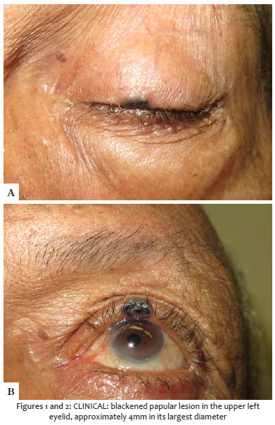
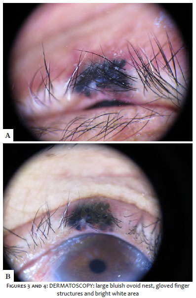
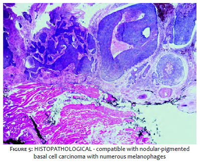
A 77-year-old man with Fitzpatrick skin phototype IV presented a blackened nodular lesion on the left upper eyelid. The lesion was approximately 4 mm (Figures 1 A and B) for six months and had slow and asymptomatic growth. Dermoscopy showed a big bluish ovoid nest, maple-leaf-like structures, and white-shiny area (Figures 2 A and B). Excision was performed, and the histopathology was compatible with nodular-pigmented basal cell carcinoma with numerous melanophages (Figure 3).
The presence of BCC in the upper eyelid region is rare. The pigmented variant is more common in higher phototypes because it has a large concentration of melanin.5
It is important to emphasize that dermoscopy is a helpful tool in identifying pigmented BCC and highly pigmented lesions that often confuse the diagnosis, such as melanoma and melanocytic nevus. The proposed treatments are similar to the BCC located in other areas, such as surgical excision, imiquimod, radiotherapy, and vismodegib.4
It is worth mentioning that as dermatologists, we must always examine the entire patient, including assessing the most difficult areas to access. Dermoscopy in pigmented BCCs may present bluish ovoid nests, maple-leaf-like structures, and white-shiny areas.
Raquel de Melo Carvalho | 0000-0002-3991-4569
Approval of the final version of the manuscript; study design and planning; intellectual participation in propaedeutic and/or therapeutic conduct of studied cases.
Thaiana Botarelli | 0000-0001-7619-7696
Approval of the final version of the manuscript preparation and writing of the manuscript; intellectual participation in propaedeutic and/or therapeutic conduct of studied cases.
Aline Corrêa | 0000-0001-8554-1911
Intellectual participation in propaedeutic and/or therapeutic conduct of studied cases; critical literature review.
Juliana Corrêa Marques da Costa | 0000-0003-3205-7020
Approval of the final version of the manuscript preparation and writing of the manuscript; active participation in research orientation; intellectual participation in propaedeutic and/or therapeutic conduct of studied cases; critical literature review.
1. Saleh GM, Desai P, Collin JR, Ives A, Jones T, Hussain B. Incidence of eyelid basal cell carcinoma in England: 2000-2010. Br J Ophthalmol. 2017;101(2):209-12.
2. Bolognia JL, Jorizzo JL, Schaffer JV. Dermatology. 3rd ed. Philadelphia, PA: Elsevier Saunders; 2012. 2827p.
3. Wu A, Sun MT, Huilgol SC, Madge S, Franzco DS. Histological subtypes of periocular basal cell carcinoma. Clin Experiment Ophthalmol 2014;42:603-7.
4. Shi Y, Jia R, Fan X. Ocular basal cell carcinoma: a brief literature review of clinical diagnosis and treatment. Onco Targets Ther. 2017;10:2483-9.
5. Totir M., Alexandrescu C, Pirvulesco R, Gradinaru S, Costache M. Clinical, histopathological and therapeutical analysis of inferior eyelid basal cell carcinomas. J med life. 2014;7(Spec Iss 4):18-22.
 All content the journal, except where identified, under the Creative Commons Attribution 4.0 International licence - ISSN-e 1984-8773


 Read in Portuguese
 Portuguese PDF
 Print
Print
 Send this article by email
 How to cite this article
 Submit a comment
 Mendeley
 Pocket