


Natacha Quezada Gaón1; Teresa De La Cerda1; Cristian Vera-Kellet1,2
Received on: 01/02/2020
Approved on: 28/02/2020
Financial support: None
Conflict of interests: None
Study conducted at the Department of Cosmiatry, Dermatology Service, Faculty of Medicine, Pontifícia Universidad Católica de Chile, Chile
Systemic sclerosis is a disease of unknown cause, is a rare pathology that, when not treated in time, produces severe damage to the facial aesthetic anatomy causing a significant impact on the quality of life. The case of a 54-year-old woman, with a history of systemic sclerosis of 25 years of evolution, and who was stable from her disease over four years ago. The patient consults for resolution of her facial deformity, which is why she proceeds to perform minimally invasive low-risk procedures such as transcutaneous blepharoplasty and fillings with hyaluronic acid obtaining aesthetically adequate results.
Keywords: Blepharoplasty; Hyaluronic acid; Scleroderma, diffuse
Systemic sclerosis (SS) is a disease of the connective tissue that is characterized by an excessive deposit of collagen and other substances in the extracellular matrix, producing cutaneous sclerosis and damage to the microvasculature of the skin and internal organs.1-4 It predominantly affects women, with a women:men ratio of 4:1.5 The most common symptoms include Raynaud's phenomenon, polyarthralgia, dysphagia, heartburn, edema, skin thickening, and contractures of the fingers.5
There are three phases of dermal involvement: initially, there is an edematous phase, which often presents stiff and swollen hands and fingers; the second phase, called indurative, is characterized by thickening and hardening of the skin (sclerodactyly and the classic expressionless face); and finally, there is an atrophic phase.5,6
SS is classified as limited (LSS), diffuse (DSS), and sine scleroderma (without scleroderma - ssSS). In cases of LSS (CREST syndrome: calcinosis, Raynaud's phenomenon, esophageal dysmotility, sclerodactyly, and telangiectasia), patients develop skin tension on the face and in the distal portion of the elbows and knees. They may also have gastroesophageal reflux disease.6,7 In DSS, there is great diffuse skin involvement; patients present Raynaud's phenomenon and gastrointestinal complications. This type of SS tends to evolve rapidly. Interstitial lung disease and scleroderma renal crisis are the main complications.8-10 In ssSS, patients have antibodies related to SS and visceral manifestations of the disease, but no skin involvement.9
Its incidence is <10 per 1 million per year, and its prevalence is <150 per 1 million in northern Europe and Japan. In the United States, Canada, southern Europe, and Australia it has an incidence of >10 per 1 million per year and prevalence estimates of >150 per 1 million.11
The term scleroderma was introduced by Gintrac in 1847 and arose from the importance of the skin's participation in vascular disease and fibrotic changes.12 Currently, the term scleroderma refers to the skin involvement of patients. Every patient with morphea has scleroderma (cutaneous fibrosis), but not every patient with SS has scleroderma, which is why the term scleroderma should not be used as a synonym for systemic sclerosis.5
The exact pathophysiology of scleroderma is unknown. It is considered to be secondary to an autoimmune reaction that causes localized collagen overproduction. In some cases, it has been linked to exposure to chemicals. Genetic and infectious factors were involved as possible causal agents.5
Facial impairment of systemic sclerosis and morphea is associated with oral complications, and aesthetic changes strongly affect the patient's self-image and quality of life.13-15
We present the case of a 54-year-old woman with a history of hypothyroidism, controlled with levothyroxine 50mg, severe depression, treated with escitalopram 10mg, and a diagnosis of SS with 25 years of evolution. She was being treated with tacrolimus 1.5mg every 12 hours, sirolimus 1.5mg per day, and methylprednisolone 4mg per day.
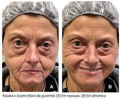
On physical examination, she presented atrophic skin, not very hard, without dyschromia, and also severe atrophy of the malar and zygomatic fats and the cheek area. We also observed, secondary to this loss of facial volume, a great herniation of the lower eyelid fat compartments, and microstomy, resulting in the classic aspect of mask-like appearance in this type of patients (Figure 1).
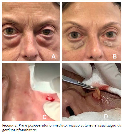
Procedure: Local anesthesia without vasoconstrictor was performed, followed by a transcutaneous incision 0.5cm in the area of greater fat extrusion in the lower eyelid. We removed part of the exuberant fat bags one centimeter below the eyelid, controlling the almost nonexistent bleeding and suturing the lesion with a single stitch using 6-0 monocryl, which was removed after seven days. We decided on this approach through the skin due to the exuberance of the bags, as it was the easiest and least complicated technique, without risk of ectropion (Figure 2).16,17
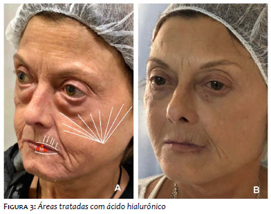
One week later, filling with high G prime or high-density hyaluronic acid (HA) (Voluma, Allergan, Guarulhos, Brazil) was applied with a cannula 21g in a linear retrograde fan pattern in the supraperiosteal plane in the malar, zygomatic, and in Bichat's fat, totaling 4ml of HA per cheek (Figure 3).19-23
Then we apply 1ml of low-density HA (Volbella, Allergan, Guarulhos, Brazil), using a with needle 30G, to perform linear retroinjection in the perioral wrinkles and lip contour. Finally, we applied medium-density HA 1ml (Voliftt, Allergan, Guarulhos, Brazil), with a needle in small points in the lip vermilion to improve the turgor. 24,25
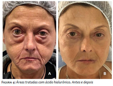
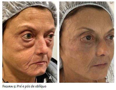
A very suitable aesthetic result was obtained, significantly improving the quality of life of this patient (Figures 4 and 5).
Recent studies cite that injections of HA, a non-sulfated anionic glycosaminoglycan widely distributed in all connective tissues, epithelial and neural, would be a possible treatment for cutaneous fibrosis.18,19 It is believed that hyaluronic acid would be a valid therapy in patients with scleroderma due to its filler properties, in addition to its ability to retain water, smoothing, and moisturizing the skin.20,21 Also, HA has been shown to induce the production of type I collagen in the dermis, which could explain its long-lasting effect.22
Recently, the group led by Guggino included ten women between 18 and 70 years old, with systemic sclerosis.
Each patient was treated with three injections of HA and platelet-rich plasma every 15 to 20 days. All patients were assessed monthly, at three and 24 months after the end of the treatment, regarding mouth's opening, freedom of movement of the lips, and skin elasticity. The group observed that eight patients (80%) showed higher mouth's opening and increased thickness of the upper lip since the first month of follow-up, maintaining these results after two years of initial control.
Another potential filler could be the autologous fat. Autologous fat lipotransfer for soft tissue filling has been described as a well-established aesthetic, and plastic surgical technique.14 Patients with stable scleroderma who have been injected with autologous fat have been reported.13 It is believed that the fat grafting may have tissue regenerative properties, not only serving as a filler, thus postulating that in the fat there could be stem cells, fibroblasts, and endothelial cells that would decrease fibrosis.13,14
There is no consensus in the literature on the application of fillers and minimally invasive surgery in patients with systemic sclerosis. Thus, we suggest and consider that the stability of the systemic condition for more than two years (clinical and laboratory stability) should be sufficient to allow the use of HA or autologous fat in these patients. Note that, before using any filler, you must confirm the stability of the disease, verify and all medications in use by the patient (immunosuppressants, anticoagulants, etc.), and update the laboratory tests (complete blood count, platelet count, hepatitis C and B, HIV, and quantiferon-TB).16
Minimally invasive procedures, such as transcutaneous blepharoplasty and facial filling with HA, can successfully improve the cutaneous cosmetic complications of SS. The appropriate aesthetic result will depend on the experience, technique, and skill of dermatologists and/or plastic surgeons.
Natacha Quezada Gaón | ORCID 0000-0003-2322-3402
Approval of the final version of the manuscript; study design and planning; preparation and writing of the manuscript; intellectual participation in propaedeutic and/or therapeutic conduct of studied cases; active participation in research orientation; critical literature review; critical revision of the manuscript.
Teresa De La Cerda | ORCID 0000-0002-18225-0097
Approval of the final version of the manuscript; study design and planning; active participation in research orientation; critical revision of the manuscript.
Cristian Vera-Kellet | ORCID 0000-0001-8697-9245
Preparation and writing of the manuscript; intellectual participation in propaedeutic and/or therapeutic conduct of studied cases; critical literature review; critical revision of the manuscript.
1. Goldblatt F, O'Neill SG. Clinical aspects of autoimmune rheumatic diseases. Lancet. 2013;382(9894):797-808.
2. Sontheimer RD. Skin manifestations of systemic autoimmune connective tissue disease: diagnostics and therapeutics. Best Pract Res Clin Rheumatol. 2004;18(3):429-62.
3. Knobler R, Moinzadeh P, Hunzelmann N, Kreuter A, Cozzio A, Mouthon L, et al. European Dermatology Forum S1-guideline on the diagnosis and treatment of sclerosing diseases of the skin, Part 1: localized scleroderma, systemic sclerosis and overlap syndromes. J Eur Acad Dermatol Venereol. 2017;31(9):1401-24.
4. Masi AT. Preliminary criteria for the classification of systemic sclerosis (scleroderma). Arthritis Rheum [Internet]. 1980;23(5):581-90.
5. Furue M, Mitoma C, Mitoma H, Tsuji G, Chiba T, Nakahara T, et al. Pathogenesis of systemic sclerosis - current concept and emerging treatments. Immunol Res. 2017;65(4):790-797.
6. Sobolewski P, Maślińska M, Wieczorek M, Łagun Z, Malewska A, Roszkiewicz M, et al. Systemic sclerosis - Multidisciplinary disease: Clinical features and treatment. Reumatologia. 2019;57(4):221-33.
7. Orlandi M, Barsotti S, Lepri G, Codullo V, Di Battista M, Guiducci S, et al. One year in review 2018: Systemic sclerosis. Clin Exp Rheumatol. 2018;36 Suppl 113(4):S3-23.
8. Denton CP, Khanna D. Systemic sclerosis. Lancet. 2017;390(10103):1685-99.
9. Kucharz EJ, Kopeć-Mȩdrek M. Systemic sclerosis sine scleroderma. Adv Clin Exp Med. 2017;26(5):875-80.
10. Muratore M, Quarta L, Raho L, Costanza D, Frisenda S, Calcagnile F, et al. Management of cutaneous discomfort in Patients With Scleroderma : a Clinical Trial. Reumatismo. 2013;65(5):240-7.
11. 1ndréasson K, Saxne T, Bergknut C, Hesselstrand R, Englund M. Prevalence and incidence of systemic sclerosis in southern Sweden: Population-based data with case ascertainment using the 1980 ARA criteria and the proposed ACR-EULAR classification criteria. Ann Rheum Di. 2014;73(10):1788-92.
12. Gintrac M. Note sur la sclerodermie. Rev Med Chir Paris. 1847; 2: 263-81.
13. Gupta K, Bhari N, Verma KK, Gupta S, Urdiales-Gálvez F, Delgado NE, et al. Use of Lipotransfer in Scleroderma. Aesthetic Plast Surg. 2017;43(2):S33-7.
14. Coleman SR. Structural fat grafting: more than a permanent filler. Plast Reconstr Surg. 2006;118(3 Suppl):108S-120S
15. 15. Oh CK, Lee J, Jang BS, Kang YS, Bae YC, Kwon KS, et al. Treatment of atrophies secondary to trilinear scleroderma en coup de sabre by autologous tissue cocktail injection. Dermatol Surg. 2003;29(10):1073-1075.
16. Mateus A PE. Cosmiatria e Láser: pratica no consultorio médico. São Paulo: AC Famacêutica. 2012;104-7.
17. 17. I. GACC. Cirurgia dermatológica. 3 ed. São Paulo: Ed Atheneu. 2017;926-46.
18. 1akhari A, Berkland C. Applications and emerging trends of hyaluronic acid in tissue engineering, as a dermal filler, and in osteoarthritis treatment. Acta Biomater. 2013;9(7):7081-92.
19. 1aurent TC, Fraser JR. Hyaluronan. FASEB J. 1992;6(7):2397-404.
20. Thareja SK, Sadhwani D, Alan Fenske N. En coupe de sabre morphea treated with hyaluronic acid filler. Report of a case and review of the literature. Int J Dermatol. 2015;54(7):823-6.
21. Choksi AN, Orringer JS. Linear morphea-induced atrophy treated with hyaluronic acid filler injections. Dermatol Surg. 2011;37(6):880-3.
22. Wang F, Garza LA, Kang S, Varani J, Orringer JS, Fisher GJ, et al. In vivo stimulation of de novo collagen production caused by cross-linked hyaluronic acid dermal filler injection in photodamaged human skin. Arch Dermatol. 2007;143(2):155-63.
23. Braz A, Sakuma TH. Atlas de anatomia e preenchimento global da face. Rio Janeiro: Guanabara Koogan. 2018;256-326.
24. Ayres EL, Sandoval MHL. Preenchedores. 2 ed. Rio Janeiro: Guanabara Koogan. 2018;70-188.
25. de Maio M1, Swift A, Signorini M, Fagien S; Aesthetic Leaders in Facial Aesthetics Consensus Committee. Facial Assessment and Injection Guide fot Botulinum Toxin and Injectable Hyaluronic Acid Fillers. Plast Recontstructive Surg. 2017;140(3):393e-404e.
 All content the journal, except where identified, is under a Creative Commons Attribution-NonCommercial 4.0 International license - ISSN-e 1984-8773


 Read in Portuguese
 Portuguese PDF
 Print
Print
 Send this article by email
 How to cite this article
 Submit a comment
 Mendeley
 Pocket