


Sérgio Schalka1; Ludmilla Coelho Donato2
Received on: 30/09/2019
Approved on: 14/12/2019
Financial support: Study sponsored by Farmoquímica S/A
Conflict of Interest: The study received financial support from Farmoquímica S/A. Coauthor Ludmilla Coelho Donato is a clinical research analyst at Farmacoquímica S/A
Research performed at Medcin Institu- to da Pele - Osasco (SP), Brazil
INTRODUCTION: Polypodium Leucatomos Extract (PLE) is a recognized topical and oral photoprotective agent that adds biological protection to physical and chemical filters.
OBJECTIVE: To evaluate the efficacy of an SPF 90 sunscreen containing physical and chemical filters, and PLE in reducing sun damage when compared to the same formulation but without the presence of PLE.
METHODS: Ten volunteers were included, each representing 4 areas (non-irradiated skin; irradiated and unprotected skin; irradiated and protected with sunscreen not containing PLE; irradiated and protected with sunscreen containing PLE) exposed to solar radiation. Colorimetric evaluations of erythema and pigmentation were performed. Samples were collected for histopathology.
RESULTS: The area treated with PLE-containing SPF 90 sunscreen, compared to the sunscreen of the same formulation but without the presence of PLE, showed lower intensity of erythema and pigmentation, lower generation of sunburn cells, p53, and MMP-1, and higher CD1-a cell positivity (lower Langerhans cell depletion).
CONCLUSIONS: The association of Polypodium Leucatomos Extract with physical and chemical filters is effective in reducing the damage caused by solar radiation. The presence of EPL in the formulation contributed to the reduction of damage when compared to the formulation without the active.
Keywords: Sunscreens; Polypodium; Solar radiation
Solar radiation is capable of triggering acute or chronic harmful effects to the skin.1 The mechanisms of sun damage result from the absorption of solar energy by different chromophores in the skin, such as melanin, DNA, RNA, proteins, aromatic amino acids such as tyrosine and tryptophan, and urocanic acid, among others. Absorption of solar radiation by the chromophores generates different photochemical reactions and secondary interactions, involving reactive oxygen species that result in harmful effects in the presence of excessive exposure.1
The four principal mechanisms of sun damage are: direct damage to the keratinocyte's DNA, generation of reactive oxygen species (ROS), suppression of innate cell immunity via depletion of Langerhans cells (photoimmunosuppression), and increased melanin production (melanogenesis).1
DNA is one of the principal targets of solar radiation. The pyrimidines undergo photochemical modifications resulting in cyclobutane dimers and other byproducts, which are repaired physiologically by specific enzymes. This system is effective. However, excessive solar exposure can make this repair less efficient.2 The spectrum of solar radiation capable of directly damaging DNA is ultraviolet radiation B (UVB).2
A second protective mechanism against the alterations to nuclear DNA is the apoptotic mechanism, by which damaged cells undergo activation of a self-destructive mechanism (apoptosis), thus avoiding the generation of new damaged cells. The principal gene involved in the apoptotic mechanism resulting from solar radiation is p53.3 The apoptotic cells (also called "sunburn cells") can be observed soon after exposure to UVB radiation.2
When DNA repair or apoptosis mechanisms are not sufficient to avoid generation of new damaged cells, the carcinogenic mechanism is initiated and can result years later in skin cancer lesions, particularly non-melanoma skin cancer.3,4
The photochemical reactions have important effects on the human skin, depending on the wavelength and amount of energy. The natural consequence of photochemical reactions is the generation of reactive oxygen species, highly reactive molecules capable of damaging cellular and extracellular structures such as fibroblasts, collagen, elastin, and glycosaminoglycans. The principal triggering pathway for oxidative alterations in collagen is via proteinases such as matrix metalloproteinases (MMPs), particularly MMP-11.
The epidermis and dermis undergo chemical and histological alterations following persistent solar exposure, which favors the accelerated emergence of wrinkles roughness, dryness, telangiectasias, and irregular pigmentation, all clinical manifestations of photoaging. All ranges of solar radiation are capable of generating ROS: ultraviolet A and B (UVA and UVB), visible light (VL), and infrared (IR)1.
Another cellular effect of solar radiation, particularly of UV radiation, is its capacity to reduce cell immunity, basically by reducing the number and activity of the Langerhans cells, with an impact on the capacity to respond to external agents such as viruses and also internal agents like neoplastic cells. Clinically, the potential effects of photoimmunosuppression include the appearance of infectious dermatoses such as herpes simplex and contribution to the development of neoplastic lesions.5
The skin's pigmentation is a photo-adaptive protective mechanism against the harmful effects of solar radiation and results from the oxidation of melanin in keratinocytes or the production of new melanin by melanocytes. Both mechanisms depend essentially on the individual's phenotypical characteristics (skin type), and time of exposure to solar radiation, with UVA radiation and visible light as the most effective in the production of immediate and persistent pigmentation.6
Regular use of sunscreens (photoprotectors) is considered an essential measure for reducing the harmful effects of solar radiation.7
Photoprotectors act essentially via organic and inorganic filters, molecules or particles capable of reflecting, dispersing, or absorbing the radiation striking the skin surface and thus inhibiting its penetration into the epidermal and dermal layers.8,9
The mechanism by which sunscreens act depends essentially on the formation of a homogeneous film on the skin's surface, and they are thus highly susceptible to insufficient applications in terms of the recommended form, frequency, or quantity.10,11
More recently, active ingredients with biological photoprotective action have been proposed. These active ingredients, rather than interacting directly with the incident radiation, act via biological mechanisms protecting against (or reducing) the radiation's effects on the cell structures and minimizing its harmful effects.1
These agents include Polypodium leucotomos extract, a plant extract resulting from a standardized extraction process from a fern species present especially in Central American countries.12
The indexed international literature includes more than 70 publications on the photoprotective effects of Polypodium leucotomos extract, demonstrating its biological effect via topical or oral use, involving, among other mechanisms, the reduction of cellular DNA damage, antioxidation, protection of Langerhans cells, and reduction of melanogenesis.12,13,14,15,16,17
The current study's objective was to conduct a comparative assessment of the efficacy of a sunscreen containing Polypodium leucotomos extract and another without PLE in the reduction of the effects from acute exposure to solar radiation.
The study was approved by the Institutional Review Board of the University of São Francisco on November 5, 2018.
The study was conducted from December 3, 2018, to February 6, 2019, and included 10 volunteers of both sexes with age ranging from 18 and 70 years of age and Fitzpatrick skin type III.
Other criteria for non-inclusion were determined and assessed, such as active disease, use of any systemic or local medication, prior exposure to solar radiation, and clinical alterations in the area of the dorsum to be irradiated.
The test product was a SPF 90 sunscreen with PLE (Heliocare Max Defense SPF 90), commercially available and duly registered with the Brazilian National Health Surveillance Agency (ANVISA), containing a combination of organic and inorganic filters and standardized 0.5% Polypodium leucotomos extract (PLE).
The comparator product, a SPF 90 sunscreen without PLE, was developed especially for the study, containing the same combination of organic and inorganic filters as the test product, but without PLE.
The study was performed with a solar simulator (Solar Light – USA), adapted to exposure to the full sunlight spectrum (UVB, UVA, visible light, and infrared).
The study methodology with regard to the determination of the minimal erythema dose, irradiation, equipment, amount and form of product's application, and visual reading of the erythema adhered rigorously to ISO 24444:2010,18 considered the reference standard by ANVISA for determination of SPF for registration of sunscreens in Brazil.19
Before the test itself, it was necessary to determine each participant's minimal erythema dose (MED).
For this, an area on the volunteer's back was determined and exposed to UV radiation in six subsites with doses in geometric progression, with a ratio of 1.12, with a resulting variation of 12% between each subsite.
Twenty-four hours later, the participants' MED was defined as the presence of evident erythema with well-defined contours on the site with the lowest dose of ultraviolet radiation (UV).
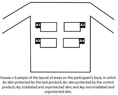
After determining the MED, we demarcated the four areas in the region on the participant's back. Figure 1 shows how the arrangements of the areas were randomized among the participants.
After the areas were demarcated, the products were applied on the respective sites, with 2mg/cm2.
After an average drying time of 15 minutes, the areas were exposed to UV irradiation in six subsites with dosses in geometric progression at a ratio of 1.12, with resulting variation of 12% between each subsite.
Importantly, according to ISO 24444:201018, the irradiation doses in the areas protected by the two investigational products was multiplied by the product's estimated SPF, which was 90 in this particular case. In other words, the irradiation applied to areas A1 and A2 was 90 times higher than area A3. This measure was necessary to eliminate the photoprotective effect (physical and chemical filters) from the analysis.
Twenty-four hours after the exposures, readings were performed of the erythema on the exposed areas, after which colorimetry was performed on all the subsites of all the study areas.
Colorimetry was performed with Chromameter CR-400 (Minolta), and the values for parameters a*, b*, and L* were used to determine the individual typology angle (ITAo).
After the colorimetry, material was collected for anatomical pathology and immunohistochemical tests, with a sample taken in each of the four irradiated areas, in the same subsite, defined as the third subsite. The samples were assessed by a specialized laboratory for the following markers: quantification of sunburn cells (apoptosis of keratinocytes and thus DNA damage), quantification of CD1-A antibody (a marker for Langerhans cells), quantification of p53 antibody (marker for DNA damage), quantification of MMP-1 antibody (marker for matrix metalloproteinases and thus collagen damage), and quantification of tyrosinase antibodies (marker of melanogenesis).
Twenty-one participants were screened, of whom 10 were selected for the study.
Participants' age ranged from 18 to 55 years, with a mean of 32 years.
Individual typology angle is a colorimetric parameter calculated from the variation in colorimetry parameters b* and L*, and determines the skin's pigmentation, according to the following equation:
ITAº = ArcTg [(L – 50)/b] x 180/3,14159
The higher the ITAº, the lighter the skin; thus, in the pigmentation process, a decrease in ITAo represents darkening of the skin.
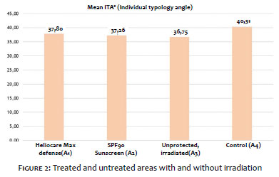
Figure 2 shows the treated and control areas with and without irradiation.
The three irradiated areas, treated or untreated, showed a variation in relation to the non-irradiated area (control). Considering that area 04 (control) did not receive treatment or irradiation, the difference in ITA between the treated areas (A1 and A2) and untreated irradiated area (A3) and the control area (A4) shows how much the skin was pigmented following irradiation, where it was expected that use of the test product (A1) would present a greater protective effect against pigmentation than the SPF 90 sunscreen product (A2), and that both would present less variation than the unprotected area (A3). Thus, the closer to zero, the smaller the difference between the irradiated and control areas, demonstrating less pigmentation of the area.
According to the results, the unprotected irradiated area showed a variation in ITA of 3.56 points in relation to baseline (non-irradiated area), while the areas protected by the test product and the SPF 90 sunscreen varied by 2.51 and 3,04 points, respectively.
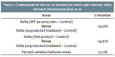
Table 1 shows the percent variation between the deltas of the irradiated areas and the control area without irradiation.
When comparing the area treated with the test product and the unprotected area, the product was 29.67% superior, while the SPF 90 product was 14.56% superior to the untreated area, practically half the response of the product containing PLE, thus demonstrating the efficacy of the latter product's formulation in relation to its comparator in inhibiting skin pigmentation following exposure to solar radiation.
The difference between the intensity of pigmentation in the areas treated with the investigational product and the areas treated with the SPF 90 sunscreen without PLE was 17.70%, where the test product was more effective in protecting against skin pigmentation from solar radiation.
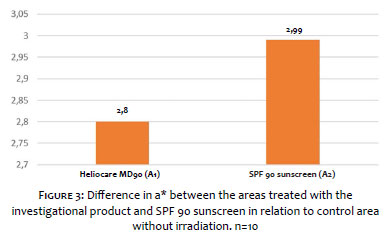
Parameter a* of colorimetry indicates the color spectrum between green (negative) and red (positive). Thus, the smaller the a*, the lower the redness or erythema of the skin.
Considering that area 04 (control) did not receive treatment or irradiation during the study, the difference in a* between the treated areas (A1 and A2) and the control area (A4) demonstrates the amount of erythema produced by the skin, where the investigational product (A1) was expected to present less intense erythema than the SPF 90 sunscreen without PLE (A2).
Figure 3 shows the differences between the areas with the investigational product (A1) and the SPF 90 sunscreen without PLE (A2) and the control area without irradiation (A4).
The difference in the intensity of erythema between the treated areas was 6.79%, that is, Heliocare MD 90 resulted in 6.79% less erythema than the SPF 90 sunscreen without PLE.
Keratinocytes are the skin's principal cell type and thus the main target of alterations mediated by different types of stress.
Solar radiation is capable of triggering morphological changes in the keratinocyte's DNA and at the same time promote a mechanism of cell apoptosis as a protective mechanism. The apoptotic cells are also called "sunburn cells". The keratinocytes that underwent apoptosis were quantified in the four study areas (area protected with the investigational product, area protected with SPF 90 sunscreen without PLE, unprotected irradiated area, and control area).
Skin samples not protected by either of the test products generally displayed more apoptotic keratinocytes per field, while the areas protected with the test products showed fewer apoptotic cells (Figure 4).
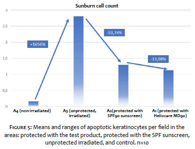
Figure 5 shows the mean numbers of apoptotic keratinocytes per field and the percent variation between the areas.
The unprotected irradiated area showed the highest keratinocyte apoptosis rate, 1,600% greater than the control area (without irradiation and without protection), demonstrating the irradiation's effect on keratinocyte apoptosis.
The SPF 90 sunscreen without PLE led to a 53.74% reduction in the number of apoptotic cells, demonstrating the efficacy of the combination of physical and chemical filters in this effect. Meanwhile, the investigational product led to a 59.79% reduction in the number of apoptotic cells.
The investigational product presented 13.08% fewer apoptotic cells than the other sunscreen with the same SPF, demonstrating greater effectiveness in protection against cellular DNA damage and thus against keratinocyte apoptosis, based on the presence of Polypodium leucotomos extract.
The p53 protein is expressed in mutated keratinocytes as a result of altered nuclear DNA with the generation of cyclobutane pyrimidine dimers (CPD), due to due to the action of UVB radiation. The expression of p53 antibody is thus a marker of cellular DNA damage and consequently of the carcinogenic effect of solar radiation. It was expected that the unprotected irradiated area would have the highest p53 antibody rate, and this was confirmed, as shown in Figure 06.
The unprotected irradiated area showed 426% higher p53 antibody expression than the control area (without irradiation and without sunscreen), demonstrating the effect of irradiation in generating cellular DNA damage and thus p53 expression.
The SPF 90 sunscreen without PLE led to a 69.10% reduction in p53 expression, demonstrating the efficacy of the combination of physical and chemical filters in this effect. Meanwhile, the investigational product led to a reduction of 75.34% in p53 expression.
The investigational product was 20.18% more effective than the comparator product in protecting against the generation of p53 and thus in protecting against carcinogenesis. This effect resulted from the presence of Polypodium leucotomos extract in the product.
CD1a antibody marks the expression of antigen-presenting epidermal dendritic cells, also known as Langerhans cells. Langerhans cells are important elements in innate cell immunity, with microbial antigen-presenting action (viruses and bacteria) as well as tumor cell antigens. Solar radiation is known to greatly reduce the activity or amount of Langerhans cells in the epidermis, triggering photoimmunosuppression and facilitating viral infections (such as herpes simplex) and reducing immune activity in protection against tumor cell clonal expansion.
As mentioned, for this antibody, irradiation would be expected to negatively affect the irradiated skin. We would thus observe a decrease in the number of Langerhans cells marked with the antibody, which our data confirmed.
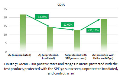
Figure 7 shows the mean amounts of CD1a antibody in the respective areas.
The results showed that exposure to solar radiation led to a 33.89% reduction in CD1a expression, demonstrating a downward effect on Langerhans cells (and thus an immunosuppressive effect) from solar radiation.
Unexpectedly, the SPF 90 sunscreen led to a 12.02% reduction in CD1a expression, showing that the combination of physical and chemical filters was not effective in protecting against photoimmunosuppression. Meanwhile, the investigational product with PLE led to a 33.01% increase in CD1a expression. We thus observed that the investigational product was effective in protecting Langerhans cells from the effects of solar radiation.
Comparing the two sunscreens, we found that Heliocare SPF 90 was 51.18% more effective in CD1a expression (and thus in Langerhans cell expression) than the product with the same formulation and same SPF, but without Polypodium leucotomos extract.
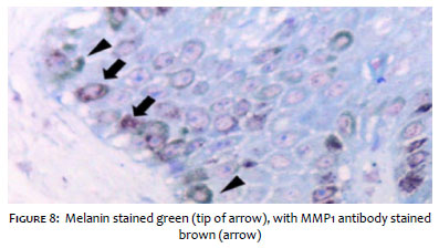
The samples were marked for matrix metalloproteinase-1 (MMP-1), counterstaining the samples with Giemsa. This meant that all the melanin in the keratinocytes' cytoplasm was stained green, while the MMP-1 produced by them was stained brown (Figure 08).
MMP-1 can be considered a marker for stimulus of collagen damage triggered by oxidative stress factors, in this case resulting from solar radiation. Solar radiation is known to promote oxidative stress, triggering increased expression of metalloproteinases, including MMP-1. These in turn are known enzymes leading to collagen breakdown and resulting in trophic changes to the dermis, leading to long- term clinical evidence of skin aging, such as wrinkles and skin laxity.
As expected, in the great majority of cases, the areas protected with the test products presented little or no positivity for MMP-1. Meanwhile, the unprotected irradiated area showed high positivity for this antibody.
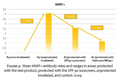
Figure 9 is a graph with the means and ranges of positive staining for MMP-1 antibody in the areas protected with Heliocare Max Defense SPF 90, protected with SPF 90 sunscreen, unprotected and irradiated, and control areas.
The unprotected irradiated area tested 100% greater positivity for the MMP-1 marker compared to the control area (without irradiation and without protection), demonstrating the effect of solar radiation in generating matrix metalloproteinase 1.
The SPF 90 sunscreen led to a 36.92% reduction in MMP-1 expression, demonstrating the partial efficacy of the combination of physical and chemical filters. Meanwhile, the investigational product led to a 70.90% reduction in MMP-1 expression.
Comparing the products, the investigational product was 53.88% more effective than the SPF 90 sunscreen without PLE in reducing MMP-1 expression, demonstrating greater capacity to protect the skin from the degenerative effects of solar radiation on collagen, based on the presence of Polypodium leucotomos extract.
The enzyme tyrosinase participates in the melanogenesis cascade, transforming tyrosine into melanin at the end of the process.
Solar radiation is known to be capable of stimulating melanogenesis, with increased tyrosinase activity. Meanwhile, sunscreens with anti-melanogenesis action should be capable of inhibiting tyrosinase activity, even partially.
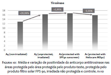
Figure 10 shows the mean positive staining rates for tyrosinase antibody in areas protected with the investigational product, with the SPF 90 sunscreen, unprotected and irradiated, and controls.
The results showed that exposure to solar radiation triggered a 26.06% increase in tyrosinase expression, demonstrating a pigmenting effect from solar radiation, as expected.
Unexpectedly, the SPF 90 sunscreen led to a 2.95% increase in tyrosinase, which demonstrates the lack of protective effect against pigmentation from the combination of physical and chemical filters. Meanwhile, the investigational product led to a 2.77% reduction in p53 expression.
We found the test product to be more effective in protecting against melanogenesis when compared to the SPF 90 sunscreen without Polypodium leucotomos extract, although the difference between the groups was slight, only 5.55%.
Polypodium leucotomos extract is a known photoprotective agent with oral and/or topical use, based on its antioxidant and immunoprotective effects and protection of cellular DNA and against the collagen-degrading effect of solar radiation.12 The scientific literature features numerous publications demonstrating its effect, based on in vitro and in vivo studies, particularly with its oral use.
As for the topical form, some studies have demonstrated PLE's action, protecting keratinocyte DNA and Langerhans cells and the action in matrix metalloproteinases on collagen breakdown.13-16
The current study's innovative aim was to demonstrate the topical action of Polypodium leucotomos extract in photoprotection, in combination with physical and chemical filters in a finished formulation with SPF 90 sunscreen.
To distinguish the action of PLE itself, the study opted to make a comparison with the same formulation but without the PLE. Thus, the changes found in the products' results (investigational versus comparator) would result exclusively from the presence of PLE in the formula.
The selected study model was clinical, in volunteers submitted to solar radiation through a solar simulator capable of emitting radiation in the UV, visible light, infrared ranges. Volunteers were exposed to increasing doses of solar radiation in a study model similar to that for the determination of SPF, including the premises of the internationally accepted method, namely ISO 24444: 2010.18
The colorimetric results were consistent, and as expected, they showed that solar radiation was capable of producing erythema and pigmentation. The use of a SPF 90 sunscreen succeeded in reducing the intensity of erythema and pigmentation, also as expected. When a sunscreen was used that contained both physical and chemical filters and Polypodium leucotomos extract in its formulation, the results in terms of reduction of erythema and pigmentation were even more evident, thus providing an unprecedented demonstration of the extract's efficacy, when associated with physical and chemical filters, reducing the two main biological events involved in acute sun damage: erythema and pigmentation.
The results of the histological and immunohistochemical markers showed that in all the situations (DNA, immunosuppression, breakdown of collagen and dermal elements due to MMP-1, and melanogenesis stimulation) the entire spectrum of solar radiation (ultraviolet A and B, visible light, and infrared) was capable of producing the biological effects. This finding shows the adequate selection of markers for the study.
When we protected the volunteers' skin with SPF 90 sunscreen without Polypodium leucotomos extract, we found that the physical and chemical filters were partially effective in protecting against DNA damage and damage to dermal structures, but that they were not effective in protecting against immunosuppression or melanogenesis. These findings may be explained by the role of long UVA radiation, visible light, and infrared in these effects, particularly in melanogenesis, not adequately protected by ultraviolet filters.
Finally, when assessing the response in the areas protected by the investigational product, a formulation containing a combination of physical and chemical filters and Polypodium leucotomos extract, we found this formulation to be effective in reducing all the histological markers, with superior results those in the area treated only with the combination of physical and chemical filters.
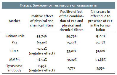
Table 2 provides a didactic demonstration of variations in the histopathological results that can be attributed to the set of UV filters, PLE, or the combination of the two.
As we can see, the most relevant data are seen in the comparison of the results in the investigational and comparator products. The differences found in the comparison result exclusively from the presence of Polypodium leucotomos extract, since it was the only ingredient in the investigational product that was not present in the comparator product.
Analysis of the individual markers highlighted the photoimmunoprotective effect (with a 51.18% proportional increase in Langerhans cells when compared to the SPF 90 sunscreen) and protection against damage to collagen structures, evidenced by the 53.88% greater reduction from the investigational product when compared to the same formulation without PLE.
In relation to p53 and sunburn cells, markers of DNA damage, the investigational product showed a better response than the comparator SPF 90 sunscreen (20.18% and 13.08%, respectively).
Finally, as for tyrosinase inhibition, the two products' responses were virtually similar, with a slight variation in favor of the investigational product. This may be due to the less acute nature of the skin pigmentation process, which depends on successive exposures to solar radiation, which was not the case in this study. The results demonstrate that Polypodium leucotomos extract was able to add several positive effects to the sunscreen formulation consisting only of ultraviolet filters, thus indicating the investigational ingredient's photoprotective biological effect.
This pilot study succeeded in showing a positive effect from the combination of physical and chemical filters and Polypodium leucotomos extract in the reduction of clinical effects (erythema and pigmentation) and biological effects (DNA damage, collagen breakdown, and immunosuppression) resulting from exposure to solar radiation.
Although these data are preliminary, considering the small sample of volunteers, they indicate biological action by Polypodium leucotomos extract when added to the combination of physical and chemical filters, enhancing the filters' effect and contributing to more complete, effective photoprotection.
Sérgio Schalka | 0000-0003-2425-7962
Principal investigator, conception and execution of the study; writing of the report.
Ludmilla Coelho Donato | 0000-0003-1838-8464
Conception and planning of the study, critical revision of the article.
1. Schalka S, Steiner D, Ravelli FN, Steiner T, Terena AC, Marçon CR, et al. Brazilian consensus on photoprotection. An Bras Dermatol. 2014; 89(6 suppl 1):1-74.
2. Sheehan JM, Cragg N, Chadwick CA, Potten CS, Young AR. Repeated ultraviolet exposure affords the same protection against DNA photodamage and erythema in human skin types II and IV but is associated with faster DNA repair in skin type IV. J Invest Dermatol. 2002;118(5):825-9.
3. Hussein MR. Ultraviolet radiation and skin cancer: molecular mechanisms. J Cutan Pathol. 2005;32(3):191-205.
4. Chen AC, Halliday GM, Damian DL. Non-melanoma skin cancer: carcinogenesis and chemoprevention. Pathology. 2013;45(3):331-41.
5. Dumay O, Karam A, Vian L, Moyal D, Hourseau C, Stoebner P, et al. Ultraviolet AI exposure of human skin results in Langerhans cell depletion and reduction of epidermal antigen-presenting cell function: partial protection by a broad-spectrum sunscreen. Br J Dermatol. 2001;144(6):1161-8.
6. Sklar LR, Almutawa F, Lim HW, Hamzavi I. Effects of ultraviolet radiation, visible light, and infrared radiation on erythema and pigmentation: a review. Photochem Photobiol Sci. 2013;12(1):54-64.
7. Seité S, Fourtanier AM. The benefit of daily photoprotection. J Am Acad Dermatol. 2008;58(5 Suppl 2):S160-6.
8. Shaat NA. The chemistry of ultraviolet filters In: Shaat NA. Sunscreens: regulation and commercial development. 3rd ed. Boca Raton: Taylor and Francis; 2005. p. 217-39.
9. Schlossman D, Sho Y. Inorganic ultraviolet filters In: Shaat NA. Sunscreens: regulation and commercial development. 3rd ed. Boca Raton: Taylor and Francis; 2005. p. 239-81.
10. Schalka S , dos Reis VM , Cucé LC. The influence of the amount of sunscreen applied and its sun protection factor (SPF): evaluation of two sunscreens including the same ingredients at different concentrations. Photodermatol Photoimmunol Photomed. 2009;25(4):175-80.
11. Schalka S, Reis VMS. Sun protection factor: meaning and controversies. An Bras Dermatol. 2011;86(3):507-15.
12. Winkelmann RR, Rosso JD, Rigel DS. Polypodium Leucotomos Extract: A Status Report on Clinical Efficacy and Safety. J Drug Dermatol. 2015;14(3):254-61.
13. González S, Pathak MA, Cuevas J, Villarrubia VG, Fitzpatrick TB. Topical or oral administration with an extract of Polypodium leucotomos prevents acute sunburn and psoralen-induced phototoxic reactions as well as depletion of Langerhans cells in human skin. Photodermatol Photoimmunol Photomed. 1997;13(1-2):50-60
14. Torricelli P, Fini M, Fanti PA, Dika E, Milani M. Protective effects of Polypodium leucotomos extract against UVB-induced damage in a model of reconstructed human epidermis. Photodermatol Photoimmunol Photomed. 2017;33(3):156-63.
15. Gonzalez S, Brieva A, Ramírez A, Domínguez M, Guerrero A, Mascaraque M, et al. Fernblock FC® inhibits the production of metalloproteinase-1. 13º Congresso de Primavera da Academia Européia de Dermatologia e venerealogia (EADV) (pôster). Grécia. Atenas; 2016.
16. Schalka S, Silva PVF, Canale C, Sufi B. Fotoprotetor com Polypodium leucotomos protege contra os efeitos da luz visível e infravermelho. 30º Congresso Brasileiro de Cirurgia Dermatológica (pôster). Brasil. Belo Horizonte; 2018.
17. Schalka S, Vitale-Villarejo MA, Agelune CM, Bombarda PCP. The benefits of using a compound containing Polypodium leucatomos extract for reducing erythema and pigmentation resulting from ultraviolet radiation. Surg Cosmet Dermatol. 2014;6(4):344-8.
18. International Standards Organization [Internet]. Cosmetics - Sun protection test methods - Determination of sunscreen Sun Protection Factor. ISO 24444 2010). [cited 2019 Dec 27]. Available from: https://www.iso.org/standard/46523.html
19. Agência Nacional de Vigilância Sanitária [Internet]. Resolução - RDC Nº 30 de 1º de Junho de 2012. [acesso em 27 Dez 2019]. Disponível em: rdc%2030-2012%20-%20protetores%20solares%20em%20cosmticos.pdf
 All content the journal, except where identified, under the Creative Commons Attribution 4.0 International licence - ISSN-e 1984-8773


 Read in Portuguese
 Portuguese PDF
 Print
Print
 Send this article by email
 How to cite this article
 Submit a comment
 Mendeley
 Pocket