


Cristina Diniz Borges Figueira de Mello1; Nilton Di Chiacchio2
Received on: 15/08/2019
Approved on: 02/09/2019
Study conducted at Hospital das Clínicas da Universidade Estadual de Campinas - Campinas (SP), Brazil, and at Hospital do Servidor Público Municipal de São Paulo - São Paulo (SP), Brazil.
Financial support: None.
Conflito de Interesses: None.
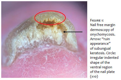
Onychomycosis is the nail disease responsible for 50% of onychopathies. The most common clinical form is distal and lateral onychomycosis. The dermoscopic finding of subungual hyperkeratosis in “ruin appearance” in the nail free margin is a relevant diagnostic clue and, when present, suggests the diagnosis of onychomycosis.
Keywords: Dermoscopy; Nail diseases; Onychomycosis
Nail dermoscopy or onychoscopy is a fundamental tool for better visualization of clinical findings and may be a key to the diagnosis of some nail pathologies. Nail free margin dermoscopy is complementary to plate examination, providing information on nail thickness, presence and pattern of subungual hyperkeratosis, and characteristic findings of nail pathologies.
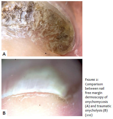
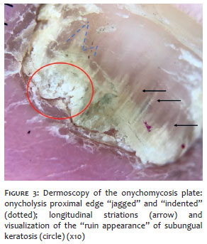
Onychomycosis is the prevalent nail disease, and it is responsible for approximately 50% of onychopathies.1The similarity between some onychopathies tends to make it challenging to obtain a definitive clinical diagnosis. Direct mycological examinations and fungal culture or nail plate biopsy (periodic Schiff staining [PAS] staining) should confirm the diagnosis. However, these tests may show false-negative results in more than 35% of cases.1 The most common clinical form is distal and lateral onychomycosis, representing over 85% of cases.2 In this clinical form, the fungal invasion occurs from the infected skin, passing through the hyponychium or lateral fold, reaching the nail bed and, finally, the ventral surface of the nail plate. As a consequence, subungual hyperkeratosis and onycholysis occur. When examining the free margins with the dermoscope, keratotic debris creates a unique appearance under the free edge of the nail that De Crignis et al. called a "ruin appearance" (Figure 1).3 The formation of this type of subungual hyperkeratosis is due to the exacerbation of the irregular indented shape of the ventral region of the nail plate (Figure 1) associated with the accumulation of debris resulting from fungal invasion. This finding can also be observed in total dystrophic onychomycosis. The absence of the "ruin appearance" in dermoscopy, common in traumatic onycholysis (Figure 2), does not necessarily exclude the diagnosis of onychomycosis. However, its presence strongly indicates the diagnosis, mainly when associated with dermoscopic findings in the nail plate (Figure 3).2 For definitive diagnosis, direct mycological examinations, nail plate culture or histology (nail clipping) should be performed.1 Nail free margin dermoscopy is a noninvasive exam that should be routinely used to assess suspected cases of onychomycosis. The dermoscopic finding of subungual hyperkeratosis showing a "ruin" pattern is a relevant detection and, when present, suggests the diagnosis of onychomycosis.
Cristina Diniz Borges Figueira de Mello | 0000-0001-7199-8451
Case submission; writing of the manuscript; general revision.
Nilton Di Chiacchio | 0000-0001-9536-2263
Case submission; general revision.
1. Gupta AK, Versteeg SG, Shear NH. Onychomycosis in the 21st Century: An Update on Diagnosis, Epidemiology, and Treatment. J Cutan Med Surg. 2017; 21(6):525-39.
2. Freedman J B, Tosti A. Distal Subungual Onychomycosis. In: Tosti A, Vlahovic TC, Arenas R, editors. Onychomycosis: An Illustrated Guide to Diagnosis and Treatment. 1st ed. Cham: Springer; 2017. p23-31.
3. De Crignis G, Valgas N, Rezende P, Leverone A, Nakamura R. Dermatoscopy of onychomycosis. Int J Dermatol. 2014; 53(2):e97-9.
 All content the journal, except where identified, under the Creative Commons Attribution 4.0 International licence - ISSN-e 1984-8773


 Read in Portuguese
 Portuguese PDF
 Print
Print
 Send this article by email
 How to cite this article
 Submit a comment
 Mendeley
 Pocket