


Francisco Macedo Paschoal1,2; Andressa Sobral Soares de Deus1; Anelise Damiani da Silva Citrin1; Gisele Gargantini Rezze2
Received on: 10/04/2019
Approved on: 01/06/2019
This study was carried out at Department of Dermatology of Centro Universitário Saúde ABC, Santo André (SP), Brasil.
Financial support: None
Conflict of interests: None
Microneedling is an ambulatory surgical procedure that can be used for different indications with the objective of stimulating the production of collagen. Five cases were evaluated in the first 72 hours after the procedure by reflectance confocal microscopy in order to evaluate the pores lifetime.
Keywords: Collagen; Wound healing; Ambulatory surgical procedures
Microneedling has been used as minimally invasive technology for the treatment of various dermatological conditions such as acne scars, stretch marks and skin rejuvenation. 1, 2 It has also been applied aimed at increasing the absorption of drugs via transdermal route, creating pores in the epidermis and papillary dermis. 3, 4 Nevertheless, few studies have evaluated its initial effects within the epidermis and dermis. Therefore, the authors of the present paper have studied a series of cases through confocal reflectance microscopy (CRM), which is an in vivo auxiliary examination that allows the visualization of different levels of the skin with histological resolution. 5
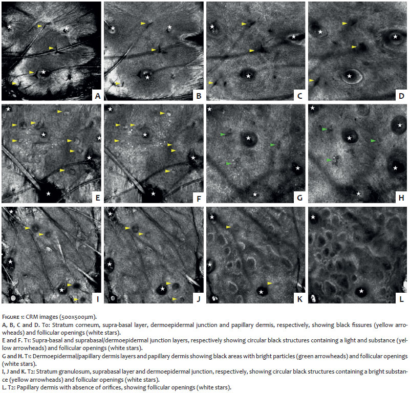
A total of five patients with acne scars and skin photoaging who have signed a Free and Informed Form of Consent were included in the present study. Confocal reflectance microscopy images were acquired through a laser scanning confocal microscope with reflection close to that of the infrared (Vivascope 3000®, Caliber I. D, Rochester, NY, US), from the stratum corneum to the papillary dermis (horizontal cuts), at the right temple. One hour after of topical anesthesia had been applied (Pliaglis®, Galderma, São Paulo, SP, Brazil), microneedling was performed with the assistance of the Derma Roller device (Fabinject Technology, Taubaté, São Paulo, Brazil). The device had 540 microneedles with 1.5mm in length and was applied in the whole face aimed at causing punctate bleeding on the total facial area. The application region was evaluated by CRM immediately after (T0), 24 hours after (T1), 48 hours after (T2) and 72 hours after (T3) the procedure. All patients were advised not to apply any topical cream to the facial skin between CRM assessments. The CRM evaluation conducted at T0 evidenced a black linear cleft extending from the top of the epidermis to the papillary dermis – being more triangular in the dermis (Figure 1) – in all cases. At T1 and T2, the cleft has become a black circular structure in the upper epidermis (stratum corneum), in the epidermis and in the dermo-epidermal junction. Some of them contained a mild and bright substance. In the dermis, these black areas presented bright particles at T1 (Figure 1).
Microneedling has been increasingly used in dermatology for cosmetic reasons due to its easy application technique and rare complications. It also appears promising for drug delivery since the stratum corneum is the major barrier for transdermal drug delivery and can be punctured by microneedles that mechanically pierce the skin layers leading to the transdermal absorption of the drug. 3, 4
Using a new technology (CRM), the present study allowed the observation of perforations in the skin resulting from microneedling. The presence of the orifices in the epidermis and dermis, possibly increasing the skin's permeability – which is essential for the concept of transdermal drug release – is noticed immediately after microneedling (T0). The presence of a mild and bright substance in the epidermis' pores at T1, T2 and T3 may correspond to local subclinical inflammation responsible for micropore occlusion. This physiological process is not yet known, however it is believed that the micropore may close in a matter of hours. 3, 4 The finding of black areas with bright particles inside the papillary dermis allows the authors of the present paper to hypothesize whether it could correspond to inflammation caused by the micro injuries, leading to neovascularization and neocollagenesis, involved in skin rejuvenation. 4
Finally, little is known about the useful life of the orifices and the injury caused by the treatment with microneedles. Therefore, the authors of the present article believe that CRM may be useful for unprecedented visualization of microneedling's mechanical and inflammatory events.
Francisco Macedo Paschoal | ORCID 0000-0002-6264-1538
Approval of the final version of the manuscript; study design and planning; preparation and Drafting of the manuscript; data collection, analysis and interpretation; research guidance; intellectual participation in propaedeutic and / or therapeutic approach of the cases studied; critical review of the literature; critical review of the manuscript.
Andressa Sobral Soares de Deus | ORCID 0000-0002-8569-4229
Study design and planning; preparation and Drafting of the manuscript; data collection, analysis and interpretation; intellectual participation in the propaedeutic and / or therapeutic approach of the cases studied; critical review of the literature.
Anelise Damiani da Silva Citrin | ORCID 0000-0002-2986-6188
Preparation and Drafting of the manuscript.
Gisele Gargantini Rezze | ORCID 0000-0001-9084-4634
Approval of the final version of the manuscript, preparation and drafting of the manuscript; data collection, analysis and interpretation; research guidance; critical review of the literature; critical review of the manuscript.
1. Andrade LE. Microneedling in facial recalcitrant melasma: report of a series of 22 cases. An Bras Dermatol. 2015; 90( 6 ):919-21.
2. Singh A, Yadav S. Microneedling: Advances and widening horizons. Indian Dermatol Online J. 2016;7(4):244-54.
3. Badran MM, Kuntsche J, Fahr A. Skin penetration enhancement by a microneedle device (Dermaroller) in vitro: dependency on needle size and applied formulation. Eur J Pharm Sci. 2009;36(4-5):511-23.
4. Vandervoort J, Ludwig A. Microneedles for transdermal drug delivery: a minireview. Front Biosci. 2008;13:1711-5.
5. Langley RG, Rajadhyaksha M, Dwyer PJ, Sober AJ, Flotte TJ, Anderson RR. Confocal scanning laser microscopy of benign and malignant melanocytic skin lesions in vivo. J Am Acad Dermatol. 2001;45(3):365-76.
 All content the journal, except where identified, under the Creative Commons Attribution 4.0 International licence - ISSN-e 1984-8773


 Read in Portuguese
 Portuguese PDF
 Print
Print
 Send this article by email
 How to cite this article
 Submit a comment
 Mendeley
 Pocket