


Ricardo Vieira1,2; André Pinho1; Ana Brinca1
Received on: 20/11/2018
Approved on: 15/12/2018
This study was performed at the Centro Hospitalar e Universitário de Coimbra - Coimbra, Portugal.
Financial support: None
Conflict of interests: None
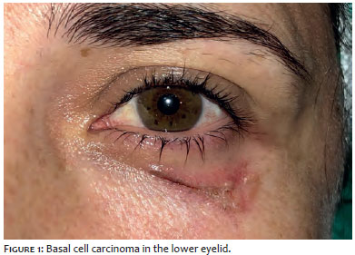
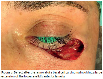
Cutaneous defects of the lower eyelid frequently require reconstructions with grafts or flaps to avoid ectropion. Removal of a basal cell carcinoma from the lower eyelid of a 39-year-old patient resulted in an exclusively cutaneous defect, with transversal and vertical diameters of 32 and 13mm, respectively. Despite the large size of the defect, the elevation of the suborbicularis oculi fascia to the periosteum of the lateral orbital border made primary closure possible. The position of the lower was also reinforced with a lateral canthopexy, with excellent aesthetic and functional results.
Keywords: Carcinoma, basal cell; Ectropion; Eyelids; Orbit; Reconstruction
Defects involving the lower eyelids anterior lamella are relatively common in dermatologic surgery, with most of them resulting from the excision of basal cell carcinomas, xanthelasmas, and several other neoplasms or benign conditions. The repair of these defects is challenging due to the relatively small amount of tissue in the region, and often require the use of skin flaps or grafts aimed at preventing eyelid retraction or vertical tension vectors, which cause postoperative ectropion with rele-vant aesthetic and functional impacts.1
Align with that, primary closure is usually limited to some small defects. Larger defects should be repaired with full-thickness skin grafts (usually taken from the upper eyelid) or with various types of flaps (Tripier flap or cheek skin advancement or rotation flaps).12
A 39-year-old female patient underwent extensive ex-cision of a basal cell carcinoma in the lower eyelid (Figure 1). The resulting defect involved the lower eyelids anterior lamella, at a distance of 4 mm from the eyelids margin, measuring 32 mm and 13 mm in its transverse and vertical diameters, respectively (Figure 2). The snap-back test did not reveal alterations, with the eyelid immediately returning to the normal position. Another test was performed by taking the suborbicularis oculi fascial tissue through the lower edge of the surgical wound using a forceps, so as to lift it towards the lateral orbital rim. This action allowed complete juxtaposition of the defects edges.
In this manner, the repair was planned in three stages:
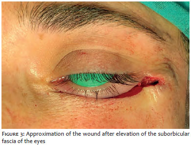
- Elevation of the suborbicular fascia of the eyes, attaching the prezygomatic area’s deep fascia to the lateral orbital border’s periosteum, using a non-absorbable 4/0 polypropylene suture with a double needle. The first needle was used to approach a generous portion of the fascia underlying the subcutaneous fat, on the lower margin of the defect. The second needle was used to reach the lateral orbital borders periosteum. After having been tied, this suture elevated the inferior margin of the defect, allowing an almost complete approximation (Figure 3).
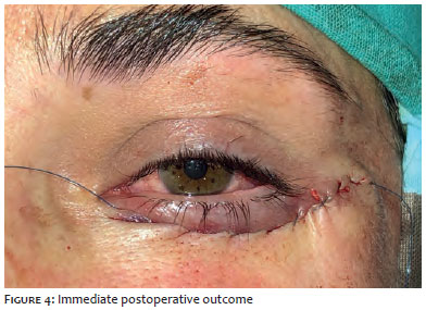
- Performance of a lateral canthopexy with the same double-needle polypropylene suture, attaching the lateral end of the inferior tarsal plate to the periosteum of the inner side of the lateral orbital border. This step was carried out observing the need to preserve the position of the lateral canthus leveled with that of the contralateral side.
- Primary closure of the skin with a continuous 5/0 polypropylene suture, without knots, fixed on each of the sides by reinforced with 6 mm wide adhesive strips (Steri-Strip, 3M) (Figure 4).
The continuous suture was removed on the sixth day.
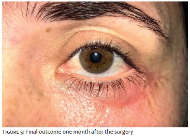
The final outcome after one month was deemed excellent by both physicians and patient (Figure 5). No ectropion or functional compromise could be observed in the lower eyelid during the follow-up.
Ectropion with aesthetic and functional compromise is a possible complication of surgical procedures in the lower eyelid.3
Even in aesthetic procedures — as well as in lower eyelid bleph-aroplasty — ectropion is probably the most serious long-lasting complication.4 It can result from excessive skin removal, however might also occur in cases where the immediate outcome appears to be adequate, especially in patients with decreased eyelid elasticity. In these cases, a modified technique combining canthopexy and the elevation of the suborbicular fascia is capable of preventing this complication.5 This modified technique can be applied to the reconstruction of eyelid defects, allowing an effective approximation of the edges, elevating the pre zygomatic soft tissues,6 completely removing the tension from the skin by exerting tension on the deep tissues. The suspension of the suborbicularis soft tissue towards a fixed structure (lateral orbital rim) avoids the creation of vertical tension vectors over the lower eyelid that would ultimately result in ectropion. In addition, since the lateral canthal ligament is reinforced by a lateral canthopexy, the proper positioning of the lower eyelid is easily obtained.
The elevation of the suborbicular fascia associated with lateral canthopexy may allow the primary closure of relatively large defects of the anterior lower eyelid lamella for which, in normal conditions, full thickness skin grafts or local flaps would be required. This procedure appears to be a relatively simple alternative to flaps and grafts, especially in defects affecting the central or lateral thirds of the lower eyelid. Meticulous analysis should be undertaken when considering this method in patients with diminished elasticity of the lower eyelid (with abnormal snapback test), as well as in defects that are located very close to the palpebral margin or in those located in the medial part of the lower eyelid.
Ricardo Vieira | ORCID 0000-0002-5914-9171
Drafting of the manuscript, surgeon responsible for the described surgical procedure.
André Pinho | ORCID 0000-0001-6433-311X
Review of the manuscript, member of the surgical team.
Ana Brinca | ORCID 0000-0002-7539-9912
Review of the manuscript, member of the surgical team.
1. Hayano SM, Whipple KM, Korn BS, Kikkawa DO. Principies of periocular reconstruction following excision of cutaneous malignancy. J Skin Cancer 2012;2012: 438502.
2. Subramanian N. Reconstructions of eyelid defects. Indian J Plast Surg. 2011;44(1): 5-13.
3. Baek JS, Kim KH, Lee JH, Choi HS. Ophtalmologic complications associated with oculofacial plastic and esthetic surgeries. J Caraniofac Surg. 2018;29(5):1208-11.
4. Oestreicher J, Mehta S. Complications of blepharoplasty: prevention and management. Plast Surg Int. 2012; 2012: 252368.
5. Khan M, Aziz K, Javed A, Gorman M, Othman D, Riaz M. Modified lower eyelid blepharoplasty improves aesthetic outcomes in patients with hypoplastic malar prominences. Plast Aesthet Res. 2017;4:228-35.
6. Amodeo CA, Casasco A, Cornaglia AI, Kang R, Keller GS. The suborbicular oculi fat (SOOF) and the fascial planes: has everything already been explained? JAMA Facial Plast Surg. 2014;16(1):36-41.
 All content the journal, except where identified, under the Creative Commons Attribution 4.0 International licence - ISSN-e 1984-8773


 Read in Portuguese
 Portuguese PDF
 Print
Print
 Send this article by email
 How to cite this article
 Submit a comment
 Mendeley
 Pocket