


Flávia Alvim Sant`Anna Addor
Received on: 01/10/2018
Approved on: 08/12/2018
This study was performed at the Institution MEDCIN Pesquisa - Osasco (SP), Brazil.
Financial support: The in vitro study was sponsored by Melora-FQM Rio de Janeiro (RJ), Brazil
Conflict of interests: The methodology, execution and analysis of results were obtained by the institution's involved researcher without any interference of the sponsoring company.
INTRODUCTION: The interaction between the skin and air pollutants has demonstrated effects in the cutaneous barrier, as well as triggering oxidative processes related to premature ageing of the skin. The aryl hydrocarbon receptor (ArH) is a transcription protein that interacts with xenobiotics, regulating the transcription of genes involved with oxidative stress, inflammation, immunosuppression and pigmentation, besides leading to processes related to ageing and carcinogenesis.
OBJECTIVE: Evaluate the anti-pollution efficacy of an antioxidant association for the prevention of the nuclear translocation of the AhR receptor.
METHODS: A in vitro model (keratinocyte culture) was exposed to cigarette smoke and the presence of AhR was measured through sanduwich ELISA assay.
RESULTS: The treated culture demonstrated inhibition of the nuclear translocation of AhR in all concentrations evaluated: ArH increase of 75.38%; 59.88% and 117.79% are seen with the concentrations of 0.316; 0.100 and 0.0316mg / mL, respectively.
CONCLUSION: The results suggest the ability of the formulation analyzed in preventing the activation of genes responsible for the damaging effects of cigarette smoke.
Keywords: DNA Damage; Air Pollution; Aryl Hydrocarbon Receptor Nuclear Translocator
There is consistent evidence that air pollution has deleterious effects on human health, with respiratory and cardiovascular repercussions. More recently, the interaction of these pollutants with the skin has also shown effects on the cutaneous barrier,1 in addition to triggering oxidative processes related to premature skin aging, as demonstrated in a cohort study in large urban centers in Germany.2
These mechanisms of damage involve the expression of proinflammatory mediators and AhR (aryl hydrocarbon receptor) translocation protein, acting on both keratinocytes and melanocytes, leading to increased production of oxygen-free species.3,4
The AhR receptor is a transcription protein that interacts with xenobiotics, which are chemical compounds foreign to the body. This interaction with xenobiotics is similar to that of hormone receptors. The binding of a xenobiotic compound activates the receptor, allowing its translocation to the nucleus, which regulates the transcription of genes involved with oxidative stress, inflammation, immunosuppression, and pigmentation, therefore being capable to lead to premature aging and carcinogenesis processes. Inhibition of AhR-mediated gene activation could significantly interfere with signs of aging and carcinogenesis.4,5
Cigarette smoke contains over 4,000 toxic compounds, including many polycyclic aromatic hydrocarbons (PAHs), dioxins and furans that exert harmful effects on the skin by the induction of genes that act on inflammation and oxidation,6,7 being able to activate the translocation of AhR.8,9
Exposure to the xenobiotic agents contained in the pollutants (e.g. cigarette smoke) can reduce (in vitro) the presence of cytoplasmic AhR, since this receptor, once activated by the agent (e.g. PAH), would migrate to the nucleus aiming at activating the genes linked to inflammation and oxidation.10
Vanzo et al.11 investigated the translocation inhibition effect in keratinocyte culture in 2015. A strategy aimed at preventing damage caused by constant exposure to exogenous agents — in particular cigarette smoke — involves the use of substances that inhibit the translocation of AhR to the nucleus, thus avoiding imbalance of cutaneous cellular homeostasis and the progression of reactions that oxidize organic substrates.
To evaluate the anti-pollutant efficacy of a nutrient association in the prevention of nuclear translocation of the aryl hydrocarbon receptor (AhR), induced in an in vitro model (culture of keratinocytes) by exposure to cigarette smoke.
This trial was carried out in HaCat human keratinocyte culture, purchased from the State of Rio de Janeiros Cell Bank, Rio de Janeiro (RJ), Brazil.
The cultures were incubated at three previously determined non-cytotoxic concentrations of the Exímia Temporize® formulation (Melora-FQM, Rio de Janeiro, Brazil), consisting of the vitamins E and C, beta-carotene, lutein, lycopene, linseed oil, zinc and selenium. The concentrations were 0.316, 0.100 and 0.0316 mg/ml, respectively, and the cells were kept in touch with the formulation for 48 hours.
After 48 hours of treatment, the cultures were exposed to cigarette smoke using an appropriate chamber that allowed the complete combustion of two cigarettes. The cells were incubated for an additional 24 hours with the evaluated formulation. After this period, they underwent extraction of cytoplasmic lysate (three cycles of freezing at -20 °C, with a 20 minute interval between each cycle, and subsequent centrifugation at 10,000 rpm for 10 minutes), followed by the quantification of the mediator AhR.
The presence of AhR was measured using the sandwich-Elisa (Enzyme Linked ImmunoSorbent Assay) using commercially available kits (Uscn Life Science Inc., Houston, TX, USA). The absorbance reading was performed using a Multiskan® GO® monochromator. AhR values were normalized based on the total protein of the sample measured using the Bradford technique.12
To analyze the results, the statistical evaluation used the ANOVA test, which allowed measuring the datas variation base on comparisons between the groups, followed by the Bonferroni post-test. A 5% significance level (GraphPad Prism v6) was used.
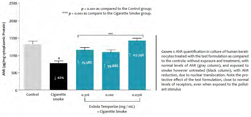
Exposure of cells to cigarette smoke promoted a 42% reduction in AhR availability in the cytoplasm of the cell when compared to baseline control (P <0.001). This result indicates a possible migration of the AhR to the cell nucleus that is coupled to the xenobiotic agent.
Pretreatment of the cultures with the evaluated formulation was shown to inhibit AhR nuclear translocation at all concentrations evaluated, with the inhibition value reaching a level higher than 100% as compared to the cells exposed to cigarette smoke. Increases of 7.38%, 59.88% and 117.79% were observed at the concentrations of 0.316; 0.100 and 0.0316 mg/ml, respectively. The results suggest the evaluated formulation has the ability to prevent the activation of the genes responsible for the damaging effects of cigarette smoke (Graph 1).
Cigarette smoke exerts harmful effects on the skin that are mediated by the AhR receptor, which is a cytosolic transcription factor found in its inactive form, which binds to the toxic agent and translocates it to the cell nucleus. In the nucleus, AhR regulates the transcription of genes involved in oxidative stress, inflammation, immunosuppression, pigmentation, premature aging and carcinogenesis.13
In addition to mediating these mechanisms at the nuclear level, AhR can also be regulated by oxidative mechanisms. Therefore, inhibiting AhR translocation would prevent damage caused by the xenobiotics in the cell nucleus.14
In order to analyze AhR’s involvement in cutaneous aging caused by cigarette smoke, in 2008 Ono et al.10 exposed primary human fibroblasts to a tobacco smoke extract, observing increased induction of mRNA for metalloproteinase 1, associated with greater expression of cytochrome P1B1 (CYP P1B1).
The present study has demonstrated that the treatment of keratinocytes with the association of nutrients avoided nuclear translocation, keeping the AhR receptors in the cytoplasm. The prevention of this translocation — characterized by the increased concentration of these receptors — reached 117.79% as compared to the area that had not received any treatment before having been exposed to cigarette smoke.
The association of nutrients contained in the evaluated formulation exerted cellular protective effect against the damage caused by xenobiotic pollutants to keratinocytes, since it protected the nuclear translocation of the AhR receptor, reaching a value in excess of 100% regarding the control. These findings demonstrate that the evaluated treatment has adjuvant action in the prevention and treatment of the skin aging process due to extrinsic factors, which is represented in the present study by cigarette smoke.
Flávia Alvim Sant'Anna Addor | ORCID 0000-0003-1851-7342
Conduction of the study, data compilation and review, oversight of the statistical evaluation, preparation of the manuscript.
1. Farage MA, Katsarou A, Maibach HI. Sensory, clinicai and physiological factors in sensitive skin. Contact Dermatitis. 2006;55(1):1-14.
2. Vierkotter A, Schikowski T, Ranft U, Sugiri D, Matsui M, Kramer U, et ai. Airborne particle exposure and extrinsic skin aging. J Invest Dermatol. 2010;130(12):2719-26.
3. Choi H, Shin DW, Kim W, Doh SJ, Lee SH, Noh M. Asian dust storm particles induce a broad toxicological transcriptional programin human epidermal keratinocytes. Toxicol Lett. 2011;200(1-2):92-9.
4. Krutmann J, Liu W, Li L, Pan X, Crawford M, Sore G, et al. Pollution and skin: from epidemiological and mechanistic studies to clinical implica-tions. J Dermatol Sci. 2014; 76(3):163-8.
5. Morita A, Torii K, Maeda A, Yamaguchi Y. Molecular basis of tobacco smoke-induced premature skin aging. J Invest Dermatol Symp Proc. 2009;14(1):53-5.
6. Kasai A, Hiramatsu N, Hayakawa K, Yao J, Maeda S, Kitamura M. High levels of dioxin-like potential in cigarette smoke evidenced by in vitro and in vivo biosensing. Cancer Res. 2006;66(14):7143-50.
7. Kitamura M, Kasai A. Cigarette smoke as a trigger for the dioxin recep-tor-mediated signaling pathway. Cancer Lett. 2007; 252(2):184-94.
8. Nakamura M, Ueda Y, Hayashi M, Kato H, Furuhashi T, Morita A. Tobacco smoke-induced skin pigmentation is mediated by the aryl hydrocarbon receptor. Exp Dermatol. 2013;22(8):556-8.
9. Ono Y, Torii K, Fritsche E, Shintani Y, Nishida E, Nakamura M, et al. Role of the aryl hydrocarbon receptor in tobacco smoke extract-induced matrix metalloproteinase-1 expression. Exp Dermatol. 2013;22(5):349-53.
10. Ono Y, Yasuda Y, Shintani Y, Sakakibara N, Abel J, Fritsche E, et al. Tobacco smoke extract induces matrixmetalloproteinase (MMP-1) expression in human skin fibroblasts through the arylhydrocarbon receptor (AhR) pathway. J Invest Dermatol. 2008;128(Suppl 1):S42.
11. Vanzo ACJR, Pereira AFC, Cascais LC, Lage R, Andrade CC, Araujo RB, et al. Uso de metodologia in vitro para o estudo de efeitos antipoluição para formulações cosméticas. Poster of II Congreso latino-americano sobre métodos alternativos em los ensayos, la investigacion, la indústria e la educacion (COLAMA); 2015 Jul 5-9; Balneario de Varadero, Cuba. Havana: Red Latino-Ibero-Americana para Métodos Alternativos; 2015.
12. Bradford MM. Rapid and sensitive method for the quantitation of microgram quantities of protein utilizing the principle of protein-dye binding. Anal Biochem. 1976; 72(1-2):248-54.
13. Abel J, Haarmann-Stemmann T. An introduction to the molecular basics of aryl hydrocarbon receptor biology. Biol Chem. 2010;391(11):1235-48.
14. R. Brigelius-Flohe R, Flohe L. Basic principles and emerging concepts in the redox control of transcription factors. Antioxid Redox Signal. 2011;15(8):2335-81.
 All content the journal, except where identified, under the Creative Commons Attribution 4.0 International licence - ISSN-e 1984-8773


 Read in Portuguese
 Portuguese PDF
 Print
Print
 Send this article by email
 How to cite this article
 Submit a comment
 Mendeley
 Pocket