


Luiza Oliveira Tocantins Álvares1; Eduardo dos Santos Martins Neto1; Gisele Moura de Oliveira Leite1; Mariana Albuquerque Dórea1; Elisa Maria Novaes Barros1; Mariseth Carvalho de Andrade2; Miguel Saraty de Oliveira3
Received on: 02/08/2018
Approved on: 24/09/2018
This study was performed at the Universidade do Estado do Pará, Belém (PA), Brazil.
Financial support: Pibic Fapespa
Conflict of interests: None
INTRODUCTION: Cutaneous flaps can be complicated by both ischemia and estrogen deficiency. In this manner, it is desirable to improve the healing process with the use of medicinal plants, such as Aloe vera.
OBJECTIVE: To evaluate the effect of Aloe vera’s glycolic extract on skin flaps performed on oophorectomized rats.
METHODS: Cutaneous flap was performed in 20 animals, distributed in four groups, submitted to differentiated procedures, with microscopic (21st day) and macroscopic analysis (seventh and 14th day).
RESULTS: Microscopic variables were not significant. Two groups showed improvement in the general condition of the wound, and only one presented significant closure of the wound.
CONCLUSIONS: The Aloe vera extract yielded promising parameters regarding the macroscopic aspects; however further studies are necessary for a better evaluation.
Keywords: Aloe; Wound healing; Estrogens
In the fields of plastic surgery and dermatology, skin flaps are widely used, especially in reconstructive surgeries. Ischemic complications, however, are of great concern and may require secondary surgical interventions, generate multiple infections and delay future treatments, due to the presence of tissue necrosis.1
In face of this, estrogen deficiency has been shown an important mechanism in healing complications. Due to the process of demographic transition and the resulting increase in the population’s average age, almost a third of a woman’s life is characterized by a state of estrogen deprivation. This is associated with numerous age-related events, including poor healing and decreased viability of cutaneous flaps.2
Current literature supports the thesis that estrogen acts as a free radical scavenger, with this effect extending beyond its hormonal role in biological systems. In this sense, Coksun et al.3 have demonstrated that oophorectomy had unfavorable effects on the skin flap viability in a rat model, with a significant increase in tissue myeloperoxidase levels.
For better healing of the flap, vasodilator drugs, including medicinal plants, can be associated. Regarding phytotherapy, Aloe vera is widely used in studies that associate the plant’s effects with wound healing, mainly due to the immunomodulatory effects, such as the activation of cytokines linked to the healing process and the formation of new vessels.5,6
It is estimated that Aloe vera (L.) has about 200 biologically active molecules that act synergistically on fibroblasts during the formation of a new epithelium, acting on the production of collagen and glycosaminoglycans, improving tissue remodeling.7 Eshghi et al. concluded that topical application containing A. vera resulted in less postoperative pain and accelerated healing with lower analgesic consumption as compared to the placebo group.8
The cicatrizing effects of A. vera were demonstrated in several animal models. Mendonça et al.9 observed a rapid rate of epithelialization and increased blood vessel formation when studying the morphological and morphometric aspects of the cicatricial process of induced surgical lesions in Wistar rats. Proteins and glycoproteins that were isolated from the A. vera gel showed anti-inflammatory activity in vitro by significantly reducing the COX-2 and lipoxygenase enzymes. When tested on animals, these proteins were able to accelerate the healing process and increase cell proliferation.10
Based on positive outcomes obtained with Aloe vera’s leaf extract in several healing models and in light of the need for therapeutic measures for improving the viability of skin flaps in the presence of estrogen deprivation, the present study was aimed at evaluating the macroscopic and microscopic aspects of cutaneous flaps in oophorectomized rats treated with Aloe vera’s glycolic extract.
To evaluate the healing effect of Aloe vera’s glycolic extract on skin flaps in oophorectomized rats.
This investigation was carried out in compliance with the Brazilian Federal Law No. 11,794 of 2008 and the Ethics Principles of the Brazilian Society of Animal Experimentation (COBEA), with prior approval of the research project by the Research Ethics Committee for Animal Use of the Universidade do Estado do Pará, Belém (PA), Brazil.
The present study was characterized as an experimental, prospective and interventional study aimed at evaluating the effect of Aloe vera’s glycolic extract on the healing of cutaneous flaps induced in oophorectomized Wistar lineage rats.
Twenty-nine female Rattus norvegicus (Wistar), of approximately 90 days old, weighing between 200g and 250g were used. The animals were kept in an environment with controlled temperature before and after the procedure, under 12-hour light-dark cycle, with water and rat specific ration offered ad libitum.
The animals were randomly assigned to four study groups, each with five individuals.
Group AV (GAV): Animals that underwent the dorsal cutaneous flap technique 2x2, followed by topical treatment with Aloe vera for 21 days.
Group SF (GSF): Animals that underwent the dorsal cutaneous flap 2x2 technique, without topical treatment of Aloe vera.
Group OOP AV (GOOPAV): Animals technique the oophorectomy technique one month before the flap was performed. At the end of this period, the 2x2 dorsal cutaneous flap technique was performed followed by topical treatment with Aloe vera for 21 days.
Group OOP SF (GOOPSF): Animals that underwent the oophorectomy technique one month before the flap was performed. At the end of this period, the 2x2 dorsal cutaneous flap technique was performed, without topical treatment with Aloe vera.
The animals were anesthetized with ketamine (70mg / kg) and Xylazine (10mg / kg), by intraperitoneal route, with the anesthetic plane evidenced through interdigital pressure and absence of paw retraction reflex, in the absence of the breech reflex.
The animals were anesthetized and harnessed with the aid of adhesive tape on a surgical board (20x30cm) in the ventral decubitus position. The trichotomy of the dorsal region and posterior antisepsis of the surgical area were performed with polyvinylpolypyrrolidone.
In the animals of OOP AV and OOP SF groups, the abdominopelvic cavity was incised, with subsequent identification of the ovaries. The ovarian pedicles were clamped and the vessels ligation was performed with 8-0 nylon. Next, the ovaries were removed bilaterally at the junction of the uterine horns, followed by revision of the hemostasis. Then the cavity was closed with 4-0 nylon monofilament thread. One month after the procedure, the skin flap was performed.
The preparation of the 2cm long and 2cm wide subcutaneous dorsal skin flap with cranial base was performed according to the Acevedo-Bogado et al.11 modified technique, observing the inferior angles of the scapulae as anatomical cranial limits. In all animals, the area was demarcated using previously prepared and sterilized molds. Next, the cutaneous flap was performed using scalpel with nº. 15 blade. Subsequently, the flap’s site divulsion was carried out with the displacement of the adjacent muscle-aponeurotic plane. This was followed by the elevation of the floor, repositioning and suture with single stitch, standardizing the same technique for all groups, using 4-0 nylon thread.11
The glycolic extract of Aloe vera was supplied as topical gel by Farmácia Personale Ltda, Belém, (PA) Brazil. Immediately after performing the dorsal cutaneous flap in the rats, the animals of AV and OOP AV groups underwent topical treatment with Aloe vera’s glycolic extract,12 by application of the gel in the skin flap with the assistance of a sterile swab.9 This procedure was repeated every 24 hours for 21 days, totaling 21 dressings.
After the surgery, the rats were placed in individual cages with sterile wood shavings for postoperative recovery, having bee observed for two hours, separated from each other in order to avoid dehiscence of the sutures.
Photographic follow-up of the development of the skin flaps was performed with a Canon EOS T3 Rebel 10 megapixels camera from a 15cm distance between the camera and the animal, on the 7th and 14th postoperative days, in all groups.12
The analyzed macroscopic aspects of the wound were: wound’s general state – (4) excellent, (3) good, (2) regular, and (1) poor; wound’s appearance – (0) closed or (1) open; presence of crusts – (1) present or (0) absent; and observations related to inflammation in the operative wound (edema and hyperemia) – (0) absent, (1) slight, (2) moderate, and (3) intense.
All animals underwent biopsy and euthanasia on the 21st day after the flap was performed, with a sample of the skin of each animal having been removed by means of a cold scalpel biopsy, leaving a 1cm margin between the scar and the collection incision.
The specimens were removed and immediately deposited in 10% formalin. After preparation of the tissues on slides for microscopic study, they were stained with hematoxylin / eosin, in addition to Masson’s trichrome staining, for evaluation of collagen fibers.
A pathologist physician, who was unaware of the group to which the slides belonged, analyzed the samples morphologically under optical microscopy.
The parameters analyzed were collagen fibers, epithelialization, as well as proliferation of vessels and fibroblasts, which were classified as (0) absent, (1) mild, (2) moderate, and (3) intense, according to the parameters of Vieira et al.13
The outcomes underwent the statistical analysis of multiple comparisons using the chi-square partition and Kruskal-Wallis tests, and in the case of a statistically significant difference, α = 0.05 was adopted as the significance level. In addition, the data were stored in Excel 2010 spreadsheets, and analyzed using the Bioestat® 5.3 software.
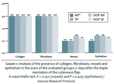
The moderate presence of collagen and fibroblasts was similar in all groups after the biopsy that was performed in the 21st postoperative day, thus not presenting statistical difference. (Graph 1)
The proportion of vessels was higher in OOP AV group, having been considered moderate in 40% of the rats, whereas in OOP SF group, 20% of the animals had such a parameter, mainly in comparison to AV and SF groups, which showed mild proliferation. Both increases, however, were not statistically significant (Graph 2).
Epithelization occurred at higher levels in GSF and GOOPAV, in 40% of the rats in both groups, whereas in OOP SF group it occurred only in 20% of the animals, nevertheless there was no statistical significance between the groups (Graph 1).
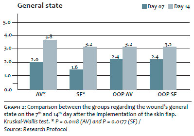
Regarding the macroscopic analysis, according to the general state of the wound (Graph 2), there was statistical significance in the comparison between each group between D7 and D14 in AV and SF groups, which showed improvement of about 90% and 100%, respectively. Regarding the medians (Table 1), AV group yielded a higher value as compared to SF group, in both D7 and D14. The rats in OOP AV and OOP SF groups also showed improvement of approximately 34% in D14; however, there was no significant support in the analyzed statistics.
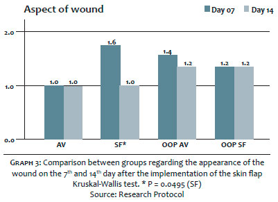
Regarding the wound’s appearance (Graph 3), only SF group presented significant closure of wounds in about 60% of the animals in D14. Meanwhile, AV and OOP SF groups showed a reduction of about 14% without significance.
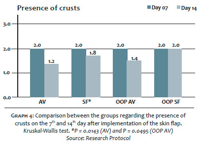
Regarding the presence of crusts (Graph 4), comparing D7 and D14, AV group showed a significant reduction of 80%, as did OOP AV group, with 30%. In addition, SF group had a 10% reduction, however without statistical significance, whereas OOP SF group did not present variation between the two experimental timepoints.
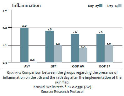
As for inflammation (Graph 5), on the 7th and 14th days, a total and significant reduction of 100% was observed in all GAV rats at D14, whereas the other groups did not yield significant results.
It is worth mentioning that when comparing the groups, the authors of the present study verified a significant difference only in the variable presence of crusts at D14, when GAV yielded the most significant improvement – of 40% – in the reduction of the operative wound’s crusts.
From a microscopic point of view, the present study did not identify differences in the presence of collagen and in the proliferation of fibroblasts between the analyzed groups. It is known that the proliferative phase of healing process can be divided into three subphases: reepithelialization, fibroplasia and neoangiogenesis. In the fibroplasia subphase, fibroblasts arise and collagen production occurs. 14 It was therefore inferred that, on the 21st day of the evaluation, all groups had also passed through the fibroplasia phase, thus presenting similar amounts of these components in their wounds. This fact is related to a limitation of the study, which corresponds to the late evaluation of the mentioned variables.
Regarding the presence of vessels in the studied groups, a higher proportion was observed in OOP AV group. It is known that estrogen deprivation is linked to slower healing of acute wounds, as well as to the loss of estrogen’s protective factors, such as prevention of ischemic lesions. 15 In this manner, it is suggested that the use of Aloe vera was beneficial in the evaluated group, allowing less inflammation and improvement of neoangiogenesis, resulting in greater tissue perfusion, which reduced the risk of ischemia and wound necrosis.
In this circumstance, the action of Aloe vera as a potentiator of angiogenesis and access to oxygen, associated with its antioxidant effects mediated by the presence of superoxide dismutase and glutathione peroxidase in its gel stands out, 16 facilitating estrogen-deficient healing.
In a comparative analysis between the 7th and 14th day of development of the healing of a cutaneous flap in rats with and without estrogenic deprivation, the present study showed that there was a macroscopic improvement in the animals treated with topical extract of algae in their wounds.
When approaching the wounds’ general state, the authors of the present study could observe the presence of improvement in the development patterns from the 14th day, in the groups that received Aloe vera – mainly in AV group, unlike in OAB AV – due to the presence of estrogen, which is crucial to the healing process, as demonstrated by Routley and Ashcroft. 15 The fact that AV group had a higher average, with a greater number of animals rated with excellent for the appearance of their wounds, is linked to the presence of phytotherapics, especially in the balance of antioxidants / pro-oxidants that act in a beneficial way in the cellular repair phases. 17
That action can also be observed in open wounds, as in the study by Mendonça, 9 which assessed the positive synergism of the association of a microcurrent and albumin in the healing process of wounds in Wistar rats. However, the positive patterns of the general state of the wound was not verified in the healing of burns, a fact that was evaluated by Dat et al., 18 who demonstrated that Aloe vera is not an incremental factor in healing when compared to the use of silver sulfadiazine.
Regarding the appearance of the wound, it is interesting to note that a significant closure was verified only in GSF due to the fact that the operative wounds of this group remained open in D7, with improvement occurring only in D14. In other groups, especially in GAV, the wounds of the vast majority of animals were already closed at the beginning of the evaluation.
It should be noted that there was closure, even if not significant, in OOP AV group, while there was no variation regarding wound closure in OOP SF group between the evaluative experimental timepoints. This comparison raises questions about the Aloe vera’s possible healing action, demonstrating that the presence of the phytotherapic allowed the closure of wounds in the presence of estrogen deprivation – although later than in the non-oophorectomized groups – a phenomenon that was not seen in the group that was not treated with the plant.
This difference in the wound closure pattern can be attributed to the positive action of Aloe vera in tissue regeneration, 19 even in oophorectomized animals, since the plant not only increases the amount of collagen in the wound’s site, but also increases the transverse connections between the collagen bands, resulting in the acceleration of the healing process. 20
Regarding the to inflammation in the operative wound criteria, all groups analyzed showed a predominance of moderate and intense degrees of edema and / or hyperemia on the 7th day. On the other hand, on the 14th day, 100% of GAV animals did not show significant inflammatory signs, a fact evidenced by the anti-inflammatory action of Aloe vera, especially regarding the percentage decrease in studies of neutrophil infiltration, angiogenesis and expression of TCD8+ lymphocytes. However, despite a 50% decrease in the GOOPABF’s phlogistic signs, which would imply in a possible beneficial action arising only from the plant’s action, the authors did not evaluate the statistical significance of this variable in any of the groups.
Regarding the presence of crusts, an indicative of better healing, it was more significant in GAV group, in which there was synergistic presence of estrogenic action and Aloe vera. Estrogen is proven to be a potentiator of the healing process, especially demonstrated in studies with genistein, a natural substance that mimics estrogenic action, improving healing parameters, even in the postoperative period of techniques that used cutaneous flaps similar to those used in the present study, resulting in the group with the greatest healing. 2 Aloe vera is also considered by other authors as having a strong positive impact on wound closure, especially in recent studies that demonstrated the biological function of accelerating the healing process in chronic wounds, in addition to reducing the chronic pain process and the persistent inflammation with reduction of lymphocytic infiltration. 21, 22
It is also interesting to note that the isolated action of Aloe vera was beneficial, especially in the oophorectomized rats in GOOPAV, since the operative process decreases the hormonal amounts of estrogen and hampers the healing of cutaneous flaps. Despite that, this group experienced a significant reduction in the size of their crusts, although it was smaller regarding the results experienced in GAV group, in which there was association of the estrogen with Aloe vera. Of the other groups compared by the authors, GSF showed a minimal, probably physiological, but not significant reduction, whereas GOOPSF showed the lowest reduction rate, possibly explained by the absence of physiological estrogen, nevertheless not experiencing significant outcomes either.
Aloe vera extract has promising effects on the healing of skin flaps in oophorectomized rats. Outcomes showed a significant improvement in the use of the plant in variables such as the animal’s wound’s general state, wound’s appearance, inflammation and presence of crusts. There was, however, no significant outcome that would confirm improvement of the microscopic parameters associated with the use of Aloe vera. In this manner, the authors encourage future studies that analyze, in different time periods, the healing quality, in order to determine the histological performance linked to the presence of Aloe vera’s leaf extract. Finally, a more reliable identification of the active compounds responsible for the healing process when there is deprivation of estrogen also depends on further investigation.
Luiza Oliveira Tocantins Álvares | ORCID 0000-0003-3803-2465
A Approval of the final version of the manuscript; Study design and planning; Structuring and drafting of the manuscript; Data collection, analysis and interpretation; Intellectual participation in the propaedeutic and / or therapeutic approach of studied cases; Critical review of the literature; Critical review of the manuscript
Eduardo dos Santos Martins Neto | ORCID 0000-0003-1089-5832
Approval of the final version of the manuscript; Study design and planning; Manuscript tructuring and drafting; Data collection, analysis and interpretation; Intellectual participation in the propaedeutic and / or therapeutic approach of studied cases; Critical review of the literature; Critical review of the manuscript
Gisele Moura de Oliveira Leite | ORCID 0000-0002-1671-1697
Study design and planning; Data collection, analysis and interpretation; Intellectual participation in the propaedeutic and / or therapeutic approach of the cases studied
Mariana Albuquerque Dórea | ORCID 0000-0002-4820-4841
Study conception and planning; Data collection, analysis and interpretation; Intellectual participation in the propaedeutic and / or therapeutic approach of studied cases
Elisa Maria Novaes Barros | ORCID 0000-0001-9406-7158
Study conception and planning; Data collection, analysis and interpretation; Intellectual participation in the propaedeutic and / or therapeutic approach of studied cases
Mariseth Carvalho de Andrade | ORCID 0000-0002-6257-5972
Statistical analysis; Data collection, analysis and interpretation
Miguel Saraty de Oliveira | ORCID 0000-0002-0971-8671
Approval of the final version of the manuscript; Study conception and planning; Effective participation in research guidance; Intellectual participation in propaedeutic and / or therapeutic approach of the cases studied; Critical review of the manuscript
1. Turin SY, Li DD, Vaca EE, Fine N. Nitroglycerin ointment for reducing the rate of mastectomy flap necrosis in immediate implant based breast reconstruction. Plast Reconstr Surg. 2018;142(3):264e-70e.
2. Faber L, Kovác I, Mitrengová P, Novotny M, Variská L, Vasilenko T, et al. Geinstein improves skin flap viability in rats: a preliminary in vivo and in vitro investigation. Molecules. 2018;23(3):1-17.
3. Coskun A, Arikan DC, Coban YK, Sayar H, Kilinc M, Ozbag D. The effect of ovariectomy on the skin flap viability and myeloperoxidase levels. Bratisl Lek Listy. 2014;115(12):766-70.
4. Estevão LRM, Medeiros JP, Scognamillo-Szabó VR, Baratella-Evêncio L, Guimarães EC, Câmara CAG, et al. Neoangiogênese de retalhos cutâneos em ratos tratados com óleo de copaíba. Pesq Agropec Bras. 2009;44(4):406-12.
5. Ramos AP, Pimentel LC. Ação da Babosa no reparo tecidual e cicatrização. Braz J Health. 2011;2(1):40-8.
6. Ahluwalia B, Magnusson MK, Isaksson S, Larsson F, Ohman L. Effects of Aloe barbadensis Mill. Extract (AVH200R) on human blood T cell activity in vitro. J Ethnopharmacol. 2016;179:301-9.
7. Oryan A, Mohammadalipour A, Moshiri A, Tabandeh MR. Topical application of Aloe vera accelerated wound healing, modeling, and remodeling: an experimental study. Ann Plast Surg. 2016;77(1):37-46.
8. Eshghi F, Hosseinimehr SJ, Rahmani N, Khademloo M, Norozi MS, Hojati O. Effects of Aloe vera cream on posthemorrhoidectomy pain and wound healing: results of a randomized, blind, placebo-control study. J Altern Complement Med. 2010;16(6):647-50.
9. Mendonça FAS, Passarini Junior JR, Esquisatto MAM, Mendonça JS, Franchini CC, Santos GMT. Effects of the application of Aloe vera (L.) and microcurrent on the healing of wounds surgically induced in wistar rats. Acta Cir Bras. 2011;24(2):150-5.
10. Das S, Mishra B, Gill K, Ashraf MS, Singh AK, Sinha M, et al. Isolation and characterization of novel protein with anti-fungal and anti-inflammatory properties from Aloe vera leaf gel. Int J Biol Macromol. 2011;48(1):38-43.
11. Acevedo-Bogado CE, Bins-Ely J, D'Acampora AJ, Neves RE. Efeito da hialuronidase na sobrevida de retalhos cutâneos em ratas. Acta Cir Bras. 2002;17(1):14-6.
12. Brandão ML, Reis PR, Araújo LA, Araújo AC, Santos MH, Miguel MP. Evaluation of wound healing treated with latex derived from rubber trees and Aloe vera extract in rats. Acta Cir Bras. 2016;31(9):570-7.
13. Vieira RC, Bombardiere E, Oliveira JJ, Lino-Júnior RS, Brito LAB, Junqueira-Kipnis AP. Influence of Copaifera langsdorffii oil on the repair of a surgical wound in the presence of foreign body. Pesq Vet Bras. 2008;28(8):358-66.
14. Xue M, Jackson CJ. Extracellular matrix reorganization during wound healing and its impact on abnormal scarring. Adv Wound Care (New Rochelle). 2015;4(3):119-36.
15. Routley CE, Ashcroft CS. Effect of estrogen and progesterone on macrophage activation during wound healing. Wound Repair Regen. 2009;17(1):41-50.
16. Hashemi SA, Madani SA, Abediankenari S. The review on properties of Aloe vera in healing of cutaneous wounds. Biomed Res Int. 2015. [Acessado em 20 de julho de 2018]. Disponível em https://www.hindawi.com/journals/bmri/2015/714216/.
17. Cock IE. The Genus Aloe: Phytochemistry and Therapeutic Uses Including Treatments for Gastrointestinal Conditions and Chronic Inflammation. Prog Drug Res. 2015;70:179-235.
18. Dat AD, Poom F, Pham KBT, Doust J. Aloe vera for treating acute and chronic wounds. São Paulo Med J. 2014;132(6):382.
19. Tabandeh MR, Oryan A, Mohammadalipour A. Polysaccharides of Aloe vera induce MMP-3 and TIMP-2 gene expression during the skin wound repair of rat. Int J Biol Macromol. 2014;65:424-30.
20. Toutain CE, Brouchet L, Raymond-Letron I, Vicendo P, Bergès H, Favre J, et al. Prevention of skin flap necrosis by estradiol involves reperfusion of a protected vascular network. Circ Res. 2008;104(2):245-54.
21. Prakoso YA, Kurniasih. The effects of Aloe vera cream on the expression of CD4+ and CD8+ lymphocytes in skin wound healing. J Tropical Medicine. 2018. [Acessado em 20 de julho de 2018]. Disponívem em https://www.hindawi.com/journals/jtm/2018/6218303/.
22. Panahi Y, Izadi M, Sayyadi N, Rezaee R, Jonaidi-Jafari N, Beiraghdar F, et al. Comparative trial of Aloe vera/olive oil combination cream versus phenytoin cream in the treatment of chronic wounds . J Wound Care. 2015;24(10):459-65.
 All content the journal, except where identified, under the Creative Commons Attribution 4.0 International licence - ISSN-e 1984-8773


 Read in Portuguese
 Portuguese PDF
 Print
Print
 Send this article by email
 How to cite this article
 Submit a comment
 Mendeley
 Pocket