


Ritha de Cássia Capelato Rocha1; Luana Bertelli Castilho2; Danielle Mechereffe do Amaral Blaas3; Reinaldo Tavares Júnior4; Ana Paula Tavares5; Mariana Isis Wanczinski6
Received on: 11/11/2017
Approved on: 23/03/2018
This study was conducted at the Dermatology Clinic Dr. Ritha Capelato - Maringá (PR), Brazil.
Financial support: None
Conflict of Interests: None
Cutaneous ischemia, caused by arterial embolism, is one of the vascular adverse events resulting from the application of hyaluronic acid. Early application of hyaluronidase will lead to the successful degradation and reversal of the condition – which might be severe and cause harm to the patient. The authors report cases with suspected arterial occlusion in which degradation was performed with hyaluronidase at different timepoints. This analysis has evidenced that the degradation time and the procedure’s effectiveness are correlated to the intervention timepoint.
Keywords: Dermatology; Ischemia; Reperfusion
Cutaneous ischemia is one of the most serious and undesirable complications following application of hyaluronic acid in aesthetic rejuvenation and facial volumization procedures. It is triggered by arterial embolism caused by hyaluronic acid or occlusion caused by compression, often with immediate manifestations. Its diagnosis is clinical. Changes range from livedo reticularis, erythema to pallor and, more rarely, necrosis. The use of hyaluronidase in the right concentration and early application to treat occlusion will most certainly lead to the successful degradation and reestablishment of local blood flow.¹
This adverse effect constitutes a potential aesthetic discomfort and risk of irreversible damage to the patient if early diagnosis and early degradation are not carried out. The authors of the present paper report two cases of ischemia after use of hyaluronic acid in aesthetic procedures, reversed with hyaluronidase at different time points, for the reversion of the picture, demonstrating that early degradation led to a considerably faster resolution of the picture.
The indications listed by the US Food and Drugs Administration (FDA) for the use of hyaluronidase are classified into three situations:2-4 1) facilitation of absorption and dispersion of other injectable drugs; 2) aid in the infusion of subcutaneous fluids; and 3) use in subcutaneous urography. In dermatology, this drug has been indicated for hair transplant and for tumescent liposuction. More recently, it has also been used in cutaneous filling procedures followed by ischemia, aiming at reducing the time of tissue revascularization and aiding in the healing process, leading to a favorable prognosis. Side effects of hyaluronidase use have a low incidence – among them is pruritus due to local application, described by Sopakar et al. in only two patients within a sample of 100 individuals.2-4 Another side effect is related to home-made bovine origin hyaluronidase, associated with the occurrence of spongiform encephalopathy.5-7
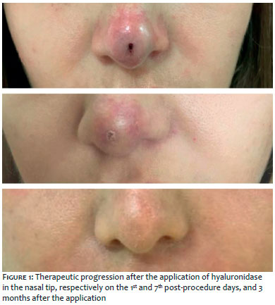
MBF, a 27-year-old female patient, sought medical attention due to dissatisfaction with the aesthetics of her nasal region, even though she had previously undergone rhinoplasty. After evaluation, 0.8 ml of hyaluronic acid (Emervel Deep®, Galderma, Otten, Switzerland) was applied in the columella and nasal tip region with a needle, aspirating before the injection, with no alteration being observed at the moment of the procedure. The patient returned to the practice 36 hours after, with intense local pain, pallor interspersed with livedo reticularis areas, compatible with arterial occlusion. Hyaluronidase Biometil® (Laboratório Biometil, São Bento do Sul, SC, Brazil) was used for reverting the picture in a single 1,600 IU application, associated with the following oral drugs: 100mg/day Aspirin® (Bayer, Barmen, Germany), 100mg 12/12 hours cilostazol (Eurofarma, São Paulo, Brazil), 40 mg/day prednisone (Eurofarma, São Paulo, Brazil), 20 mg/day rivaroxaban (Bayer, Barmen, Germany), 500mg 12/12 hours ciprofloxacin (Eurofarma, São Paulo, Brazil), and 300mg 12/12 hours clindamycin (Teuto-Brasileiro S/A, Anápolis, GO, Brazil), for 15 days. Doppler ultrasonography of the site was performed, showing normal arterial and venous blood flow. Three hyperbaric chamber sessions were carried out in the first three days, and warm compresses were applied several times a day for 7 days. The patient evolved with progressive improvement and complete resolution of the condition after 3 weeks.
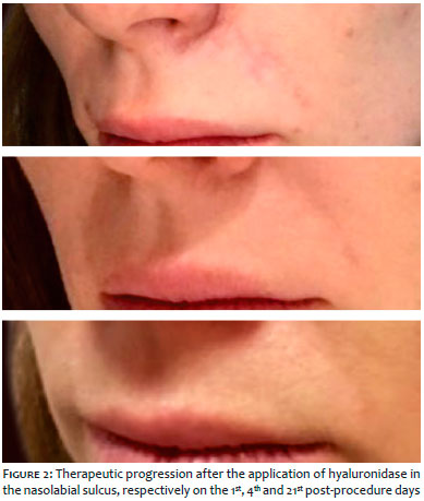
MSM, a 31-year-old female patient, sought medical attention aimed at undergoing facial harmonization treatment. Hyaluronic acid was applied to several sites of the face, among them in the deep proximal nasogenian sulcus (piriform fossa) – 0.1ml Emervel Deep® (Galderma, Otten, Switzerland), with a needle positioned at 90º and aspiration prior to the injection. An erythematous-purplish area emerged instantly, extending from the left lateral region of the nasal wing to the medial region of the ipsilateral nasogenian sulcus. In face of the persistence of the picture and hypothesis of arterial occlusion for 12 hours, the authors decided for the early degradation using 400 IU of hyaluronidase (Biometil®) and 100 mg/day sildenafil, 40 mg/day Clexane (Sanofi-Aventis Farmarcêutica Ltda, Suzano, São Paulo, Brazil), 40 mg/day prednisone (Eurofarma, São Paulo, Brazil), 500mg 12/12 hours ciprofloxacin (Eurofarma, São Paulo, Brazil), and 500mg 12/12 hours clarithromycin (Eurofarma, São Paulo, Brazil) for 15 days. Doppler ultrasonography of the site was also performed, showing normal blood flow. Improvement in the coloration was observed 2 days after the degradation, with complete resolution after 4 days.
With the increased numbers of hyaluronic acid based esthetic procedures, knowledge of the possible adverse reactions and respective handling is necessary. The pathophysiology of the embolic process caused by these procedures has not yet been elucidated, nevertheless the decrease in blood flow may be linked to the volume injected and the site of injection of high viscosity fillers.8 In both cases reported, alterations in the blood flow resulting from application of hyaluronic acid were observed, with typical manifestations at different time points. The chronology of the dermatological manifestations of embolic processes usually follows the sequence bleaching, emergence of vesicles, bedsores and necrosis, meaning it is crucial for the practitioner to recognize them early.1 The use of hyaluronidase is recommended for the degradation of the material, which leads to the normalization of the blood flow and prevents sequelae. The supportive therapy is based on increased perfusion, decreased inflammatory process and prophylaxis of associated infections, and may rely on vasodilators, corticosteroids, antimicrobials and antiaggregants.2
Comparing the cases reported and observing their development in Figures 1 and 2, it is possible to observe that the time for degrading hyaluronic acid had a significant impact on the prognosis and normalization of the local blood flow. In addition, it was possible to see that hyaluronidase only degrades injectable hyaluronic acid and does not interfere with what exists in the body, given that a large amount was used in Case 1 with absence of alterations whatsoever in the patient’s previous physiognomy.
Ritha de Cássia Capelato Rocha | ORCID 0000-0001-9792-758x
Main author, responsible for reviewing the manuscript and providing guidance to the other authors.
Luana Bertelli Castilho | ORCID 0000-0003-0680-8337
Responsible for reviewing articles on the subject and writing the report.
Danielle Mechereffe do Amaral Blaas | ORCID 0000-0002-3350-1405
Responsible for reviewing articles on the subject and writing the report.
Reinaldo Tavares Júnior | ORCID 0000-0003-2140-7160
Responsible for reviewing articles on the subject and for writing part of the report. Responsible for the imaging tests that prove revascularization after the procedure employing hyaluronidase.
Ana Paula Tavares | ORCID 0000-0001-7432-6730
Responsible for reviewing articles on the subject and writing part of the report.
Mariana Isis Wanczinski | ORCID 0000-0003-3118-6138
Responsible for reviewing articles on the subject and writing part of the report.
1. DeLorenzi C. New High Dose Pulsed Hyaluronidase Protocol for Hyaluronic Acid Filler Vascular Adverse Events. Aesthet Surg J.2017;37(7):814-25.
2. Glaich AS, Cohen JL, Goldberg LH. Injection necrosis of the glabella: protocol for prevention and treatment after use of dermal fillers. Dermatol Surg. 2006;32(2):276- 81.
3. Lee A, Grummer SE, Kriegel D, Marmur E. Hyaluronidase. Dermatol Surg. 2010;36(7):1071-77.
4. Rzany B, Becker-Wegerich P, Bachmann F, Erdmann R, Wollina U. Hyaluronidase in the correction of hyaluronic acid-based fillers: a review and recommendation for use. J Cosmet Dermatol. 2009;8(4):317-23
5. Brody HJ. Use of hyaluronidase in the treatment of granulomatous hyaluronic acid reactions or unwanted hyaluronic acid misplacement. Dermatol Surg. 2005;31(8 Pt 1):893-7.
6. Hirsch RJ, Brody HJ, Carruthers JD. Hyaluronidase in the office: a necessity for every dermasurgeon that injects hyaluronic acid. J Cosmet Laser Ther. 2007;9(3):182-5
7. Jones D, Tezel A, Borrell M. In vitro resistance to degradation of hyaluronic acid dermal fillers by ovine testicular hyaluronidase. Dermatol Surg. 2010;36(Suppl 1):804-9.
8. Cohen JL. Understanding, avoiding, and managing dermal filler complications. Dermatol Surg. 2008;34(Suppl. 1):S92-9.
 All content the journal, except where identified, is under a Creative Commons Attribution-NonCommercial 4.0 International license - ISSN-e 1984-8773


 Read in Portuguese
 Portuguese PDF
 Print
Print
 Send this article by email
 How to cite this article
 Submit a comment
 Mendeley
 Pocket