


Caroline da Silva Alves Palheta1,2; Wescley Miguel Pereira da Silva2; Rodrigo Paracampo Couteiro2; Paulo Ricardo Garcia da Silva2 ; Raíra Martins Trindade Souza2; Daniela Vale Dias2; Bianca Caroline do Nascimento Alho2; Andressa Miléo Ferraioli Silva2; Nara Macedo Botelho3; Francisca Regina Oliveira Carneiro4
Introduction: Microneedling is a technique aiming at stimulating the production of collagen as well as serving as drug delivery. Copaiba oil has healing and anti-inflammatory effects that have already been demonstrated in several animal models.
Objective: To evaluate the effect of copaiba oil associated with microneedle removal on the skin of rats.
Methods: Thirty rats were distributed in six groups of five animals each, subsequently undergoing: isolated microneedling, microneedling associated to mineral oil, and microneedling associated with copaiba oil. Biopsies were carried out in all animals at 14 and 30 days after the procedure. The parameters evaluated were: presence of collagen, fibroblasts and vessels, according to the following ratings: absence (0), mild (1), moderated (2) or intense (3).
Results: There was a statistically significant difference between the groups regarding the production of collagen at 14 days (p = 0.0091) and 30 days (p = 0.0357); and fibroblasts at 30 days (p = 0.0357). the group that used microneedling and copaiba oil, presented, after 30 days, a greater production of collagen and fibroblasts.
Conclusions: Copaiba oil associated with microneedling was capable of stimulating a greater production of collagen and fibroblasts in the skin of rats.
Keywords: dermatology; plants, medicinal; wound healing; collagen
Ablative techniques such as chemical and surgical peels have been employed for the treatment of scars; however, a current trend of the indication of less invasive procedures, in isolation or in combination, with the aim of reducing complications and allowing a faster return to work activities has been observed, taking into consideration the cost/benefit relationship.1,2 In this context, new techniques for the treatment of scars such as microneedling, that can stimulate the production of collagen without completely removing the epithelium, has gained more emphasis.3,4
Microneedling performed with the appropriate device has gained popularity as a technique that can treat scars, particularly acne sequelae.4 This device emerged from concepts described in 1995 by Orentreich and Orentreich, who reported the restoring effect of subcision or dermal needling in scars.5 In 1997, Camirand and Doucet used a “tattoo gun” for the treatment of scars.6 However, only in 2005 Desmond Fernandes developed the percutaneous collagen induction therapy (PCI) with a microneedling device.7
Drugs or active substances associated to microneedling (drug delivery) have been more frequently used, which can act in the skin through channels created by the tissue trauma with the microneedles, with the aim of treating different types of der-matological issues, such as melasma, periorbital melanosis, atrophic facial scars and others.8-13 Among the most used substances is vitamin C, due to its antioxidant action.3 However, in the literature, there are no reports of the use of copaiba oil as drug delivery associated to the microneedling technique.
Copaiba oil, extracted from trees of the genus Copaifera, family Leguminosae-Caesalpinioideae, stands out because of its importance in Brazilian natural medicine. Its healing and anti-inflammatory effects were demonstrated in many animal models. One of those studies observed an increase in the lesion’s crust, granulation tissue and number of blood vessels when studying the morphological and morphometric aspects of the healing of open cutaneous wounds in rats treated with Copaifera reticulata oil.14,15
Despite presenting anti-inflammatory, bactericidal, antitumor and healing effect, among many others scientifically proven,15-18 the evaluation of the effects of the use of copaiba oil associated to microneedling in rats’ skin has not yet been studied.
This way, the objective is to perform a histopathological examination of the effect of copaiba oil associated to microneedling in rats’ skin of the species Rattus norvegicus.
Type of study and sample selection
The study is characterized as experimental, prospective, longitudinal and comparative. Thirty male rats of the species Rattus novergicus, Wistar lineage, weighting between 350g and 400g, approximately 120 days old were used.
Ethical aspects
The animals in this study were evaluated according to the current national legislation for the raising and use of animals (Federal law n. 11.794, 2008) and the norms of the Colégio Brasileiro de Experimentação Animal (Cobea). The study was approved by the committee of ethics in the use of animals (Ceua) of the Centro de Ciências Biológicas e da Saúde of the Universidade do Estado do Pará (Uepa) on 12/08/2015, protocol n. 18/2015.
The animals were randomly distributed in to six groups.
• Group microneedle 14 days (group MAG14), with 5 animals submitted to microneedling and euthanasia on the 14th day (D14).
• Group microneedle 30 days (group MAG30), with 5 animals submitted to microneedling on D0 and euthanasia on the 30th day (D30).
• Group microneedle + copaiba 14 days (group MAOC14), with 5 animals submitted to the microneedling technique on D0 and application of 1ml of copaiba oil, with euthanasia on the 14th day (D14).
• Group microneedle + copaiba 30 days (group MAOC30), with 5 animals submitted to the microneedling technique on D0 and application of 1ml of copaiba oil, and euthanasia on the 30th day (D30).
• Group microneedle + mineral oil 14 days (group MAOM14), with 5 animals submitted to the microneedling technique on D0 and application of 1ml of mineral oil, and euthanasia on the 14th day (D14).
• Group microneedle + mineral oil 30 days (group MAOM30): with 5 animals submitted to the microneedling technique on D0 and application of 1ml of mineral oil, and euthanasia on the 30th day (D30).
Surgical procedure
The procedure was initiated with the intraperitoneal injection of the anesthetics ketamine and xylazine in doses of 70mg/ kg and 7mg/kg, respectively. The rats were positioned in prone position. Trichotomy was then performed in an area of 5 x 10cm on the left lateral region and disinfection with chlorhexidine.
Subsequently, microneedling with a cylinder with 192 1.5 mm needles (Dr. Roller® MTO, Porto Alegre, RS, Brazil) was performed, which was positioned between the index finger and the thumb, with moderate pressure, applied in back and forth movements 10 times, in 4 directions (vertical, horizontal and diagonal) until a uniform pattern of petechiae was achieved.
In the Maoc group, immediately after the microneedling, the topical application of 1ml of copaiba oil (Copaifera reticulata) was also performed, which was supplied by the Empresa Brasileira de Pesquisa Agropecuária (Embrapa).
The animals were kept with food and water ad libitum, in individual cages with a safety net at the back to allow for anesthetic recovery.
According to the research protocol, skin samples were collected from each animal for histopathological Analysis with a 5 mm punch biopsy 14 (D14) and 30 (D30) days after the microneedling.
Painless induced death was performed by intraperitoneal administration of ketamine chlorate (210mg/kg) and xylazine chlorate (21mg/kg).
Copaiba oil associated with microneedling on rats 291
Histopathological examination
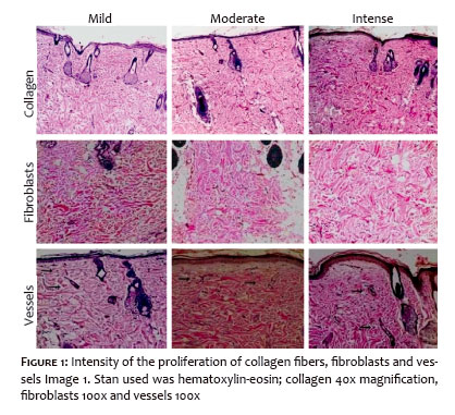
The sample collected was stored in a jar with formalin 10%, sent for histopathological processing and stained by hematoxylin-eosin (HE) and by Masson’s trichrome (MT) for the evaluation of collagen fibers. The slides were processed and analyzed by an experienced dermatopathologist using optical microscopy for examination and classification of the findings. The parameters analyzed were collagen fibers, proliferation of vessels and fibroblasts (Figures 1 and 2) that were proposed by Vieira et al in 2008; these parameters were then classified as absent (0), mild (1), moderate (2) and intense (3).19
Statistical analysis
The data obtained in the study were inserted into a Microsoft Excel 2010® spreadsheet and subsequently submitted to statistical analysis. For the analysis of the groups 14 and 30 days, test G was used. For the comparison between the three treatment groups MAG, Maom and Maoc the partitioning chi-square test, adopting as significance level < = 0.05 for both. The software BioEstat 5.3® was used.
On histology, all groups had some degree of collagen, fibroblast and vessel production after the studied procedure.
Regarding the production of collagen (Graph1), a statistically significant difference was observed between the groups MAG, Maom and MAO with 14 days (p = 0.0091) and 30 days (p = 0.0357).
Comparing the 14 days groups two by two, a difference between MAG and Maom (p = 0.0365) and MAG and Maoc (p = 0.0154) was seen and between the 30 days groups a statistically significant difference was seen between MAG and Maoc (p = 0.0036). the group submitted to microneedling and application of copaiba oil with biopsy in 30 days had the most significant presence of collagen, which was considered intense.
Regarding fibroblasts (Graph 2) a statistically significant difference was seen between the groups MAG, Maom and MAO after 30 days (p = 0.0357).
Comparing the 30 days groups two by two, a difference between Maom and Maoc (p = 0.0033) was seen. The group submitted to microneedling followed by the application of copaiba oil with biopsy in 30 days had the highest frequency of fibroblasts, which was considered intense.
When assessing neovascularization (Graph 3), no statistically significant difference was seen for vessels between the 14 days (p = 0.2873) and 30 days (p = 0.4060) groups.
Microneedling or percutaneous collagen induction (PCI) has been shown to be effective in the stimulation of collagen production and therefore has been used for the treatment of acne scars, rejuvenation and stretch marks.4,8,11,13 Among its advantages are: quick execution, low cost and easy approach for areas of difficult access.7
Besides, microneedling has been associated to the application of drugs with the objective of increase permeation of the skin and penetration of many active substances in order to improve the results of the procedure.4,8
With this perspective, an experimental study observed an increase in the expression of type I collagen and in the thickness of the epidermis in the skin of rats after microneedling with 1 mm roller, with the most obvious findings in the groups also submitted to the topical application of retinol 1% and vitamin C 10%.20
The length of the microneedles was also subject of research. One of them used 0.25 mm microneedles to increase epidermal penetration of secretory factors released by human embryonic stem-cell -derived endothelial precursor cells (hES-C-EPC) in 25 Asian women, with a global improvement of the pigmentation and wrinkles compared to microneedling only.21
Copaiba oil (Copaífera reticulata) has been studied in many fields and its healing and anti-inflammatory effects were already demonstrated in many animal models; however, its effect associated to PCI14-17 was not evaluated.
In the present study, we observed an increase in the amount of collagen fibers, fibroblasts and vessels in the skin of rats in all groups studied, what is probably related to the injury caused by the microneedles, which are capable of triggering a healing process with the release of growth factors, resulting in
the proliferation of fibroblasts, capillary formation and increased collagen synthesis.4,8,11,22
The group in which microneedling was performed with the application of copaiba oil and that was submitted to biopsy after 30 days showed a higher production of collagen and fibroblasts. This finding supports what was demonstrated by the study of Estevão et al, published in 2013 regarding topical application of copaiba oil on skin graft, where large fibroblasts and more collagen fivers were seen in skin flaps of rats treated with copaiba oil 10% ointment compared to controls.23
Giesbrecht et al, in an experimental study with 31 rats evaluated the effect of copaiba oil 1% ointment in burns and demonstrated an intense presence of fibroblasts and more organized collagen fibers.24
Regarding neovascularization, there was no statistically significant difference between the groups studied, even though all showed some degree of neoangiogenesis. This finding is in accordance with a study by Brito et al. who, in an experiment of healing of cutaneous wounds in rats using copaiba oil (C. multijuga), reported that the oil was capable of increasing the vascular network.25
Eurides et al also observed an increased formation of granulation tissue and blood vessels during the process of tissue repair in wound healing in rats, demonstrating that copaiba oil (Copaifera langsdorffii) is capable of increasing vascularization.26
Microneedling proposes a stimulation of the collagen without removing the epithelium completely, since it preserves an area of the skin that will aid in the repair process.4 In contrast, published studies with copaiba oil and healing frequently evaluate surgical wounds that alone can cause more damage to the skin with the formation of granulation tissue on which copaiba oil proved to be able to stimulate neovascularization.17,19,26
The mechanism of action of the active ingredients in copaiba oil is not completely clear. Most of its therapeutic properties is attributed to diterpenes. It is believed that from its chemical characteristics of one or more of the oil’s components and through its synergistic action, the effects found by the use of copaiba oil become evident.23,27
Copaiba oil associated to microneedling was capable of stimulating a higher collagen and fibroblasts production in the skin of rats, although no influence on neovascularization was shown when compared to microneedling alone.
We would like to thank the technical and scientific contribution of the dermatologist Maraya de Jesus Semblano Bittencourt in the preparation of the histopathological images of this study.
Caroline Da Silva Alves Palheta:
Conception and design, collection of data, interpretation of data, dermatological procedures, statistical analysis, elaboration and writing of the article
Wescley Miguel Pereira Da Silva:
Aid in the performance of the dermatological procedures, collection of data, interpretation of data, statistical analysis and writing of the article
Rodrigo Paracampo Couteiro:
Aid in the performance of the dermatological procedures, collection of data, interpretation of data, statistical analysis and writing of the article
Paulo Ricardo Garcia Da Silva:
Aid in the performance of the dermatological procedures, collection of data, interpretation of data, statistical analysis and writing of the article
Raíra Martins Trindade Souza:
Aid in the performance of the dermatological procedures, collection of data, interpretation of data, statistical analysis and writing of the article
Daniela Vale Dias:
Aid in the performance of the dermatological procedures, collection of data, interpretation of data, statistical analysis and writing of the article
Bianca Caroline Nascimento Alho:
Aid in the performance of the dermatological procedures, collection of data, interpretation of data, statistical analysis and writing of the article
Andressa Miléo Ferraioli Silva:
Aid in the performance of the dermatological procedures, collection of data, interpretation of data, statistical analysis and writing of the article
Nara Macedo Botelho:
Active scientific and intellectual contribution for the study, conception and design, evaluation of the training, critical review and final approval
Francisca Regina Oliveira Carneiro:
Aid in the performance of the dermatological procedures, collection of data, interpretation of data, statistical analysis and writing of the article
1. Mandebelbaum SH, Di Santis EP, Mandelbaum MHS. Cicatrization: current concepts and auxiliary resources - Part I. An Bras Dermatol. 2003; 78(4):393-410
2. Lima AA, Souza TH, Grignoli LCE. The benefits of microneedling in the treatment of aesthetic dysfunction. Revista Científica da FHO-Uniara-ras. 2015; 3(1):92-9.
3. Doddaballapur S. Microneedling with dermaroller. J Cutan Aesthet Surg. 2009; 2(2): 110-1.
4. Lima EVA, Lima MA,Takano D. Microneedling experimental study and clas- sification of the resulting injury. Surg Cosmet Dermatol. 2013; 5(2): 110-4.
5. Orentreich DS, Orentreich N. Subcutaneous incisionless (subcision) surgery for the correction of depressed scars and wrinkles. Dermatol Surg. 1995;21(6):543-9.
6. Camirand A, Doucet J. Needle dermabrasion. Aesthetic Plast Surg. 1997;21(1):48-51
7. Fernandes D. Minimally invasive percutaneous collagen induction. Oral Maxillofac Surg Clin North Am. 2005;17(1):51-63
8. Kalil CLPV, Frainer RH, Dexheimer LS, Tonoli RE, Boff AL. Treatment of acne scars using the microneedling and drug delivery technique. Surg Cosmet Dermatol. 2015; 7(2): 144-8.
9. Budamakintla L, Loganathan E, Suresh DH, Shanmugam S, Suryanarayn
S, Dongare A, et al. A randomised, open-label, comparative study of Tranexamic Acid microinjections and Tranexamic Acid with microneedling in patients with melasma. J Cutan Aesthet Surg. 2013; 6(3): 139-43.
10. Sahni K, Kassir M. DermaFracT ™: an innovative new treatment for periorbital melanosis in a dark skinned male patient. J Cutan Aesthet Surg. 2013; 6 (3): 158-60.
11. Majid I. Microneedling therapy in atrophic facial scars: an objective assessment. J Cutan Aesthet Surg. 2009; 2(1): 26-30.
12. Liebl H, Kloth LC. Skin Cell Proliferation Stimulated by Microneedles. J Am Coll Clin Wound Spec. 2012; 4(1):2-6.
13. El-Domyati M, Barakat M, Awad S, Medhat W, El-Fakahany H, Farag H. Microneedling therapy for atrophic acne scars: an objective evaluation. J Clin Aesthet Dermatol. 2015;8(7):36-42.
14. Brito NMB, Kulay JR. L, Simões MJ, Mora AO, Ramalho LNZ, Novo NF et al. Aspectos morfológicos, morfométricos e imunohistoquímicos pelo PCNA, do colo uterino de ratas ooforectomizadas, após aplicação do óleo de copaíba. Acta Cir Bras. 2000; 15(suppl 1): 1-7.
15. Montes LV, Broseghini LP, Andreatta FS, Sant'Anna MES, Neves VM, Silva AG. Evidências para o uso da óleo-resina de copaíba na cicatrização de ferida - uma revisão sistemática. Natureza online. 2009; 7(2): 61-7.
16. Francisco SG. Uso do óleo de Copaíba (Copaifera officinalis) em inflamação ginecológica. Femina. 2005; 33(2): 89-93.
17. Yamaguchi MH, Garcia RF. Copaiba oil and its medicinal properties: a bibliographical review. Revista Saúde e Pesquisa. 2012; 5(1): 137-46.
18. Yasojima EY, Teixeira RK, Hoaut Ade P, Costa FL, Yamaki VN, Feitosa-ju-nior DJ, et al. Copaiba oil influences ventral hernia repair with vicryl® mesh? Arq Bras Cir Dig. 2015; 28(3): 186-189.
19. Vieira RC, Bombardiere E, Oliveira JJ, Lino-Júnior RS, Brito L., Junqueira--Kipnis AP. Influence of Copaifera langsdorffii oil on the repair of a surgical wound in the presence of foreign body. Pesq Vet Bras. 2008;28(8), 358-366.
20. Zeitter S, Sikora Z, Jahn S, Stahl F, Straub S, Lazaridis A, et al. Micro-ne- edling: matching the results of medical needling and repetitive tre- atments to maximize potential for skin regeneration. Burns. 2014; 40(5)966-73.
21. Lee HJ, Lee EG, Kang S, Sung J-H, Chung H-M, Kim DH. Efficacy of micro-needling plus human stem cell conditioned medium for skin rejuvenation: a randomized, controlled, blinded split-face study. Ann Dermatol. 2014;26(5):584-91
22. Liebl H, Kloth LC. Skin cell proliferation stimulated by microneedles. J Am Coll Clin Wound Spec. 2012;4(1)2-6.
23. Estevão LR, Medeiros JP, Baratella-Evêncio L, Simões RS, Souza MF, Evêncio Neto J. Effects of the topical administration of copaiba oil ointment (Copaifera langsdorffii) in skin flaps viability of rats. Acta Cir Bras. 2013;28(12)863-9.
24. Giesbrecht PCP. Efeitos da pomada de óleo de copaíba em queimadura cutânea em rato.Tese de Mestrado apresentada ao Programa de Mestrado em Ciência animal do Centro Universitário de Vila Velha. 2 011. 22-45.
25. Brito NMB, Simoes MJ, Gomes PO, Pessoa AF, Melo MCF. Effect of copaíba oil in the healing process of openskin wounds in rats. Rev Para Med. 1998; 12(1):28-32.
26. Eurides D, Mazzanti A, Gonçalves GF, Belleti ME; Silva LAF, Fioravanti MCS, et al. Aspectos morfológicos, morfométricos e histológicos da reparação tecidual de feridas cutâneas de camundongos tratadas com óleo de copaíba (Copaifera langsdorffii). Vet Not. 1988;4(1):77-82.
27. da Silva AG, Puziol PF, Leitao RN, Gomes TR, Scherer R, Martin ML, et al. Application of the essential oil from copaiba (Capaifera langsdorffii Desf.) for acne vulgaris: a double-blind, placebo controlled clinical trial. Altern Med Rev 2012; 17(1) 69-75.
This study was performed at the Laboratory of Experimental Surgery da Universidade do Estado do Pará (Uepa) - Belém(PA), Brasil.
 All content the journal, except where identified, under the Creative Commons Attribution 4.0 International licence - ISSN-e 1984-8773


 Read in Portuguese
 Portuguese PDF
 Print
Print
 Send this article by email
 How to cite this article
 Submit a comment
 Mendeley
 Pocket