


Sergio Schalka1; Wagner Vidal Magalhães2; Camila Cazerta3; Danielle Shitara4; Bianca da Silva Sufi5; Ananda Quadros6
Introduction: The use of nutraceutical products in order to attenuate signs of skin aging has been proposed in the literature. The stimulus to the synthesis of substances that support the dermis is one of the mechanisms linked to this effect.
Objective: To evaluate the effectiveness of a nutraceutical compound containing lutein, lycopene, vitamin C and manganese for the synthesis of collagen, elastin and hyaluronic acid.
Methods: An in vitro study was carried out based on the culture of human fibroblasts treated with the investigated product in different non-cytotoxic concentrations. The quantification of the elastin and collagen was performed with the assistance of spectrophotometric measurements. Hyaluronic acid was measure using a immunoenzymatic method.
Results: Cell cultures treated with the different concentrations of the product showed a significantly higher amount of synthesized collagen, elastin and hyaluronic acid as compared to the untreated culture (p <0.05).
Conclusions: The use of the nutraceutical compound containing lycopene, vitamin C, lutein and manganese has shown in vitro efficacy for stimulating the synthesis of collagen, elastin and hyaluronic acid, components that are crucial for providing the dermis' supporting structure, being responsible for the skin's firmness and elasticity.
Keywords: DERMATOLOGY; DIETARY SUPPLEMENTS; SKIN AGING
The interest of dermatology for oral nutrients supplementation aimed at controlling the cutaneous aging process has been increasing substantially.
Modern life, especially in large urban centers, imposes a condition that favors inadequate ingestion of nutrients, with an impact on the skin’s and adnexa’s health.1 Excessive work, stressful routines, inadequate sleeping periods, and diets based on industrialized foods with high levels of carbohydrates and fats, as well as reduced content of vitamins and trace elements, are conditions that lead to the development of a picture described as “hidden hunger”, meaning a borderline deficiency of certain nutrients, nevertheless without the clinical evidence of malnutrition.2
In the process of skin aging, in addition to the intrinsic cellular functional decline that is common to all organs, there are extrinsic factors, such as ultraviolet radiation, which can intensify the aging process via a complex biological mechanism that affects the various skin layers, is special the dermis’ connective tissue.3 Alterations in the components of the extracellular matrix responsible for the supporting structure of the dermis 4 – elastin, collagen and hyaluronic acid – induce the loss of viscoelasticity in the cutaneous tissue, with reduction of firmness and elasticity, clinically culminating with the emergence and accentuation of wrinkles, furrows and flaccidity.5
In the pathogenesis of photoaging, reactive oxygen species have a central role, consuming and damaging enzymatic and non-enzymatic antioxidant systems of the skin, destabilizing molecules and triggering chain reactions, causing damage to membranes and structural proteins.3 One of the primary events in reactive oxygen species (ROS) induced photodamage is the activation of transcription factors – such as the nuclear factor kappa B (NFkB) and the activator protein-1 (AP-1). These factors are involved in the regulation of the expression of several genes responsible for inflammation, tissue remodeling, oncogenesis, apoptosis and many degenerative processes associated with aging.6,7 Baseline levels of matrix metalloproteinases (Matrix Metalloproteinases - MMPs) are higher in aged skins as compared to younger skins, with the activation of AP-1 leading to an increase in the levels of MMPs, with greater collagen and elastin degradation.5
Recent evidence has shown that diets with a high content of vegetables, fruits and grains may reduce the risk of numerous diseases, with this benefit being linked to the presence of antioxidant substances.8
Antioxidants act on different levels protecting organisms against free radicals and are the first defense mechanism aimed at preventing their formation, particularly by inhibiting chain reactions with iron and copper.
In addition, antioxidants are capable to intercept free radicals generated by the cellular metabolism or exogenous sources, preventing their action on lipids, amino acids, polyunsaturated fatty acids’ double bond, and DNA bases, avoiding cellular structural damage.9
Carotenoids and vitamin C stand out among nutrients with antioxidant action intended for the prevention and treatment of cutaneous aging. They are not synthesized by the body and should be acquired through diet or oral supplementation.10
Vitamin C is a powerful free radicals neutralizer. Its use, either topically or orally, has been proposed in programs aimed at mitigating the aging process.5
Previous studies have demonstrated the benefit of using carotenoids in the prevention and treatment of damages caused by sunlight and photoaging.10 Oral supplementation with lycopene and lutein has been evaluated within a program of aging prevention and treatment, with encouraging results.11
In this way, the objective of the present study was to evaluate the efficacy of a nutraceutical product containing lycopene, lutein, vitamin C and manganese in the synthesis of the skin’s structural supporting elements, using human fibroblast culture models.
Ethical aspects
The experimental conditions adopted – the use of human cells under optimum cultivation conditions – are in line with the current methodologies applied, accepted and validated by the international scientific community. The human cell cultures used in the present study were commercially acquired from qualified and certified international companies.
Methodological procedures Culture of human fibroblastsHuman fibroblasts (Clonetics, Cambrex / Lonza, USA) were commercially obtained, grown and expanded in culture medium containing 90% RPMI-1640 and 10% fetal bovine serum (GIBCO Life Technologies, Baltimore, USA), plus antibiotic solution 0.02 µg/mL gentamicin (Sigma Chemical St. Louis, USA) and 0.25 µg/mL amphotericin B (GIBCO Life Technologies, Baltimore, USA), having been seeded in 75cm2 vials (Nunc, USA) and kept in a humidified incubator (Thermo Fisher) with 5% CO2 atmosphere at 37 °C. The culture medium was changed every 48 hours up until the cells had 70-80% confluence, being subsequently trypsinized and counted in Neubauer’s chamber for determination of cell density. After having been counted, the fibroblasts were established by the sowing of 1.5x10⁵ cells/well or 1x10⁴ cells/well in plates containing 6 or 96 wells (Nunc, USA), respectively.
For the determination of the non-cytotoxic concentrations of the nutraceutical compound, a preliminary cytotoxicity trial was performed using the XTT method (data not shown). Twenty-four hours after the initial seeding, the cell cultures were treated with three non-cytotoxic concentrations of the nutraceutical compound (0.065 mg/mL, 0.0325 mg/mL and 0.0163 mg/mL) for 72 hours. Subsequently, the culture’s supernatant was collected for evaluation of the proposed parameters.
Evaluation of the collagen, elastin and hyaluronic acidThe extracellular matrix elements were measured in the fibroblast culture’s supernatant using commercially available kits. The levels of collagen and elastin were determined by colorimetric trial (Biocolor, Belfast, Ireland), while hyaluronic acid levels were quantified by immunoenzymatic trial (ELISA sandwich) (R&D Systems, USA). The data obtained from the quantification of collagen, elastin and hyaluronic acid were expressed in pg/mL, mg/mL and ng/mL, respectively, and computed based on the standard curve’s reference values.
Statistical analysisFor the characterization of the statistical data, a parametric method for analysis of variance (P) (ANOVA) was applied followed by a multiple comparison test, termed Dunnett. In all groups studied, those whose P values were less than 0.05 were considered statistically significant.
Studied productThe test product contains lycopene, vitamin C, manganese and lutein.
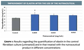
The nutraceutical product led to an increase in the synthesis of elastin in culture of human fibroblasts, incubated for 72 hours with the product, when applied at concentrations of 0.065, 0.0325 and 0.0163 (mg / mL), at 27%, 22% and 21%, respectively, when compared to the Basal Control group (Graph 1). The result was statistically significant (p <0.05) at the concentration of 0.065 mg/mL.
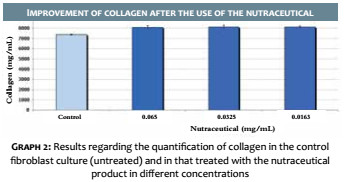
Evaluation of collagen synthesisThe nutraceutical product was capable to significantly increase (p <0.05) collagen levels at concentrations of 0.0650, 0.0325 and 0.0163 (mg/mL), yielding an increase of 9.49, 9.75 and 10%, 12%, respectively, vis a vis the Basal Control, when applied in culture of human fibroblasts for a period of 72 hours (Graph 2).
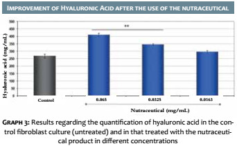
Evaluation of hyaluronic acid synthesisThe nutraceutical product increased the concentration of hyaluronic acid in culture of human fibroblasts, incubated for 72 hours with the product, when applied at concentrations of 0.065, 0.0325 and 0.0163 (mg/mL), at 53%, 29% and 11%, respectively, when compared with the Basal Control group. The result was statistically significant (p <0.05) for the 0.065 and 0.0325 mg/mL concentrations. (Graph 3)
The increase in life expectancy has raised concerns about the development of preventive and restorative measures for the aging process.17 Measures already recognized as effective for the prevention and treatment of cutaneous aging include the adequate use of sunscreens18 and cosmetic products with therapeutic action (cosmeceuticals)10,19 acting primarily through antioxidation and hydration.
More recently, evidence of the nutritional impact on the skin has been presented.4
A previous study19 evaluated the clinical efficacy of an oral supplement based on marine protein, concentrated acerola, concentrated grape seed extract, and tomato extract. After 360 days of use, the volunteers treated with the product had clinical evidence of improvement in the signs related to skin aging. These signs included the improvement of wrinkles, solar melanoses, hydration, lushness and improvement of the general appearance of the skin, associated to the ultrasonic improvement in the density of both photoprotected and photoexposed skin.
Among the agents studied, vitamin C and carotenoids – in particular lycopene and lutein – have been linked, especially in associations, to benefits for improving the quality of the dermis’ supporting structure.9, 10 In 2006, Heinrich et al.11 evaluated the use of oral supplementation with carotenoids containing lycopene and lutein, demonstrating improvement in clinical parameters such as density, skin thickness, roughness and scaling. The mechanism by which this action takes place is not yet completely established, however it is believed to involve these elements’ strong antioxidant properties, reducing the oxidative stress and its destructive effects on cellular structures, such as fibroblasts.5 Nevertheless, the effects of the association of vitamin C, lycopene, lutein and manganese assessed in the present study seems not to be restricted to the inhibition of the degradation of ROS-induced collagen and elastin. The results showed that there was a significant increase in the synthesis of collagen, elastin and hyaluronic acid, three essential components of the extracellular matrix that are responsible for the dermis’ supporting structure.
The present study’s findings may derive from the associated effects of lutein and lycopene, which are important agents with anti-inflammatory and antioxidant action;12 of manganese, a necessary element for optimal activity of the enzymes involved in the synthesis of glycosaminoglycans,13 and vitamin C, a cofactor in the hydroxylation of proline and lysine, essential amino acids in the process of collagen synthesis and capable to inhibit the accumulation of degraded elastin.14
Lutein is able to modify the extracellular matrix’s remodeling that occurs after exposure to ultraviolet radiation, through a beneficial effect on the regulation of metalloproteinases, in addition to inhibitory effects on cell loss, cell membrane damage and elastin expression.5
In addition, lutein increased the synthesis of hyaluronic acid in in vitro studies by increasing the expression of hyaluronan synthetase,3 which can justify the improvement in parameters such as skin roughness, since hyaluronic acid plays an important role in the maintenance of cutaneous hydration, anti-oxidation, as well as acting as a signaling molecule in response to skin damage.15,16
It is inferred from the stimulus to the synthesis of collagen and elastic fibers, and hyaluronic acid that the nutritional association presented is capable of positively interfering in the skin aging process, particularly in maintaining the dermis’ viscoelasticity properties, thus allowing a greater firmness and elasticity of the skin.
The use of an association containing vitamin C, lutein, lycopene and manganese has demonstrated the ability to stimulate the synthesis of collagen, elastin and hyaluronic acid in a fibroblast culture model, contributing to the improvement of the dermis’ supporting structure. As a result, it can slow down the skin aging process.
1. Viana V. Psicologia, saúde e nutrição: Contributo para o estudo do comportamento alimentar. Análise Psicológica. 2002;20(4):611-24.
2. Angelis RC. Fome oculta: bases fisiológicas para reduzir seus riscos através de alimentação saudável. São Paulo: Atheneu; 2001.
3. Wlaschek M, Tantcheva-Poór I, Naderi L, Ma W, Schneider LA, Razi-Wolf Z, et al. Solar UV irradiation and dermal photoaging. J Photochem Photobiol B. 2001;63(1-3):41-51.
4. Fisher GJ, Kang S, Varani J, Bata-Csorgo Z, Wan Y, Datta S, et al. Mechanisms of photoaging and chronological skin aging. Archives of dermatology. 2002;138(11):1462-70.
5. Philips N, Keller T, Hendrix C, Hamilton S, Arena R, Tuason M, et al. Regulation of the extracellular matrix remodeling by lutein in dermal fibroblasts, melanoma cells, and ultraviolet radiation exposed fibroblasts. Arch Dermatol Res. 2007;299(8):373-9.
6. Rittié L, Fisher GJ. UV-light-induced signal cascades and skin aging. Ageing Res Rev. 2002;1(4):705-20.
7. Pillai S, Oresajo C, Hayward J. Ultraviolet radiation and skin aging: roles of reactive oxygen species, inflammation and protease activation, and strategies for prevention of inflammation-induced matrix degradation - a review. Int J Cosmet Sci. 2005;27(1):17-34.
8. Pujol AP. Nutrientes no envelhecimento cutâneo. Nutrição aplicada à estética. Rio de Janeiro: Rubio; 2011. p. 265-76.
9. Yamamoto Y. Role of active oxygen species and antioxidants in photoaging. J Dermatol Sci. 2001;27 Suppl 1:S1-4.
10. Anunciato TP, da Rocha Filho PA. Carotenoids and polyphenols in nutricosmetics, nutraceuticals, and cosmeceuticals. J Cosmet Dermatol. 2012;11(1):51-4.
11. Heinrich U, Tronnier H, Stahl W, Béjot M, Maurette JM. Antioxidant supplements improve parameters related to skin structure in humans. Skin Pharmacol Physiol. 2006;19(4):224-31.
12. Addor FAS. Abordagem nutricional do envelhecimento cutâneo: correlação entre os efeitos em fibroblastos e os resultados clínicos. Surg Cosmet Dermatol 2011;3(1):12-6.
13. Schalka S, Steiner D, Ravelli FN, Steiner T, Terena AC, Marçon CR, et al. Brazilian consensus on photoprotection. An Bras Dermatol. 2014;89(6 Suppl 1):1-74.
14. Costa A, Pereira ESP, Fávaro R, Pereira MO, Stocco PL, Assumpção EC, Ota FS, Langen SSB. Resultado de 360 dias de uso de suplemento oral à base de proteína marinha, acerola concentrada, extrato de semente de uva e extrato de tomate em mulheres portadoras de envelhecimento cutâneo. Surg Cosmet Dermatol. 2011;3(4):302-11.
15. Lee EH, Faulhaber D, Hanson KM, Ding W, Peters S, Kodali S, et al. Dietary lutein reduces ultraviolet radiation-induced inflammation and immunosuppression. The J Invest Dermatol. 2004;122(2):510-7.
16. Leach Jr RM. Role of manganese in mucopolysaccharide metabolism. Fed Proc. 1971;30(3):991-4.
17. Shami NJIE, Moreira EAM. Lycopene as an antioxidant agent. Revista de Nutrição. 2004;17(2):227-236.
18. Palombo P, Fabrizi G, Ruocco V, Ruocco E, Fluhr J, Roberts R, et al. Beneficial long-term effects of combined oral/topical antioxidant treatment with the carotenoids lutein and zeaxanthin on human skin: a double-blind, placebo-controlled study. Skin Pharmacol Physiol. 2007;20(4):199-210.
19. Li R, Turner SD, Brautigan DL. Xanthophylls lutein and zeaxanthin modify gene expression and induce synthesis of hyaluronan in keratinocyte model of human skin. Biochemistry and Biophysics Reports 2015;4:52-8.
This study was carried out at Medcin Instituto da Pele - Osasco (SP), Brazil and at Laboratório Chemyunion Sorocaba (SP), Brazil
 All content the journal, except where identified, under the Creative Commons Attribution 4.0 International licence - ISSN-e 1984-8773


 Read in Portuguese
 Portuguese PDF
 Print
Print
 Send this article by email
 How to cite this article
 Submit a comment
 Mendeley
 Pocket