


Agnes Mayumi Nakano Oliveira1; Tatiana Cristina Pedro Cordeiro de Andrade2; Tabata Yamasaki Martins2; Gustavo Longhi Bedin3; Jaison Antonio Barreto4; Adauto José Ferreira Nunes5
Cutaneous metastases of internal malignancy are rare, and their incidence ranges from 0.7 to 9% among all cancers. They account for 2% of all skin tumors. They occur due to the growth of cancer cells in the dermis or subcutaneous cellular tissue, originating from internal neoplasia. Cutaneous metastasis arising from squamous cell carcinoma of the esophagus is rare, accounting for less than 1% of cases. We report a case of squamous cell carcinoma of the esophagus diagnosed after metastatic cutaneous manifestation in the abdomen that evolved to death due to disseminated metastatic tumor invasion.
Keywords: CARCINOMA, SQUAMOUS CELL; SKIN SEOPLASMS; NEOPLASM METASTASIS
Esophagus cancer is an extremely lethal neoplasm, with most patients diagnosed with local tumor invasion or distant metastasis. Cutaneous metastases of internal malignancy are rare, and their incidence has varied between 0.7% in a series of 865 autopsies reported by McWhorter and Cloud, to 9% in another study with 7,518 cases reported by Spencer and Helm; in a previous evaluation of five comprehensive studies, Rosen demonstrated a global incidence of approximately 2%.1 In a 2003 metaanalysis, 1,080 cases were found to have cutaneous metastasis out of 20,380 cases of cancer patients, implying an incidence of 5.3%. Melanoma, leukemia and lymphoma cases were excluded.1 In the same study it was observed that the tumor with greatest incidence of cutaneous metastasis was the breast adenocarcinoma, which was found in 24% of the cases.1 Cutaneous metastasis occur due the growth of cancer cells on the dermis or subcutaneous cell tissue, originating from internal neoplasms, and may happen due to hematogenous, lymphatic, or contiguous dissemination, and, in rare cases, iatrogenic implantation.2, 3 Neoplasms with lymphatic dissemination, such as that of the breast, commonly lead to regional cutaneous metastases, and cancers of the lung and colon lead to distant hematogenous cutaneous metastasis.2 According to a meta-analysis, the most common location are the thorax (28.4% of cases), followed by the abdomen (20.2% of cases).1 Cutaneous metastases are classified as synchronous and metachronous, depending on the time elapsed between diagnosis of the primary site and its emergece.2 Synchronous metastases occur when the they are diagnosed simultaneously with the primary tumor; and metachronous ones, when they develop months or years after the appearance of the primary cancer. Most cases of cutaneous metastases are metachronous, and in 0.5% of the cases they are the first sign of the primary neoplasm.2 They may arise in various appearances: sclerodermiform, alopecia, zosteriform, inflammatory, telangiectasia, cicatricial, pseudomyxomatous, among others.3, 4 The most common form is the nodular one, which appears as painless, round or oval, and firm nodules.3 These nodules may arise singly or in sets and, in this last case, they occur in different anatomical sites. The histological examination of the lesions allows that the diagnosis be directed to the origin of the primary site.2, 3 Epidermoid carcinoma is associated with tumors of the lung, the esophagus and the oral cavity.3 When cutaneous metastasis has an unknown origin, immunohistochemical markers can be requested.3 Some studies have shown that half of the patients with cutaneous metastases die within the first six months of diagnosis.3 Cutaneous metastases in esophageal squamous cell carcinoma are rare and represent less than 1% of all cases.4 The authors of the present study report a rare case of synchronous cutaneous metastasis in the abdomen originating from esophageal squamous cell carcinoma, which aided to direct the diagnosis of the primary site.
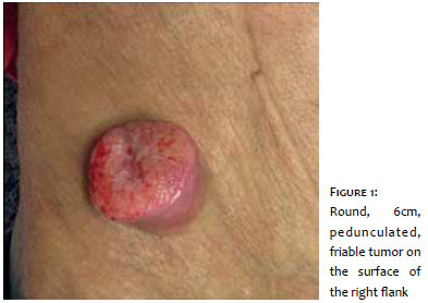
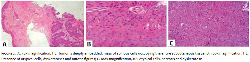
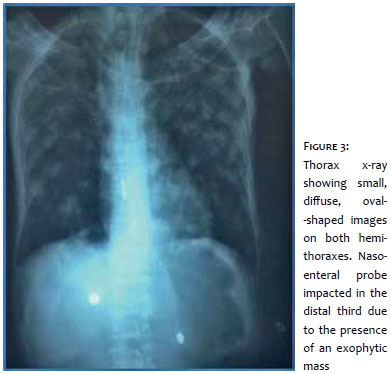
A 59-year-old male patient, smoker (50 packs/year) and former alcoholic, sought help at the Dermatology Service of the Instituto Lauro de Souza Lima, in Bauru (São Paulo, Brazil), reporting the appearance of nodules on his abdomen two months before. He also reported a gradual loss of weight of 5kg, dysphagia and anorexia during this period. During the physical examination, it was observed that the abdomen was depressed with a 6cm tumor, pedunculated, friable and oval on the surface of the right flank, and another 1cm erythematous and oval nodule on the left flank (Figure 1); Bilateral inguinal lymph node enlargement was also observed, ranging from 1cm to 3cm, with hardened lymph nodes adhered to deep planes. A biopsy was performed, and an abdomen ultrasound, chest x-ray, tumor markers and general tests were requested. The examinations showed: squamous cell carcinoma (SCC) in both lesions, evidenced in the anatomopathological examination (Figure 2), GGT = 383, ALP = 494, AST = 60, ALT = 21, total bilirubin of 5.17 (direct 2.32, and indirect 2.85), CA 15-3 = 101U/ml, Ferritin = 798, Serum Iron = 18, ESR = 109, HGB = 10.5 and chest x-ray with diffuse and small circular opacities in both hemithoraxes (Figure 3). The patient progressed with significant dysphagia, and a nasoenteral tube was implemented, which did not progress. Endoscopy was then performed, which revealed an exophytic mass that needed to be investigated. It was then hypothesized the presence of an esophageal SCC with cutaneous, pulmonary and hepatic metastases, with the patient passing away in a short period of time.
Esophageal cancer is an extremely lethal neoplasm with an insidious onset, leading to progressive and late obstruction. Most patients are diagnosed based on a local or metastatic tumor invasion, being no longer able to undergo curative treatment. The cutaneous metastases originating from the esophageal tumor represent less than 1% in their totality, and appear only in 1% of all patients with metastatic esophageal carcinoma. In the case reported, the authors observed a rare type of cutaneous metastasis, which preceded the diagnosis of the primary tumor, thus highlighting the importance of the knowledge that the dermatologist must have in all different forms of this entity.
1. Krathen RA, Orengo IF, Rosen T. Cutaneous metastasis: a meta-analysis of data. South Med J. 2003;96(2):164-7.
2. Azcune R, Spelta MG, Moya J, Lado Jurjo ML, Fontana,MI, Barbarulo AM, et al. Metástasis cutáneas de carcinomas internos, nuestra experiencia a propósito de 94 casos. Dermatol Argent 2009; 15:117-24.
3. Casimiro LM, Corell JJV. Metástasis cutáneas de neoplasias internas. Med Cutan Iber Lat Am. 2009;37(3):117-129.
4. Baijal, Rajiv, et al. "Cutaneous metastasis in esophageal squamous cell carcinoma." Indian J Med Paediatr Oncol. 2013; 34(1): 42–3.
This study was performed at the Instituto Lauro de Souza Lima (ILSL) Bauru (SP), Brazil.
 All content the journal, except where identified, under the Creative Commons Attribution 4.0 International licence - ISSN-e 1984-8773


 Read in Portuguese
 Portuguese PDF
 Print
Print
 Send this article by email
 How to cite this article
 Submit a comment
 Mendeley
 Pocket