


Jayme de Oliveira Filho1; Mariana Lacerda Junqueira Reis2; Andrea Penhalber Frange3; Nilceo Schwery Michalany4
Case report of a 16-year-old female patient with diagnosis of subungual exostosis in the right hallux with clinical and histopathological diagnosis, submitted to total excision of the lesion and follow-up for five years with excellent aesthetic result.
Keywords: EXOSTOSES; BONE NEOPLASMS; DERMATOLOGIC SURGICAL PROCEDURES
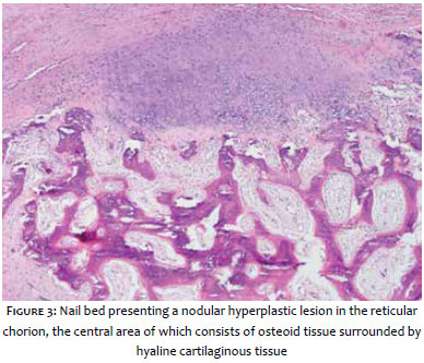
Subungual exostosis is a benign bone tumor, encapsulated by fibrocartilage, which mainly affects the distal hallux phalange, with a higher occurrence in adolescents and young female adults.1 Its etiology remains unknown, with a probable association with previous traumas, which would explain its greater occurrence in the first toe. Clinically, it presents as a painful nodule or painful hardened tumor at the distal end that produces lifting and deformity of the nail. Among the differential diagnoses, malignant tumors, viral wart, fibroma, pyogenic granuloma or subungual osteochondroma can be cited. Performing an imaging examination, such as an ultrasound or radiography allows for the visualization of abnormal bone growth with opacity and soft tissue involvement. Histologically, the tumor consists of a trabecular bone nucleus surrounded by a fibrocartilage capsule.2-4 Treatment is based on surgical removal and follow-up to avoid local recurrences.5-6
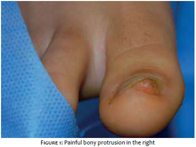
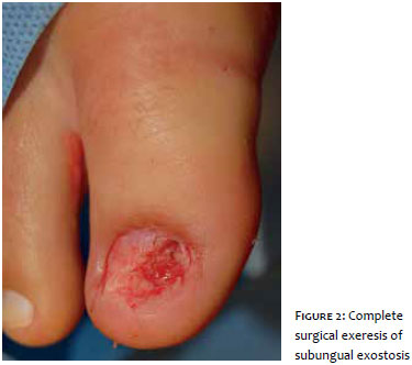
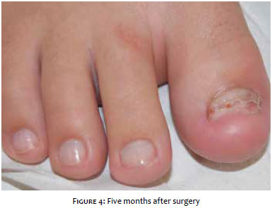
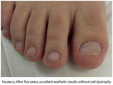
A 16-year-old patient, female, phototype II, presented a painful bone excrescence in the right hallux (Figure 1) during a dermatologic examination. An ultrasound of the preoperative lesion was performed, which elucidated the irregularity of the bone contour of the distal phalanx of the first toe. A complete marginal excision of the lesion was performed with the material that had been submitted for histopathological analysis, which confirmed the diagnosis of bone exostosis (Figures 2 and 3). Five months after surgery (Figure 4) both the radiography and ultrasound were unaltered, showing normal nail plates. During the postoperative period the patient was already saying how satisfied she was by the absence of pain and by the good appearance of the region. After five years, it was possible to observe an excellent aesthetic result, without nail dystrophy and absence of functional impairment of the affected hallux (Figure 5).
Subungual exostosis is a rare benign tumor, however it represents the bone condition most frequently associated with lesions in the nail, with probable traumatic etiology. The diagnosis is clinical and may be paired with radiography. In the case reported, the patient presented alteration on the distal phalanx with irregularity of the bone contour, characteristic of the disease. Pain is a very common symptom because it is a bone alteration. The presence of this symptom becomes important when considering differential diagnoses, such as malignant tumors, viral wart, fibroma, pyogenic granuloma or subungual osteochondroma.7 Surgical treatment with the resection of the whole tumor area is the recommended therapy, aiming to minimize damage to the nail bed and ungual matrix, and to avoid onychodystrophy, a common complication of the treatment. The patient in this case, after five years of the exeresis, presented excellent aesthetic result, without nail dystrophy, absence of functional impairment of the affected hallux, and, most importantly, no local recurrence.
1. Larralde, M; Boggio, P; Abad, ME; Pagotto, B; Castillo, A. Exostosis subungueal en un adolescente de 14 años.Arch Argent Pediatr. 2009;107(4): 349-52.
2. DaCambra MP; Gupta SK; Ferri-de-Barros F. Subungual exostosis of the toes: a systematic review. Clin orthop Relat Res.2014;472(4): 1251-9.
3. Small, O. Exostosis subungueal: a propósito del manejo quirúrgico / Subungueal exostosis: surgical management. Dermatol peru. 2009;19(2): 144-9.
4. Sampaio SAP, Rivitti EA. Onicoses."In": Sampaio SAP, Rivitti EA. Dermatologia. 3ªed. São Paulo: Artes Médicas; 2007. p.441-53.
5. Simon MJ; Pogoda P; Hövelborn F; Krause M; Zustin J; Amling M; Barvencik F. Incidence, histopathologic analysis and distribution of tumours of the hand. BMC Musculoskelet Disord. 2014; 15: 182.
6. Damron TA. Subungual exostosis of the toes: a systematic review. Clin Orthop Relat Res.2014; 472(4):1260-1
7. Valero J; Gallart J; Gonzalez D; Deus J; Lahoz M. Subungual squamous cell carcinoma and exostosis in third toe--case report and literature review. J Eur AcadDermatol Venereol. 2014; 28(10): 1292-7
This study was performed at the authors' private practices in São Paulo (SP), Brazil.
 All content the journal, except where identified, under the Creative Commons Attribution 4.0 International licence - ISSN-e 1984-8773


 Read in Portuguese
 Portuguese PDF
 Print
Print
 Send this article by email
 How to cite this article
 Submit a comment
 Mendeley
 Pocket