


Juliana Ribeiro Fernandes1; Elizabeth Leocadia Fernandes2; Denise Steiner3
Juvenile xanthogranuloma is a form of non Langerhans cell histiocytosis that mainly affects children. It usually emerges as asymptomatic yellow-brownish papules. The diagnosis is clinical and confirmed by histology. Due to its trend to involute, treatment is usually not recommended. Dermoscopy arises as a noninvasive diagnostic tool that reveals a typical pattern for this condition.
Keywords: XANTHOGRANULOMA, JUVENILE; DERMOSCOPY; HISTIOCYTOSIS, NON-LANGERHANS-CELL
Juvenile xanthogranuloma (JXG) is a common form of non-Langerhans histiocytosis. It comprises benign tumors of histiocytes, usually with spontaneous regression and that mainly affects children. It emerges as small-yellowish or brownish, single or multiple papules, most often asymptomatic. Visceral changes are rarely observed. The diagnosis is clinical and in doubtful cases, confirmation is carried out by histology. Treatment is usually not recommended in light of the self-limiting nature of this entity. It is worth to note that regression is capable of generating atrophy and hyperpigmentation.1 Dermoscopy is useful in assisting the diagnosis, since this condition has a typical pattern, characterized by an orange color background – described as the "setting sun" pattern, the presence of clouds of deposits that are pale yellow in color, and linear or arboriform vessels disposed from the periphery to the lesion's center.2-4
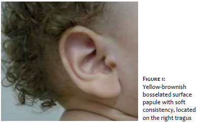
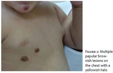
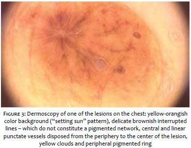
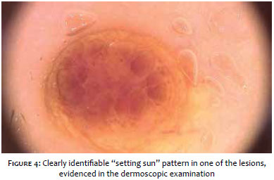
A one year-old child born and living in the city of Mogi das Cruzes (SP, Brazil), Fitspatrick's skin phototype III, presented at the consultation with a history of emergence of brownish papular lesions with onset at three months of age. The papules were asymptomatic, initially in the upper thoracic region, later on evolving to the abdomen, dorsum and face (mentum, and paranasal and preauricular regions). These lesions did not show signs of inflammation or itchiness, except for when the child accidentally traumatized them. The patient had no comorbidities or was in use of any medication, was up to date with the required vaccination, and there was no reference to similar cases in the family. The dermatological examination evidenced yellowbrownish papules with soft consistency and bosselated surface, measuring roughly 10mm in its longest diameter, located on the face (mentum, and paranasal and right preauricular regions), upper chest, dorsum and scalp's occipital region. The thoracic lesions showed a discrete yellow-orangish halo (Figures 1 and 2). There was no compromise of the patient's general condition. The dermoscopic examination allowed observing a yelloworangish color background ("setting sun" pattern), presence of delicate brownish interrupted lines (which do not constitute a pigmented network), punctate central vessels and other linear vessels disposed from the periphery to the center of the lesion, yellow-pale clouds and peripheral pigmented ring (Figures 3 and 4). After requesting an ophthalmologic evaluation, a decision was made for maintaining an expectant approach with a series of clinical follow-ups.
Juvenile xanthogranuloma (JXG) is a common form of non-Langerhans histiocytosis, arising as yellowish, sometimes pinkish asymptomatic papules, measuring between5 and 20mm in diameter, that may become yellow-brownish in color and display telangiectasias with time.1,3 About 70% of cases arise in early childhood,1 but children of all ages can be affected. Single lesions are more common, however children with less than six months often have multiple lesions. There is a predilection for the head and upper trunk segment. They tend to regress spontaneously around a year after the onset. Although uncommon, extra-cutaneous involvement, with lesions in the subcutaneous tissue, eyeball, liver and spleen is possible. The histological examinations of these lesions evidence a diffuse and dense pleomorphic histiocytic infiltrate, with the predominance of vacuolated cells in early stages and xanthomatous cells later on. The Touton cell (xanthomatous multinucleated, with nuclei arranged in the form of a ring is the most typical, nevertheless non-specific element of this condition.1,2,5 Dermoscopy is a useful tool in the diagnosis, in special when a decision is made for not to perform the lesion biopsy, as is the case in the present paper. Initially described by Palmer and Bowling,2 the dermoscopic pattern of juvenile xanthogranuloma consists of a yellow-orangish background with an erythematous halo (described as "setting sun" pattern), with pale yellow clouds corresponding to the to dermal xanthogranulomatous infiltrate (similar to the one found in sebaceous hyperplasia) and arboriform vessels disposed from the periphery to the lesion's center.1-5 In the present case, all these aspects were seen, confirming the clinical and dermoscopic diagnosis of JXG. It is possible to conclude that the dermoscopic examination provides valuable additional information for the diagnosis,3 thus avoiding invasive procedures, such as skin biopsy, in the pediatric population.
1. Belda Junior W., Chiacchio Di N.,Criado, P.R. Tratado de Dermatologia. 2 ed. São Paulo: Atheneu, 2014. p.2095-106.
2. Pretel M, Irarrazaval I, Lera M, Aguado L, Idoate MA. Dermoscopic ''setting sun'' pattern of juvenile xanthogranuloma. J Am Acad Dermatol. 2015;72(1 Suppl):S73-5.
3. Palmer A, Bowling J. Dermoscopic Appearance of Juvenile Xanthogranuloma. Dermatology. 2007;21593):256-9.
4. Rubegni P, Mandato F, Fimiani M.Juvenile Xanthogranuloma: Dermoscopic Pattern. Dermatology. 2009;218(4):380
5. KIM JH, Lee SE, KIM SC. Juvenile xanthogranuloma on the sole: Dermoscopic findings as a diagnostic clue. J Dermatol. 2011;38(1):84-6.
The present study was performed at the Dermatology Service of the Universidade de Mogi das Cruzes (UMC) - Mogi das Cruzes (SP), Brazil.
 All content the journal, except where identified, under the Creative Commons Attribution 4.0 International licence - ISSN-e 1984-8773


 Read in Portuguese
 Portuguese PDF
 Print
Print
 Send this article by email
 How to cite this article
 Submit a comment
 Mendeley
 Pocket