


Eloisa Leis Ayres1; Adilson Costa2; Adriana Chaib Ferreira Jorge3; José Euzébio Gonçalves Júnior4; Miriam Szrajbman5; Beatriz Sant'Anna6
Introduction: Melasma is a common pigmentary condition that affects exposed body areas, especially in the frontal and malar regions. Hydroquinone is an effective active principle in the treatment of hyperpigmentation, however, due to issues linked to its tolerability, many studies are being conducted aimed at developing alternative therapies with equivalent effectiveness.
Objective: To evaluate the efficacy and tolerability of a cosmeceutical formulation containing ellagic acid, hydroxyphenoxy propionic acid, yeast extract and salicylic acid in Brazilian patients with mild to moderate melasma.
Methods: Forty patients with mild to moderate melasma on the face used the cosmeceutical twice daily, combined with sunscreen for 90 days. Subjective assessments of efficacy and tolerability were carried out. Measurements of the MASI grade and the application of the MELASQoL-BP questionnaire were also performed. The evaluation of the skin's brightness and the colorimetric characteristics were obtained by colorimetry.
Results: After 90 days of treatment, a significant improvement could be observed in the clinical and colorimetric parameters evaluated, and in the quality of life questionnaire. In addition, the MASI score improved by 43%. The treatment was effective without causing adverse events.
Conclusions: The evaluated cosmeceutical formulation was proven as an effective alternative to hydroquinone for the treatment of melasma, with excellent cutaneous tolerability profile.
Keywords: HYPERPIGMENTATION; MELANOSIS; BLEACHING AGENTS
Melasma is a frequent pigmentary disease that manifests as symmetrical hyperpigmented macules on the skin,1 and affects exposed body areas, especially the frontal and malar regions.2,3
With a 90% prevalence in women, it mainly occurs during their reproductive age.4,5 It has higher prevalence in individuals of East Asian and Hispanic origins, as well in patients with high Fitzpatrick skin phototypes (IV to VI), and particularly in individuals who live in locations where incidence of UV radiation is intense.4,6-8
Melasma can still be classified according with its clinical and histologic characteristics.9 Regarding the body site, the pigment may be located in the epidermis, dermis or in both (mixed location).10-12 The relevance of this classification dwells is the fact that it can be instrumental in determining the most adequate treatment and prognosis.1,13 In fact, the dermal compartment is capable of controlling cutaneous pigmentation, since the latter is regulated by a complex melanogenic network in which both keratinocytes and fibroblasts synthesize growth factors and cytokines, such as the hepatocyte growth factor (HGF), the keratinocyte growth factor (KGF) and the stem cell factor (SCF), which in turn directly influence pigmentation.14,15
There are several therapeutic options for the treatment of melasma that act at different stages of melanogenesis. Among the inhibitors of the tyrosinase enzyme is hydroquinone, and azelaic and kojic acids. Topical corticosteroids act as non-selective suppressors of melanogenesis. The action of azelaic acid inhibits the reactive oxygen species, since some studies suggest that free radicals lead to increased production of melanin by melanocytes. There are also options of direct removal of melanin using procedures such as peels.2
Hydroquinone is still deemed as the most effective active principle in skin whitening treatments. Nevertheless, several studies are being conducted aimed at developing skin hyperpigmentation treatment alternatives with similar effectiveness, since many patients have poor tolerability and experience adverse events – such as ochronosis – and public health agencies – such as the FDA – has indicated the presence of issues involving hydroquinone's safety. An alternative to increase the whitening effect of these other molecules is to combine them in a single formulation.16,17
In a 12-week investigator-blind study, Draelos et al. demonstrated that a cosmeceutical formulation containing ellagic acid, hydroxyphenoxy propionic acid, yeast extract and salicylic acid was as effective as the combination of 4% hydroquinone and 0.025% tretinoin in improving the skin's hue, decreasing the spots' intensity and size and improving pigmentation in general.17 The tolerability problems experienced with the combination of 4% hydroquinone and 0.025% tretinoin (dry skin, for instance) were not observed with the new cosmeceutical.17
Another study by Draelos et al. demonstrated that the same cosmeceutical formulation was also effective in maintaining the results obtained with the combination of 4% hydroquinone and 0.025% tretinoin during the summer. The patients also had significant improvement in the skin's hue (p < 0.001), intensity and size of spots (p < 0.001 and p < 0.05, respectively) and overall hyperpigmentation (p = 0.002).18
Hydroxyphenoxy propionic acid led to a significant decrease in melanin production by melanocytes in an in vitro model, without affecting its viability.19 A diverse depigmenting mechanism involving this active principle is the transfer of melanin from the melanocyte cell to the keratinocyte.17 Ellagic acid is also an inhibitor of melanin production, and is found in many fruits, such as strawberries and raspberries. This active principle has a powerful antioxidant in plants and an antioxidant and anti-inflammatory in humans.17 A randomized study involving 54 patients showed that after 12 weeks, the treatment combining 0.5% ellagic acid with 0.1% salicylic acid is as effective as 4% hydroquinone.16
The yeast extract is obtained from cells of Saccharomyces cerevisiae, and its action mechanism consists in stimulating lysosomal degradation in the keratinocytes, which can assist in the degradation of melanin.17 Moreover, the yeast extract stimulates dermal fibroblasts, lending resistance to pigmentation recurrence to he skin.18
Salicylic acid acts increasing the skin penetration of the active principles described above, facilitating cutaneous exfoliation and the desquamation of keratinocytes containing melanin pigments.17
The present study was aimed at evaluating the effectiveness and tolerability after 90 days of treatment with a cosmeceutical formulation containing ellagic acid, hydroxyphenoxy propionic acid, yeast extract and salicylic acid (Advanced Pigment Corrector, SkinCeuticals, New York, United States) in Brazilian patients with melasma.
A monocentric, prospective, open clinical study was carried out with 40 patients (men and women, aged 18 to 55 years) clinically diagnosed with mild to moderate facial melasma for at least 12 months, with absence of uniformity in the skin's hue. The participants were instructed to use the investigated product twice a day (in the morning and in the evening), associated with sunscreen SPF 50 in the morning (Physical Fusion UV Defense SPF 50, SkinCeuticals) and neutral cleansing soap. The study lasted 90 days, with evaluations at 30, 60 and 90 days of treatment.
The study was conducted according to the standards of Good Clinical Practice (GPC), the international research standards for research with humans (Declaration of Helsinki), the resolution No 196 (10/10/1996) of the Brazilian National Health Council and amendments, having been approved by the Research Ethics Committee of the Universidade São Francisco (Bragança Paulista, SP, Brazil).
The selected patients underwent a dermatological clinical evaluation on the first visit for confirmation of the inclusion and exclusion criteria. All patients were instructed to use the investigated product according to the study protocol's recommendations, and answer the effectiveness subjective evaluation questionnaire and melasma patients quality of life questionnaire (MelasQoL-BP).20,21
Participants also underwent a standardized frontal view photograph (Visia® device), for the measurement of the main spot with the colorimeter Konica Minolta model CR400.
MASI – Melasma Area and Severity Index
In order to compute the MASI score, the evaluation of the hyperpigmented areas of the face was firstly carried out. For that end, the face was subdivided into four areas: frontal (F), right malar (RM), malar left (LM) and jaw (M), corresponding to 30%, 30%, 30% and 10% of the total area, respectively. The evaluation was preformed using standardized photographs obtained by the Visia® device.
The melasma in each of the four areas received numerical ratings ranging from 1 to 6: 1 (<10%), 2 (10-29%), 3 (30-49%), 4 (50-69%), 5 (70-89%) and 6 (90-100%). The pigment's intensity as compared to that of the normal skin (D) was evaluated in each area on a scale of 0 (absent) to 4 (severe). Likewise, the pigment's homogeneity (H) was evaluated on a scale from 0 (minimum) to 4 (maximal). The computation of the MASI score corresponded to the sum of the severity ratings for D and H, multiplied by the numerical value of the involved area (A). The maximum score was 48, and the minimum was 0.
Standardized photographs
The Visia® device was used on D0, D30, D60 and D90 to perform photographic records of the participants' face (frontal view) and evaluate the following attributes: total spots, spots visualized under UV radiation and brown spots.
Colorimetry
A Konica Minolta CR400 colorimeter was used on D0, D30 D60 and D90 for gauging the participants' facial skin coloration parameters. The device's light source generates different incidence angles, and the internal sensor receives the light reflected vertically by the surface in the color spectrum (Cielab, 1976). The present study evaluated the following parameters: L* (luminosity, ranging from 0 to 100, with values close to 0 representing darker colors and values close to 100 representing lighter/white colors); and ITA, which is the individual typological angle obtained by the formula ITA° = [Arc Tangent ((L*-50) / b*)] 180 / π. The angle ITA° is proportionally related to the skin's pigmentation, with narrower angles indicating greater pigmentation and wider angles, lesser pigmentation. The evaluated area was selected and properly recorded by dermatologist physicians in the D0 visit, with three points from this area being evaluated in all visits. The value recorded was the average of the three points.
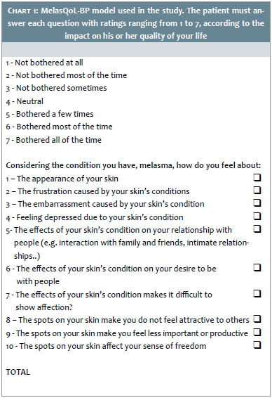
Melasma Patients Quality of Life questionnaire - MelasQoL-BP
Skin conditions like melasma can lead to a significant impact on the bearer's social, family and professional life, therefore quantifying this influence on the patient's quality of life is highly relevant. The MelasQoL was designed and validated in the English language and assists in the collection of valuable information on the impact of the pigment alterations on the quality of life. It consists of systematic form with 10 questions (Chart 1). The answer to each question ranges from 1 to 7, according to the melasma's impact on the patient's quality of life. In the present study, the participants were instructed to answer the questionnaire's Brazilian Portuguese language version (MelasQoL-BP, validated and published on the British Journal of Dermatology), on D0 and D90.2
Subjective assessments
The subjective evaluations were based on the physician's and patient's perceptions, and comprised effectiveness and safety evaluations of the investigated product. The subjective safety evaluations were performed at D0, D30, D60 and D90, when the physician focused on the following attributes of the patients' facial skin: erythema, edema, dryness and desquamation. The self-assessment carried out by the participants evaluated the following attributes: stinging, tingling, itching and burning sensation.
The subjective clinical assessment consisted in completing a comprehensive evaluation questionnaire on the clinical response after the dermatological analysis of the photographs taken at D0, D30, D60 and D90. The investigator dermatologist physician answered to the questionnaire evaluating the following attributes: hyperpigmentation, intensity of spots, coloration homogeneity, imperfections, texture, lushness, luminosity, hydration, smoothness, general appearance, erythema, edema, dryness and desquamation. The dermatologist physician's subjective evaluations were based on the photographs taken with the Visia® device.
For the assessment of the perceived effectiveness, the participants were asked to answer the questionnaire, which was aimed at capturing their opinion on the initial condition of their skin, as well as their perception regarding the improvement or worsening of the attributes during and after using the product. The questionnaire was applied on D0, D30, D60 and D90, with the following attributes: imperfections, texture, hydration, smoothness, general appearance, amount of spots and intensity of spots.
Statistical analysis
The effectiveness evaluations were performed on the 34 participants who completed the treatment, with the results from the subjective questionnaires and objective analyzes being assessed.
Descriptive statistics (i.e. mean, median, standard deviation, minimum and maximum values) were drawn from the participants' data (age, skin phototype, ethnicity). Regarding the categorical variables, the number of and percentage of individuals were provided for each answer category.
The Kruskal-Wallis paired test and the Dunn's multiple comparisons test were used for the analysis of the clinical questionnaire.
The marginal homogeneity test was used for the analysis of the questionnaires answered by the patients.
The Wilcoxon signed rank test for paired data was used for the analysis of the MelasQoL-BP.
For the assessment of the results obtained from the objective analysis and the MASI questionnaire, the ANOVA for repeated measures was used, followed by the contrast profile test, aiming at analyzing the development between visits. The data were converted into ranks due to the absence of normal distribution.
The adopted significance level was 5%.
Study patients' profile
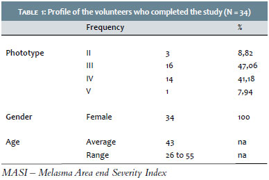
Forty patients were included and evaluated at baseline. After the 90 days, 34 patients completed the study; most of them were skin phototype III and IV women, with an average age of 43 years (min = 26 years, max = 55 years) (Table 1).
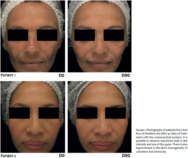
Figure 1 shows standardized photographs of patients #007 and #022, obtained with the Visia® device at T0 and T90 experimental timepoints. A significant improvement in the skin coloration's uniformity and a decrease in the spots' intensity can be observed.
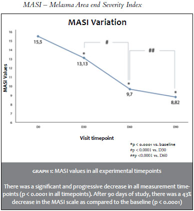
MASI – Melasma Area and Severity Index
The analysis of MASI scores showed statistically significant reductions both throughout the study period and between the experimental timepoints. At the end of the 90th day of the study, a statistically significant decrease of 43% in the MASI grade of melasma as compared to the baseline. Graph 1 depicts the values obtained from the Masi measurements.
Colorimetry
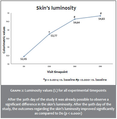
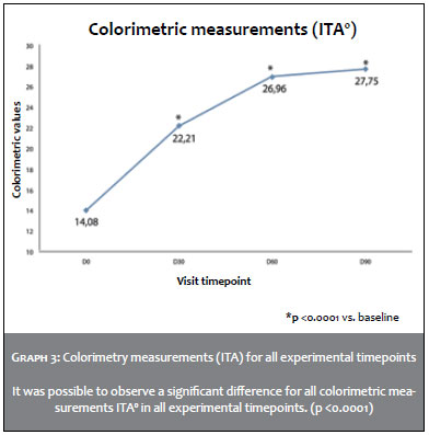
The analysis of colorimetric measurements showed a statistically significant increase in the skin's luminosity immediately after the 60 days of treatment (p < 0.0001 vs. baseline) and after 90 days of treatment (p < 0.0001 vs. baseline). The same could be observed for the visual typological angle (ITA), where there was a statistically significant increase immediately after 60 days of treatment (p < 0.0001 vs. baseline) and after 90 days of treatment (p < 0.0001 vs. baseline). Graphs 2 and 3 summarize the luminosity and ITA values for all experimental timepoints.
Visia®
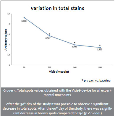
The score provided by the Visia® device offers an arbitrary global measure of the impact that the occurrence of a specific characteristic has on the patient's skin, taking into account, for this measurement, the size, total area and intensity of the characteristic in question – which in the present study were brown spots, total spots and spots visualized under UV radiation.
The results regarding brown spots evidenced a significant reduction in the score after 30 days of treatment (p < 0.0001 vs. baseline), 60 days (p < 0.0001 vs. D30) and 90 (p < 0.0001 vs. D30). Regarding the total spots, there was a significant reduction in the score immediately after 30 days of treatment (p < 0.012 vs. baseline) and 90 days of treatment (p < 0.0252 vs. baseline). Graph 4 and 5 summarize the values obtained with the Visia® device for brown and total spots.
For the spots visualized under UV radiation, there was no significant difference in the results during the study period.
Melasma Patients Quality of Life questionnaire - MelasQoL-BP
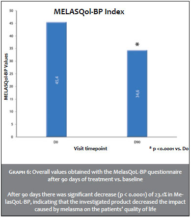
After 90 days of study, there was a significant reduction of 23.1% (p < 0.0001) in the overall MelasQoL-BP index (Graph 6), indicating that the investigated product provided a reduction of the impact on the patients' quality of life, observed in 79.4% of participants.
Subjective clinical evaluation
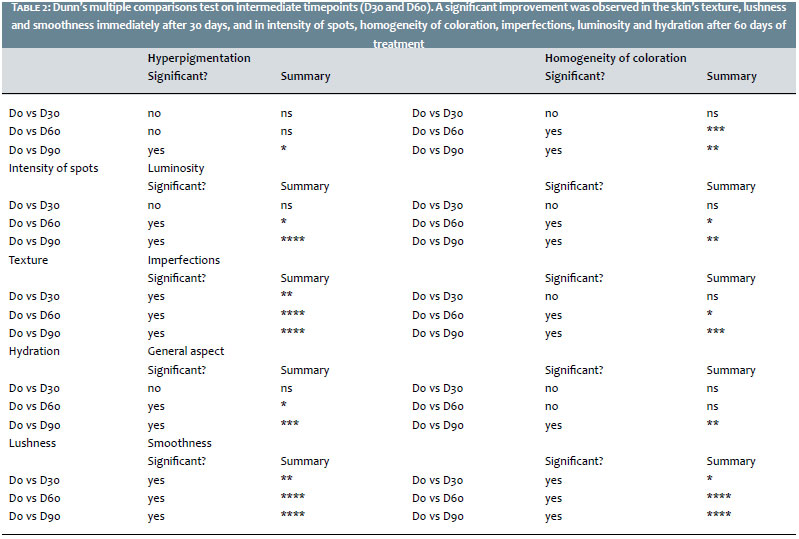
After 90 days using the investigated product, it was possible to observe a statistically significant improvement in hyperpigmentation (p = 0.0237), intensity of spots (p < 0.0003), coloration homogeneity (p = 0.0005), imperfections (p = 0.0004), general appearance (p = 0.0006), luminosity (p = 0.0003), lushness (p < 0.0001), texture (p < 0.0001), smoothness (p < 0.0001) and hydration (p = 0.0016). Table 2 shows the results of the Dunn's test for multiple comparisons at intermediate timepoints. There was significant improvement immediately after 30 days in texture, lushness and smoothness, and after 60 days in intensity of spots, coloration homogeneity, imperfections, luminosity and hydration.
Patients' perceived effectiveness
After 90 days of product use, the patients reported statistically significant improvement in the intensity of spots (p = 0.0001), imperfections (p = 0.0016), general appearance (p = 0.0025), and texture (p <0.0001). These results were statistically significant.
Tolerability
The tolerability to the studied cosmeceutical was deemed excellent by all participants in all visits. No erythema, edema, dryness or desquamation was observed during the treatment with the investigated cosmeceutical. Furthermore, during the 90 days of study, there were no reports of stinging, tingling, itching or burning sensation.
Of the various options for the treatment of melasma, hydroquinone is still the gold standard therapy. However, there are many adverse events related to its use, such as redness, skin dryness and photosensitivity. Its cytotoxicity is linked to the inhibition of DNA and RNA synthesis, abnormal melanosome formation and metabolic suppression of melanocytes.22
The present study was aimed at evaluating the effectiveness and tolerability profile of a cosmeceutical formulation containing ellagic acid, hydroxyphenoxy propionic acid, yeast extract and salicylic acid (Advanced Pigment Corrector, Skin-Ceuticals, New York, United States of America) in the treatment of mild to moderate melasma.
A treatment's depigmenting effectiveness requires the combination of active principles capable of acting on different stages of melanogenesis – including the dermal compartment – since it interferes in the pigmentation's physiology via growth factors and cytokines, as described by Kovacs et al.14 The cos-meceutical formulation has a combination of active principles that will act on different stages of the melanogenesis: tyrosinase inhibition and transfer of melanocyte melanin to keratinocytes (ellagic and hydroxyphenoxy propionic acids), cell renewal stimulation and stratum corneum exfoliation (salicylic acid), and fibroblast stimulation in the dermis (yeast extract), lending resistance to pigmentation recurrence to the skin.
The present study evidenced significant outcomes in the clinical and instrumental evaluations, as well as in the effectiveness perceived by the patients. A statistically significant reduction of 43% was obtained in the MASI score after 90 days of treatment, which could also be observed in the standardized photographs.
Colorimetry data also showed interesting outcomes linked to increases in the cutaneous luminosity and the ITA angle, instrumentally confirming the decrease in skin pigmentation. Further studies should evaluate other parameters, such as a*, which quantifies the reddish hues of the skin linked to the vascular component of melasma, since it was possible to observe a clinical reduction of skin redness in some patients, which is interesting to consider when treating pigmentary conditions such as melasma. A previous study conducted by Kin et al. showed an increase in both the number and the caliber of blood vessels, as well as increased expression of the pro-angiogenic factor VEGF (vascular endothelial growth factor) in the sites affected by melasma, confirming the relevance of future research in this field.23
The data obtained from the MelasQoL- BP questionnaire indicate a significant decrease in the impact on the patients' quality of life after 90 days, meaning a better adherence to daily treatment using the studied product. It is also worth noting that the decrease of the impact on the patients' quality of life was observed in almost 80% of the study patients.
Another important factor that should be taken into consideration in the treatment of hypermelanoses is the formulation's tolerability. The cosmeceutical formulation presented a good safety profile throughout the study's duration, with absence of reports of adverse events, therefore arising as an interesting alternative to be used as a monotherapy or maintenance treatment during the summer.
The treatment of melasma remains a major challenge in dermatology. At the same time, a great number of new active principles emerge, with possible whitening action. As a result, it is necessary to carry out studies that demonstrate these substances' therapeutic effectiveness and safety profile. According to instrumental and clinical data, as well as the patients' perception, the cosmeceutical formulation evaluated in the present study containing ellagic acid, hydroxyphenoxy propionic acid, yeast extract and salicylic acid was proven an effective alternative to hydroquinone for the treatment of melasma, with an excellent cutaneous tolerability profile.
1. Sarcar R, Arora P, Garg VK, Sonthalia S, Gokhale N. Melasma Update. Indian Dermatol Online J. 2014;5(4):426-35.
2. Steiner D, Feola C, Bialeski N, Silva FAM. Tratamento do Melasma: revisão sistemática. Surg Cosmet Dermatol. 2009;1(2):87-94.
3. Wanitphakdeedecha R, Manuskiatti W, Siriphukpong S, Chen TM. Treatment of Melasma Using Variable Square Pulse Er: YAG Laser Resurfacing. Dermatol Surg. 2009;35(3):475-82.
4. Gupta A, Gover M, Nouri K, Taylor S. The treatment of melasma: A review of clinical trials. J Am Acad Dermatol. 2006;55(6):1048-65.
5. Rendon M, Berneburg M, Arellano I, Picardo M. Treatment of melasma. J Am Acad Dermatol. 2006;54(5):S272-81.
6. Pandya A, Berneburg M, Ortonne J, Picardo M. Guidelines for clinical trials in melasma. Br J Dermatol. 2007;156(suppl. 1):21-8.
7. Grimes PE. Melasma. Etiologic and therapeutic considerations. Arch Dermatol. 1995; 131(12):1453-7.
8. Hexsel D, Lacerda DA, Cavalcante AS, Machado Filho CA, Kalil CL, Ayres EL, et al. Epidemiology of melasma in Brazilian patients: a multicenter study. Int J Dermatol. 2014;53(4):440-4.
9. Manstein D, Herron GS, Sink RK, Tanner H, Anderson RR. Fractional Photothermolysis: A New Concept for Cutaneous Remodeling Using Microscopic Patterns of Thermal Injury. Lasers Surg Med. 2004; 34(5):426-38.
10. Rokhsar CK, Fitzpatrick RE. The Treatment of Melasma with Fractional Photothermolysis: a pilot study. Dermatol Surg. 2005;31(12):1645-50.
11. Naito SK. Fractional photothermolysis treatment for resistant melasma in Chinese females. J Cosmet Laser Ther. 2007; 9(3): 161-3.
12. Lee HS, Won CH, Lee DH, An JS, Chang HK, Lee JH, et al. Treatment of Melasma in Asian Skin Using a Fractional 1,550-nm Laser: An Open Clinical Study. Dermatol Surg. 2009; 35(10):1499-504.
13. Rokhsar CK, Fitzpatrick RE. The Treatment of Melasma with Fractional Photothermolysis: a pilot study. Dermatol Surg. 2005; 31(12):1645-50.
14. Kovacs D, Cardinali G, Aspite N, Cota C, Luzi F, Bellei B, Briganti S, Amantea A, Torrisi MR, Picardo M. Role of fibroblast-derived growth factors in regulating hyperpigmentation of solar lentigo. Br J Dermatol. 2010; 163(5):1020-7.
15. Imokawa G. Autocrine and Paracrine Regulation of Melanocytes in Human Skin and in Pigmentary Disorders. Pigment Cell Res. 2004;17(2): 96-110.
16. Dahl A, Yatskayer M, Raab S, Oresajo C. Tolerance and efficacy of a product containing ellagic and salicilic acid in reducing hyperpigmentation and dark spots in comparison with 4% hydroquinone. J Drugs Dermatol. 2013; 12(1):52-8.
17. Draelos Z, Dahl A, Yatskayer M, Chen N, Krol Y, Oresajo C. Dyspigmentation, skin physiology, and a novel approach to skin lightening. J Cosmet Dermatol. 2013; 12(4): 247-53.
18. Draelos Z, Raab S, Yatskayer M, Chen N. Krol Y, Oresajo C. A method for maintaining the clinical results of 4% hydroquinone and 0,025% tretinoin with a cosmeceutical formulation. Journal of Drugs in Dermatology. 2015; 14(4): 386-30.
19. Cognis Patent US8247447 B2 Use of derivatives of 4-hydroxyphenoxy acetic acid.
20. Cestari TF, Balkrishann R, Weber MB, Prati C, Menegon DB, Mazzzotti NG. Translation and cultural adaptation to Portuguese of a quality of life questionnaire for patients with melasma. Med Cut Iber Lat Am. 2006;34(6):270-4.
21. Cestari TF, Hexsel D, Viegas ML, Azulay L, Hassun K, Almeida ART. Validation of a melasma quality of life questionnaire for Brazilian Portuguese language: the MELASQoL-BP study and improvement of QoL of melasma patients after triple combination therapy. Br J Dermatol. 2006 Dec;156 (Suppl 1):13-20.
22. Draelos ZD. Skin lightening preparations and the hydroquinone controversy. Dermatol Ther. 2007; 20(5):308-13.
23. Kim EH, Kim YC, Lee E, Kang HY. The vascular characteristics of melisma. J Dermatol Sci. 2007; 46(2):111-6.
This study was conducted at Kolderma Instituto de Pesquisa Clínica - Campinas (SP), Brazil
 All content the journal, except where identified, under the Creative Commons Attribution 4.0 International licence - ISSN-e 1984-8773


 Read in Portuguese
 Portuguese PDF
 Print
Print
 Send this article by email
 How to cite this article
 Submit a comment
 Mendeley
 Pocket