


Priscila Wolf Nassif1; Soraia Martos2; Neide Saturnino2
In search of a perfect aesthetic, facial filling procedures have been widely used. The presence of animal proteins or synthetic substances in some cutaneous fillers can cause serious allergic reactions, especially when performed by untrained professionals. There are not ideal, pure and free of side effects substances available in the marketplace. The present article is aimed at reporting a case where a facial filling was performed by a non-medical professional, resulting in foreign body reaction and severe skin infection, leading to deformities caused by the procedure carried out. The complications entailed invasive procedures for removal of the material used, and treatment with corticosteroids.
Keywords: MOHS SURGERY; CARCINOMA, BASAL CELL; NOSE NEOPLASMS; SURGICAL FLAPS; NASAL CARTILAGES
The use of cutaneous filling techniques has been growing, and the procedure's success is closely related to the chosen substance and the application method. 1 The cutaneous filling technique is included among the most commonly performed non-surgical procedures. Currently there is no commercially available ideal substance, pure and free of side effects. 2 There are several reports of complications with the use of fillers, such as inflammatory reactions, edema, hematoma, formation of nodules due to uneven distribution of the product or hypersensitivity and infection. 3 From an aesthetics standpoint, both the training of the applicator professional and the product's origin should be carefully verified. The establishments where the procedures will be performed must have sound conditions and be properly equipped. 4 The professional responsible for the procedure should have experience with the selection of the appropriate product and use of application techniques for each specific anatomical site, a capability that also requires extensive knowledge of facial anatomy. 5 The present study is aimed at describing a case of facial filling carried out by a non-medical professional that resulted in severe foreign body reaction and skin infection, leading to permanent deformities in the patient.
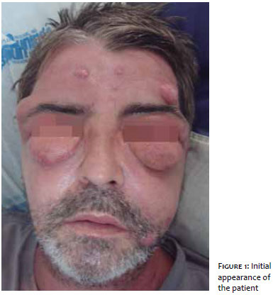
A 47-year-old male, was admitted to hospital complaining of allergic reaction to a bee sting occurred 20 days before the appearance of the symptoms. On examination the patient was febrile, tachycardic, with bilateral upper and lower eyelid edema, and erythematous-violet nodules with floating appearance in the nasogenian fold and forehead (Figure 1). Initial tests revealed leukocytosis (24,900 thousand/mm3) and the introduction of clindamycin and oxacillin based antibiotic therapy. On the second day of hospitalization, the oxacillin was replaced by ciprofloxacin, as recommended by the Infectious Diseases Clinic. In addition, the aspiration of the facial lesions was performed with a needle, yielding a purulent secretion, which was sent for culture. The culture for fungi and bacteria and Anti-HIV test came out negative. On the sixth day of hospitalization, with absence of improvement and after persistent questioning, the patient admitted to have undergone a procedure with polymethylmethacrylate injection (PMMA) in the face 20 days before, carried out by a non-medical professional.
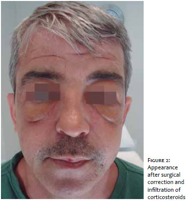
The histopathology of the facial nodule's biopsy showed granulomatous infiltrate throughout the dermis, observing microcysts amid the infiltrate, besides pseudoepitheliomatous acanthosis associated with micro-abscesses, corroborating the clinical proposition of foreign body granuloma. The patient was then referred to the plastic surgery department, where the surgical removal of the product was performed, as well as a monthly treatment with injectable corticosteroids for one year. There was partial improvement, nevertheless the patient remained with sequelae resulting from the procedure (Figure 2).
Currently there are different types of cutaneous fillers that are classified into temporary, semi-permanent (which should remain at least 18 months in the tissue) and permanent. Among the latter, are the PMMA and silicone. 6, 2 PMMA is composed of microspheres suspended in bovine collagen solution, carboxymethylcellulose or hydroxyethylcellulose. 7, 8 The silicone based filler is constituted by silicon derived polymers, and may be presented in the form of a gel, foam or liquid, depending on the degree of polymerization.9 The use of permanent fillers implies medical responsibility and requires precise injections. Mastering how to manage possible complications, the careful planning of the injection plans, as well as having the knowledge of the most indicated areas are imperative. The application of these substances can cause some undesirable side effects including local edema, inflammation, telangiectasia, hypertrophic scars, allergic reactions and granuloma formation. The latter generally arise between 6 and 24 months after the treatment, with a rate of occurrence of 0.6%, however they may also occur several years after the injections. 10
The possibility of the formation of biofilms, with the use of materials foreign to the host for implantation in soft tissues, is also noteworthy. Biofilms consist of gram-positive and gram-negative bacterial communities; nevertheless they may contain fungi, algae and protozoa. It is important to note that the biofilm hampers the action of antimicrobials, as it provides protection mechanisms for the bacteria against these agents. This may justify the infection observed in the site where the procedure was performed in the studied patient, entailing difficulty for the therapeutic response. Rosa and Macedo 1 offer some important and prudent recommendations on the use of filling substances: a) avoid carrying out these procedures on under-age patients, b) start performing filling procedures with absorbable substances before applying permanent substances, c) individually select patients and the correct indication of the procedure, since the substances are difficult to remove, d) adopt a judicious stance regarding the product manufacturer's recommendations, e) provide sterile conditions and limit the number of needle penetrations, thereby minimizing the risk of bacterial contamination, f) monitor patients scheduling return visits for the week following the application, since most bacterial infections occur within 8 to 10 days after the procedure, g) perform antiviral prophylaxis in patients with history of herpes labialis, h) pay attention to the amount injected into the corners of the mouth due to the fact that filling substances migrate easily, i) avoid performing the procedure close to the location where the supratrochlear arteries surface, during the correction of supratrochlear wrinkles.
In the present case, the patient underwent the application of the permanent filler PMMA in the face by a professional who was not qualified in the field of health sciences. Studies of the analysis of the histopathological reactions caused by PMMA revealed the presence of inflammatory infiltrates and a reduction in the quantity of the product according to the time elapsed after the performance of the procedure. In a proportion inverse to the amount of PMMA, the fibrosis and inflammatory reaction were increased with the passing of of time, leading to the formation of foreign body granuloma, as evidenced in the studied patient's pathology. Contrary to the literature, which quotes granulomas as a delayed reaction, 3, 10 the patient reported the symptoms 20 days after the procedure, linked to secondary infection of the lesions caused by inadequate asepsis techniques in the product application sites.
According to the researched literature, the isolated use of the substance could already cause adverse reactions.3 In the present case, where the procedure was performed by a non-medical professional, there were signs of exacerbation of the reactions, leading to sequelae resulting from the flaws in the technique employed and the lack of knowledge of the facial anatomy, in addition to the absence of minimum conditions of hygiene. The indiscriminate use of certain substances by unqualified professionals can lead to important consequences, resulting in social and aesthetic damage to the patient.
1. Rosa SC, Macedo JLS. Reações Adversas a Substâncias de Preenchimento Subcutâneo. Rev Soc Bras Cir Plást. 2005;20(4):248-52.
2. Crocco EI, Alves RO, Alessi C. Adverse events in injectable hyaluronic acid. Surg Cosmet Dermatol. 2012;4(3):259-63.
3. Lowe NJ, Maxwell CA, Patnaik R. Adverse Reactions to Dermal Fillers: Review. Dematol Surg. 2005;31(11 PT 2):1616-25.
4. Brasil. Agência Nacional de Vigilância Sanitária. [Internet]. Alertas de Tecnovigilância. [Acesso em 2014 Fev 15]. Disponível em: http://www.anvisa.gov.br/sistec/alerta/RelatorioAlerta.asp?NomeColuna=CO_SEQ_ALERTA&Parametro=1136
5. Funt D, Pavicic T. Dermal fillers in aesthetics: an overview of adverse events and treatment approaches. Clinical, Cosmetic and Investigatinal Dermatology. 2013:6.
6. Farahani SS, Sexton J, Stone JD, Quinn K, Woo SB. Lip Nodules Caused by Hyaluronic Acid Filler Injection: Report of Three Cases. Head and Neck Pathol. 2012;6(1):16-20.
7. Castro ACB, Collares MVM, Portinho CP, Dias PC, Pinto RA. Necrose facial extensa após infiltração com polimetilmetacrilato. Rev Bras Otorrinolaringol. 2007;73(6):850.
8. Silva MTT, Curi AL. Blindness and total ophthalmoplegia after aesthetic polymethyl-methacrylate injection: case report. Arq Neur Psiquiatria. 2004;62(3b):873-4.
9. 9 Sukhjit S, Burgett RA. Dermal filler agents: a pratical review. Curr Opin Ophthalmol. 2006;17(5):471-9.
10. Requena C, Requena L, Sanmart_ın O, Botella R. Histopathologic findings of granuloma caused by polymethylmethacrylate microspheres. Arch Dermatol. 2003;139(11):1505.
The present study was carried out at Faculdade Ingá - Maringá (PR), Brazil.
 All content the journal, except where identified, under the Creative Commons Attribution 4.0 International licence - ISSN-e 1984-8773


 Read in Portuguese
 Portuguese PDF
 Print
Print
 Send this article by email
 How to cite this article
 Submit a comment
 Mendeley
 Pocket