


Daiane Garcia Mercurio1; Patricia Maria Berardo G. Maia Campos2
Introduction: Confocal laser reflectance microscopy has been regarded as a tool with extensive application in dermatology, emerging as a revolutionary factor in the diagnosis of skin disorders and the evaluation of cutaneous characteristics. This is due to the fact that it allows cellular level visualization with almost histological resolution of cellular and tissular features through a non-invasive method, in real time. Objective: To evaluate the morphological and structural characteristics of skin aging in the epidermis and papillary dermis using confocal laser reflectance microscopy. Methods: Forty female volunteers with II, III, and IV skin phototypes were selected and divided into two age groups: young skin (18-35 years old) and aged skin (40-65 years old). The evaluation of the cellular characteristics of the different cutaneous layers was performed using a confocal laser reflectance microscope Vivascope 1500. Results: Using the confocal microscopy analysis, it was possible to observe an irregular pigmentation pattern, irregularly distributed keratinocytes, flattening of the dermal-epidermal junction, presence of damaged collagen fibers, and solar elastosis in the aged skin. Conclusions: Confocal laser reflectance microscopy is a technique that is highly useful for assessing morphological and structural characteristics of cutaneous aging at the epidermis and papillary dermis levels.
Keywords: MICROSCOPY, CONFOCAL; SKIN AGING; EPIDERMIS; DERMATOLOGY
Confocal laser reflectance microscopy (CRM) has been considered a tool of extensive application in the dermatological practice, arising as a revolution in the diagnosis of skin diseases and evaluation of cutaneous characteristics, by allowing cellular visualization with an almost histological resolution of cell and tissue characteristics using a non-invasive, real time method.1-3
Many studies describe CRM as a proper and reliable technique for the description and quantification of structural characteristics of the epidermis and upper dermis, overcoming the disadvantages of histological evaluation. The images obtained by the confocal microscope enable the evaluation of cutaneous characteristics such as the thickness of the different layers of the epidermis, the organization of keratinocytes, changes in the pigmentation pattern, the number of dermal papillae per area, the shape of the dermal papillae's contours, the size of sebaceous glands, the structure of the collagen network, the count and size of pores and microcomedones, and the evaluation of primary signs of cutaneous irritation.4-8
Due to the highly diverse possibility of technical applications, the elucidation and interpretation of CRM images related to skin aging provide subsidies for its application in the dermatology clinic, aiming at evaluating skin alterations resulting from the aging process.
The objective of the present study was to evaluate the morphologic and structural characteristics of skin aging in the epidermis and papillary dermis through the laser assisted CRM.
A prospective comparative study was carried out with 40 volunteers with skin phototypes II, III and IV, who were selected and divided into two groups, according to their age: 20 volunteers with young skin (18-35 years of age), and 20 volunteers with aged skin (40-65 years of age). 9 The study was approved by the Research Ethics Committee Involving Human Beings of the Faculdade de Ciências Farmacêuticas de Ribeirão Preto of the Universidade de São Paulo - USP.
The volunteers were instructed on the goals and methods of the study, having agreed to participate and signing the Free and Informed Term of Consent (CEP/FCFRP - Protocol number 273/2012). The study was conducted at the Faculdade de Ciências Farmacêuticas de Ribeirão Preto - USP.
The evaluation of the cell characteristics of the different layers of the skin was conducted using the confocal reflectance laser microscope VivaScope 1500®, which uses a 830nm wavelength laser source and an immersion objective lens capable of detecting 20 images per second. 4
The microscopic images were acquired using the Vivastack system, consisting of multiple confocal images at successive depths in a certain location of the tissue, with images obtained at every 1.5µm up to a depth of 37.5µm, at every 3.0µm up to a depth of 112.5µm and at every 4.5µm up to a depth of 132.5µm. 10, 11 The images were taken in the periorbital area of the volunteers' faces.
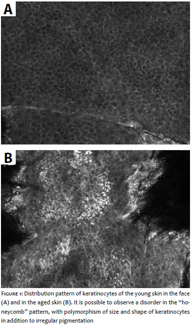
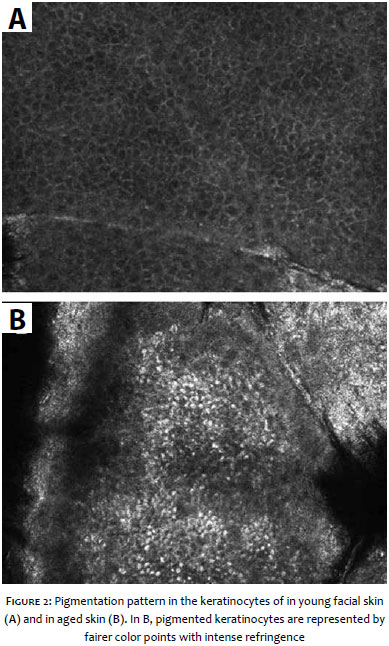
Regarding the pattern of the epidermis, there is a disorder in the "honey comb" pattern, with size and shape polymorphism in keratinocytes and also irregular pigmentation in the aged skin when compared to the normal aspect of the young skin (Figure 1). Regarding the pigmentation, it is possible to notice that there is an accumulation of melanin in the keratinocytes, which characterizes a pattern of irregular pigmentation (Figure 2).
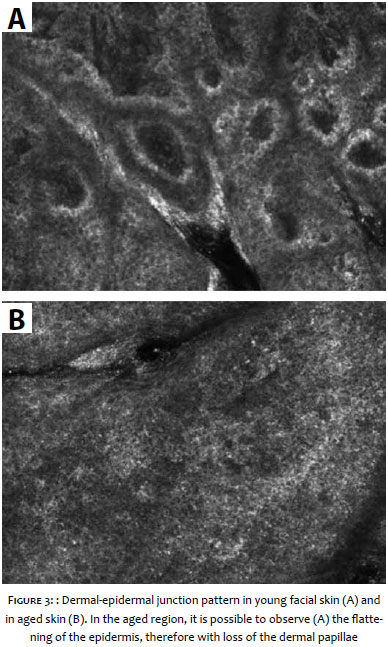
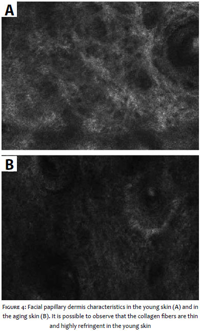
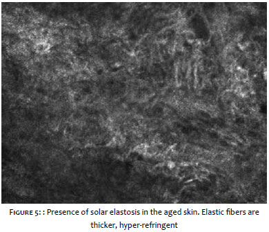
The dermal-epidermal junction's pattern is completely altered in the aged skin (Figure 3). It is possible to observe that a loss of dermal papillae takes place, entailing a flattening in the epidermis. In the papillary dermis, the young skin presents thin and highly refringent collagen fibers, whereas in aged skin, the fibers are hardly visible and have a shrunken and amorphous appearance (Figure 4). In some cases, it is possible to observe the presence of solar elastosis in the aged skin (Figure 5).
Based on the confocal microscopy analysis, it was possible to observe a uneven pigmentation pattern, keratinocytes unevenly distributed, flattening of the dermoepidermal junction, presence of deteriorated collagen fibers and severe solar elastosis in the aged skin.
The aged skin presents distinct morphological and structural characteristics, with disorganized keratinocytes resulting from lower cell renewal in this skin type. In addition, the technique has excellent application in the evaluation of uneven pigmentation in the epidermis given that based on the technique's principle, the melanin provides great contrast, facilitating the analysis and interpretation of images. 12
The morphological and structural characteristics of the dermal-epidermal junction are also related to the aging process, and this fact results from the intrinsic aging, being worsened with prolonged exposure to the sun. 13 The Laser CRM has proven a very useful tool in the evaluation of the integrity and morphology of the dermal-epidermal junction and its alterations resulting from the aging process. Moreover, it makes possible to evaluate the depth of the dermal papillae, through analysis of the sequences of images.
The images obtained by CRM of the papillary dermis in the present study were also observed by other research groups, in which thick and amorphous collagen fibers in the dermis were observed in older individuals, with loss of the fine reticulated collagen fibers, more common in younger individuals. 6, 7, 14
With the aging process, the differences between chronological aging and photoaging are observed in the extracellular matrix, revealing reduced expression of interstitial collagen genes in intrinsic aging and increased gene expression in the elastic tissue in photoaging. 15
The laser confocal reflectance microscope allows the assessment of clinical and subclinical characteristics of photoaging, being a very useful tool for the early detection of undesired cutaneous characteristics and providing subsidies for the prescription of safe and effective treatments in the prevention and improvement of the aging skin. It is therefore a useful technique to assist in the analysis of cutaneous characteristics that bother patients for their unaesthetic nature, and also to detect possible morphological and structural features that can be related to cutaneous dysfunctions.
The laser CRM is a technique that has extensive application in the assessment of morphological and structural characteristics of the aging skin in the epidermis and papillary dermis.
1. Branzan AL, Landthaler M, Szeimies RM. In vivo confocal scanning laser microscopy in dermatology. Lasers Med Sci. 2007;22(2):73-82.
2. González S, Gilaberte-Calzada Y. In vivo reflectance-mode confocal microscopy in clinical dermatology and cosmetology. Int J Cosmet Sci. 2008;30(1):1-17.
3. Nouveau-Richard S, Yang Z, Mac-Mary S, Li L, Bastien P, Tardy I, Bouillon C. Skin ageing: a comparison between Chinese and European populations. A pilot study. J Dermatol Sci. 2005;40(3):187-93.
4. Sauermann K, Clemann S, Jaspers S, Gambichler T, Altmeyer P, Hoffmann K, et al. Age related changes of human skin investigated with histometric measurements by confocal laser scanning microscopy in vivo. Skin Res Technol. 2002;8(1):52-6.
5. Sugata K, Nishijima T, Kitahara T, Takema Y. Confocal laser microscopic imaging of conspicuous facial pores in vivo: relation between the appearance and the internalstructure of skin. Skin Res Technol. 2008;14(2):208-12.
6. Longo C, Casari A, Beretti F et al. Skin aging: In vivo microscopic assessment of epidermal and dermal changes by means of confocal microscopy. J Am Acad Dermatol. 2013;68(3):e73-82..
7. Wurm EM, Longo C, Curchin C, Soyer HP, Prow TW, Pellacani G. In vivo assessment of chronological ageing and photoageing in forearm skin using reflectance confocal microscopy.Br J Dermatol. 2012;167(2):270-9.
8. Andrade, JP, Mercurio, DG, Maia Campos PMBG. Avaliação celular das estruturas cutâneas por meio da Microscopia Confocal de Reflectância. RBM: Rev Bras de Med. 2015;72:4-13.
9. Pathak, MA, Fitzpatrick, TB, Preventive treatment of sunburn, dermatoheliosis, and skin cancer with sun-protective agentsIn: Fitzpatrick TB, Eisen AZ, Wolff K, Freeberg IM, Austen KF (editors) Dermatology in General Medicine. McGraw-Hill: New York; 1993. p.1689-1717.
10. Bielfeldt S, Böhling A, Wilhelm PK. Bioengineering Methods to Assess Aging Parameters in the Depth of the Skin. SOFW-Journal. 2011;3:1-8.
11. Mercurio DG, Maia Campos PMBG, Simao JC. Biophysical, Morphological and clinical characterization of photodamaged skin for the development of effective cosmetic formulations. IFSCC CONFERENCE; 2013, Rio de Janeiro (Brazil). Book of Abstracts IFSCC Conference, 2013. p. 35-38.
12. Scope A, Benvenuto-Andrade C, Agero AL, Malvehy J, Puig S, Rajadhyaksha M, et al. In vivo reflectance confocal microscopy imaging of melanocytic skin lesions: Consensus terminology glossary and illustrative images. J Am Acad Dermatol. 2007;57(4):644-58.
13. Pinnell SR. Cutaneous photodamage, oxidative stress, and topical antioxidant protection. J Am Acad Dermatol. 2003;48(1):1-19; quiz 20-2.
14. Imayama S, Braverman IM. A hypothetical explanation for the aging of skin. Chronologic alteration of the three-dimensional arrangement of collagen and elastic fibers in connective tissue. Am J Pathol. 1989;134(5):1019-25.
15. McGrath JA, Robinson MK, Binder RL. Skin differences based on age and chronicity of ultraviolet exposure: results from a gene expression profiling study. Br J Dermatol. 2012;166(l2):9-15.
The present study was conducted at the Faculdade de Ciências Far- macêuticas de Ribeirão Preto da Universidade de São Paulo (FCFRP/USP) - Ribeirão Preto (SP), Brazil.
 All content the journal, except where identified, is under a Creative Commons Attribution-NonCommercial 4.0 International license - ISSN-e 1984-8773


 Read in Portuguese
 Portuguese PDF
 Print
Print
 Send this article by email
 How to cite this article
 Submit a comment
 Mendeley
 Pocket