


Thaís Abrão Cardoso1; Jeane Jeong Hoon Yang1; Ed Wilson Tsuneo Rossoe2
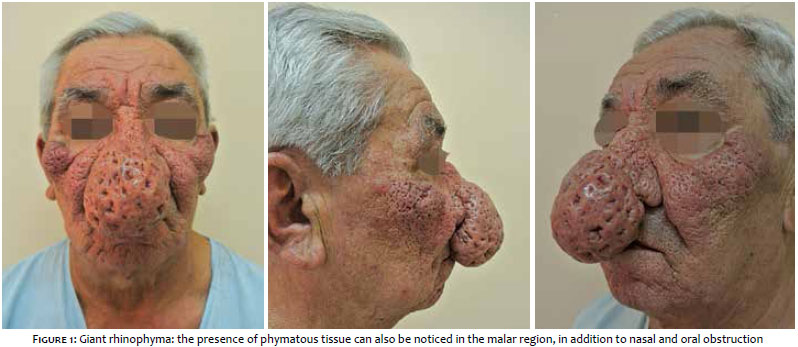
Rhinophyma is characterized by the hyperplasia of sebaceous glands in the nasal region, associated with a nodular thickening of the skin, enlargement of the pores, and fibrosis in the later stages. It occurs most commonly in male patients bearing rosacea, and may be associated with phymas in the region of the mentum (gnatophyma), ears (otophyma) and forehead (metophyma). The present article describes a case of giant rhinophyma with significant impact on the patient's quality of life, hampering breathing, sleeping, and eating. The lesion was successfully treated with electrosurgery.
Keywords: ELECTROSURGERY; RHINOPHYMA; DERMATOLOGIC SURGICAL PROCEDURES
Rosacea is a common condition in adults and is characterized by a variety of clinical manifestations. Its pathogenesis is multifactorial, nevertheless it is clearly related to vascular hyperactivity.1, 2 Rhinophyma is a late manifestation stage of rosacea and presents with hyperplasia of the sebaceous glands and the connective tissue of the nasal region in its progressive pathophysiology.3 It is possible to clinically observe swelling and subsequent lobulation, dilation of the pores, and unevenness in the surface of the nose, which may also increase in size over time. Patients with large-dimension rhinophyma can present with nasal obstruction accompanied by respiratory distress and feeding difficulty, as well as a disfiguring appearance and social isolation.1, 4
There are different treatment modalities described in the literature, such as electrosurgery, carbon dioxide laser, cryosurgery and dermabrasion. There is no consensus in the literature regarding the best technique, and all have advantages and disadvantages.4, 5
Case report
A 60-year-old male patient with a history of progressive modification of the nasal contour over the previous 10 years and with a worsening of the condition during the last three years and a significant enlargement of the nose. (Figure 1)
Electrosurgery was chosen as a therapy to approach the lesion. After asepsis and antisepsis were carried out with povidone-iodine, a bilateral anesthetic block was performed in the infraorbital nerve, with additional injection in the surface of the nasal skin.
The procedure was performed with a monopolar electrocautery (model SS500, Wem Equipamentos Eletrônicos®, Rio de Janeiro, Brazil), set at Cut Blend 1 mode, 2W potency, using the small round handle tip (0.3 mm thick and 1.2 cm in diameter). Due to profuse bleeding that occurred during the first surgery, two similar surgical steps were necessary at intervals of three months. An occlusive dressing with an ointment containing 0.6U/g collagenase and 0.01g/g chloramphenicol was applied, with daily changes for ten days. The total epithelization of the surface of the nose took place over 15 days.
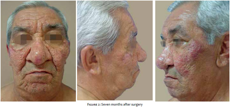
The result was very satisfactory from an aesthetic and functional point of view (Figure 2), and the patient had a significant improvement in his quality of life.
Rhinophyma is a part of late-stage rosacea. The invasive approach to treatment - either by conventional surgery, electrosurgery, carbon dioxide laser, or cryosurgery--is mandatory in the treatment and cure of the lesion.5, 6
Giant rhinophyma is not a common presentation, but is one that causes significant impact on a patient's quality of life, interfering with vital functions such as breathing and eating. A compromise of self-esteem and the entailed social isolation can also occur due to the grotesque appearance that it causes, or due to the myth of association with alcoholism.6 There is no consensus in the literature regarding the best therapeutic approach to rhinophyma.4, 5 Any therapeutic technique adopted should preserve the normal tissue, sparing the hair follicles of the dermis underlying the lesion, in order that reepithelialization can start with these follicles, lessening the risk of unsightly scars.4
In the present case, electrosurgery has proved to be a safe, effective, fast, and low cost therapeutic modality.
In addition to providing a satisfactory cosmetic result, the patient experienced a relief of respiratory distress in the immediate post-operative period. In addition to functional improvement, there was also a significant improvement in the patient's self-esteem and hence social life.1
1. Seiverling EV, Neuhaus IM. Nare obstruction due to massive rhinophyma treated using the Shaw scalpel. Dermatol Surg. 2011;37(6):876-9.
2. Sampaio SAP, Rivitti EA.. Dermatologia. 3ª ed.São Paulo Artes Médicas. 2007. p 400-1.
3. Webster GF. Rosácea e alterações relacionadas. In: Bolognia JL,Jorizzo JL,Rapini RP. Dermatologia. Rio De Janeiro: Elsevier ; 2011.
4. Lazzeri D, Agostini T, Spinelli G. Optimizing Cosmesis with Conservative Surgical Excision in a Giant Rhinophyma. Aesth Plast Surg. 2013;37(1):125-7.
5. Rohrich RJ, Griffin JR, Adams WP Jr. Rhinophyma review and update. Plast Reconstr Surg. 2002;110(3):860-9.
6. Curnier A, Choudhary S. Rhinophyma: dispelling the myths. Plast Reconstr Surg. 2004.114(2):351-4.
*This study was carried out at Complexo Hospitalar Padre Bento de Guarulhos (CHPBG) - Guarulhos (SP), Brazil.
 All content the journal, except where identified, under the Creative Commons Attribution 4.0 International licence - ISSN-e 1984-8773


 Read in Portuguese
 Portuguese PDF
 Print
Print
 Send this article by email
 How to cite this article
 Submit a comment
 Mendeley
 Pocket