


Maria Helena Lesqueves Sandoval1; Clarice Martins Caixeta2; Nathalia Meireles Ribeiro3
Introduction: Skin aging is a complex biological process that is characterized not only by clinical but also cellular alterations. The use of effective topical formulations for reducing and preventing oxidative stress is of utmost importance to delay cellular senescence. Objective: To evaluate the in vitro and in vivo efficacy of a formulation containing vitamin C, fragmented hyaluronic acid and mannose, in the prevention of skin aging. Methods: Clinical, subjective, and instrumental evaluations were carried out in 37 women (mean age: 46 years) and in vitro comparative evaluations of a dermal equivalent model contraction. Results: Statistical improvement was observed in the clinical parameters of hydration, sagginess, brightness, uniformity of skin color and amount of wrinkles after 3 months of treatment. A reduction by 12.6% in the volume of wrinkles 30 minutes after application and by 17.8% after three months of use was also evidenced by instrumental evaluation. There was greater contraction of the dermal equivalent model when compared to a commercial product for wrinkle reduction containing the same active principle. Conclusions: The product was proven effective in skin rejuvenation, by significantly reducing the amount of wrinkles. The in vitro study showed an increase in collagen synthesis, suggested by the greater activation of fibroblasts in the dermal equivalent model.
Keywords: SKIN AGING; ASCORBIC ACID; HYALURONIC ACID; MANNOSE
According to the Brazilian Society of Dermatology, aged skin is characterized as "thin, inelastic, presenting wrinkles and a deepening of expression lines." 1 In addition, it is possible to observe decreased hydration, loss of brightness, increased sagging, and uneven hue.1 Aging occurs both due to genetics or environmental factors and lifestyle habits, such as smoking, unhealthy diet, and lack of physical exercise.2-4 During intrinsic cutaneous aging a change in the genetic material takes place, with a decrease in cellular proliferation, which results in the loss of elasticity and the ability to regulate metabolism, in addition to a loss in the replication efficiency of the tissues.2 Chemical and enzymatic oxidations involving the formation of free radicals accelerate this phenomenon, generating oxidative stress, an effect having as its greatest damage the peroxidation of fatty acids in the cell membrane's double lipid layer, leading to apoptosis. 5 The skin has its own defense mechanism aimed at avoiding this process. Nevertheless, this mechanism's protective ability decreases with aging, and exogenous compounds can enhance the natural protection.6 Anti-aging products are employed in an attempt to minimize these characteristics of aging skin.7
Many antioxidants are currently available in cosmetic formulations and medications. The most common are: vitamins C and E, retinoids, resveratrol, coenzyme Q-10, idebenone, lipoic acid and flavonoids, among others.2
Pure vitamin C, also known as L-ascorbic acid (AA), is vital for the formation of collagen and elastin, increasing the skin's firmness , and is considered to be highly tolerable.8 AA is able to stimulate cell proliferation and collagen synthesis by dermal fibroblasts, regardless of patient age.9 Data indicate that topical application of vitamin C partially restores the anatomical structure of the dermal-epidermal junction in young skin and increases the number of nutritional capillary loops in the papillary dermis (near the epidermal tissue) in the skin of post-menopausal women.9 Haftek et al. have found significant clinical improvement in the clinical rating of superficial and deep wrinkles, sagging, firmness, uneven texture, and moisture levels in the skin of 20 women after six months of treatment with a dermocosmetic containing AA combined with madecassoside (5% stabilized vitamin C and 0.1% madecassoside-Redermic, La Roche-Posay Laboratoire Pharmaceutique, La Roche-Posay, France).10 These results were obtained by instrumental evaluations of the skin's elasticity and semiquantitative histological evaluations of the elastic fiber network of the papillary dermis.10, 11
Monosaccharides are ingredients widely used in anti-aging products as well. Among them is mannose,12 a monosaccharide of vegetal origin with a high water retention capacity, which is responsible for the synthesis of glycoproteins.12 It is able to alter the skin's light reflection properties, causing an optical smoothing effect.12
Another active principle that acts on skin rejuvenation is fragmented hyaluronic acid (HA). It is a polysaccharide produced by fibroblasts and keratinocytes, consisting of glucuronic acid and N-acetylglucosamine.13 It is one of the main substances in the extracellular matrix, in which collagen and elastin fibers are immersed. 14 It has a high hygroscopicity, and is responsible for maintaining the extracellular space and tissular hydration. 14
Farwick et al. performed tests with HA of different molecular weights (between 800kda and 20kda), having found that 50 kda is ideal for topical use, given that fragmented HA permeates the skin at three times the rate of non-fragmented HA. 14 Due to fragmentation, this active principle is able to act on the skin's rejuvenation, as well as to provide greater skin hydration, reducing the unwanted effects mediated by Toll-like receptors (TLR), which activate proinflammatory mediators. 14 Thus, it is clear that the topical application of fragmented HA can improve the skin's hydration and rejuvenation functions due to its ability to penetrate, which is achieved with the decreased size of its molecules. 14
The active principles vitamin C, mannose, and fragmented HA have a proven efficacy in reducing skin wrinkles, homogenization, and hydration. 10, 12, 14 The present study is aimed at investigating whether the association of these active principles can provide an improvement in outcomes and an increase in a product's anti-aging benefits for patients.
The study evaluated - in vivo and in vitro - the effects of a product containing the combination of these three active principles (5% pure vitamin C, 5% mannose, and fragmented HA) for the reduction of wrinkles and fine lines, in the immediate impression of skin uniformity, in the improvement of skin's firmness and hydration, and its comparative effectiveness in the contraction of dermal equivalents.
In vivo evaluation
A monocentric, blind study was carried out via clinical, subjective, and instrumental evaluations at the Allergisa Pesquisa Dermato-cosmética Ltda's research center, in Campinas (SP), Brazil, from February 14, 2013 to May 22, 2013. It was conducted in accordance with the Declaration of Helsinki's principles. Having been approved by the Research Ethics Committee of the Irmandade de Misericórdia de Campinas - Hospital Irmãos Penteado, 45 Brazilian women aged 40 to 50 years (mean = 46 years +/- 4.6) were recruited to take part in the evaluation. The volunteers were healthy and did not have active dermatoses in the facial area. They had mild or moderate signs of facial aging, and fell into Groups 2 and 3 according to the Glogau aging scale.15 They had fine lines, wrinkles, and signs of facial hyperpigmentation corresponding to Fitzpatrick skin phototypes I and III.16 All volunteers expressed their willingness to participate in the study by signing the Term of Free and Informed Consent (FICT) prior to undergoing any procedures provided by the protocol.
The volunteers used the test product during the study, applying it twice daily to the face for 12 weeks. In order to ensure that the product was being used appropriately, the product containers were weighed at each experimental visit. In addition, daily applications of sunscreen were carried out.
The test product contained 5% pure vitamin C, 5% mannose, and fragmented HA (Redermic Hyalu C, La Roche-Posay Laboratoire Dermatologique, La-Roche Posay, France). The associated sunscreen was Anthelios AC Cream Gel Dry Touch SPF 30 E PPD 15 (La Roche-Posay Laboratoire Dermatologique, La Roche-Posay, France).
Visits to the research center took place at baseline (T0) and after 28 (T28), 56 (T56) and 84 (T84) days of product use by the volunteers, when clinical assessments were performed by a dermatologist in addition to perceived efficacy and instrumental tests.
The clinical characteristics analyzed with a standardized visual scale (VAS - Visual Analog Scale)17 were: wrinkles, hydration, texture, sagging, brightness, and hue homogeneity. The perceived efficacy criteria were determined using a standardized questionnaire with categories for: tonicity, comfort, softness, and dryness. The devices used in the instrumental analysis were: Optical 3D Skin Measuring Device PRIMOS Compact 5.075 (GFMesstechnik GmbH, Teltow, Germany) and Visia CR (Canfield Imaging Systems, Farfield, United States). They were used for evaluating wrinkles and fine lines in the frontal and lateral region, and for taking standardized photographs, respectively.
Exploratory data analysis was performed (summary tables, graphs, frequencies and percentages). Results from experimental visits were compared using the paired Student's t-test, with unilateral hypothesis for the instrumental data and via ordinal logistic regression for the efficacy data. The confidence level considered in the comparative analysis was 95%. Software: XLSTAT 2013, STATA 10 and MINITAB 14.
In vitro evaluation
Evaluation of contraction of the dermal equivalents
The in vitro evaluation of the contraction of the dermal equivalents was carried out following the application of the test product, in addition to another commercial product for wrinkle reduction purposes also containing fragmented HA, and the control product.
The method used followed the ISO 10.993-518 international standards, based on the reduction of cellular growth caused by the exposure to a cytotoxic agent, reflected in the number of cells. The degree of growth inhibition, related to the concentration of the test sample, provided an indication of toxicity. The cells were kept in a culture, having been exposed to various concentrations of the test substance. The cultures were visually examined 24 hours after, with the highest tolerated dose (HTD) being estimated and the number of viable cells and/or total cell content determined. The number of cells present in the test substance was compared to that observed in the control, and the percentage of growth inhibition calculated. The concentration of the test sample, which showed a 50% inhibition of cell growth (IC50), was determined and expressed in mg/ml. This value allowed the comparison of the analyzed compound's relative cytotoxicity. 18
In order to assess the contraction of the dermal equivalent, the following methodologies were also implemented:
preparation of the dermal equivalent without the presence of glycosaminoglycans (GAGs); preparation of the dermal equivalent in the presence of GAGs.
The cell viability calculation - expressed in NRU (Neutral Red Uptake - the measure of the viability of cells in culture) - was performed for each concentration of the test substance by using the average NRU computed from the values of six replicates per test concentration. This value was compared with the average NRU of all values of the negative control (NC). The relative cell viability was then expressed as a percentage of the untreated control. 19
In vivo evaluation
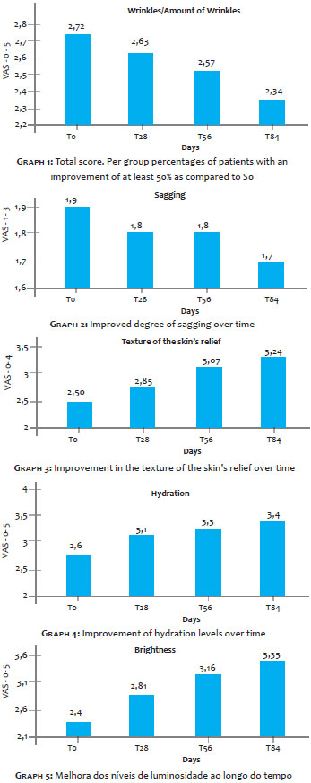
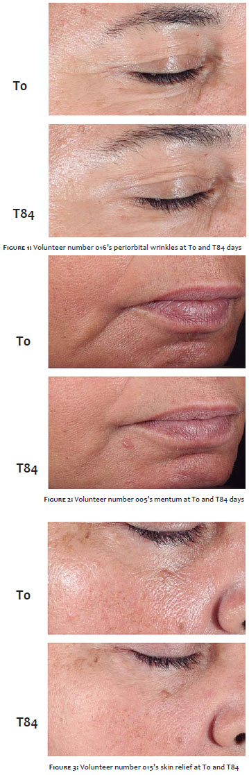
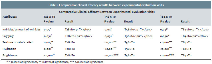
The results of the clinical trial showed that, according to the evaluation performed by dermatologists, the criteria wrinkle/amount of wrinkles, sagging, texture, hydration, and brightness improved significantly over time (Graphs 1 to 5 and Figures 1 to 3). The comparative results between experimental stages are shown in Table 1.
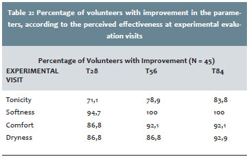
According to the evaluation of the perceived efficacy, volunteers reported improvement in tonicity, comfort, smoothness, and dryness as can be seen in Table 2.
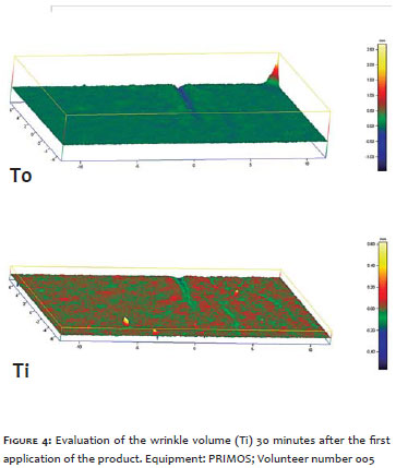
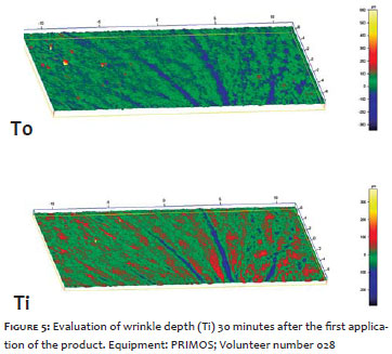
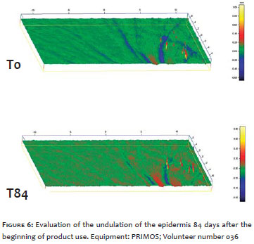
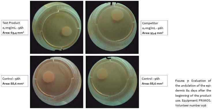
Based on the instrumental evaluation with the PRIMOS device, it was possible to observe reductions of 12.6% in the volume and 8.3% in the depth of wrinkles 30 minutes after the product's application. At the T84 measurement (after 84 days of use), there were reductions of 17.8% in the volume of wrinkles and of 10.2% in the epidermis' undulation (p<0.05) (Figures 4, 5 and 6).
In vitro evaluation
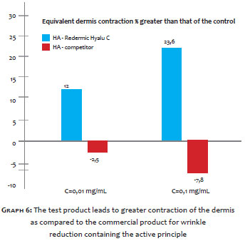
The in vitro analysis assessed the percentage of retraction (or contraction) of the dermal equivalent that was exposed to the test product and to the HA-containing commercial product for reducing wrinkles. Measurements were taken at 24, 48, and 96 hours of exposure, after an incubation time of four hours (the baseline for the start of the contraction). After 24 hours of exposure, the greatest contraction observed for the dermis treated with 0.01mg/ml test product was that of the control sample. Nevertheless, it was possible to observe 8.36% more contraction than in the control for the dermis treated with 0.1 mg/ml. The contraction difference, as compared to the control (untreated dermis), was best seen after 96 hours of exposure. For the dermis treated with 0.01 mg/ml, it was possible to observe a contraction 11.99% greater than that of the control, and the dermis treated with 0.1 mg/ml, a 23.55% greater contraction. These numbers suggest that the test product increased the contraction rate in the treated equivalent dermis as compared to the untreated dermis.
In the dermis treated with the wrinkle-reducing commercial product containing HA, an increase in the contraction rate was observed after 24 hours of exposure. When compared to the untreated control, the dermis treated with 0.01 mg/ml had 3.02% more contraction, while the one treated with 0.1 mg/ml had 7.87% more contraction. On the other hand, less contraction (as compared to the untreated control) could be better observed after 96 hours of incubation. The dermis treated with 0.01 mg/ml had 2.51% less contraction than the control, while the dermis treated with 0.1 mg/ml had 7.77% less contraction. These results suggest that the HA-containing commercial product for wrinkle reduction has a slower contraction rate than that of the control.
Thus, the test product showed greater contraction of the dermis when compared to the control and to the commercial product for wrinkle reduction containing the active principle, which is linked to the increased collagen production (as a result of the increased activation of fibroblasts) (Gráfico 6 and Figure 7).
In vivo evaluation
In the in vivo study, in light of the methodology used to assess the test product's efficacy, a significant improvement in the hydration, texture of the skin's relief, and brightness was observed after 28, 56, and 84 days of use. The product promoted the reduction of wrinkles immediately after the application. An increase in the skin's firmness and a reduction of transepidermal water loss were observed after 28, 56, and 84 days of use. The results observed in the present study confirm the already widely proven action of AA in increasing cell proliferation and collagen synthesis by dermal fibroblasts. 9, 20 AA also figures as an essential cofactor in the hydroxylation of proline and lysine, which are necessary for collagen structure and function. 21 The findings on the action of vitamin C in the skin described in the literature corroborate the results obtained in the present study, and can be further supported by the cutaneous effects of fragmented HA and mannose.
Hyaluronic acid is one of the main substances of the extracellular matrix, where the collagen and elastin are soaked. 14 Another HA characteristic is its high binding capacity with the water, contributing to the maintenance of the extracellular space and tissular hydration. 14 These properties make HA a valuable component in anti-aging cosmetic applications. 14 Mannose also has a high hygroscopic capacity due to its molecular structure, preventing transepidermal water loss and, therefore, a mitigating effect on wrinkles and fine lines. 12 In the literature, there is an absence of reports on the associated effect of these three active principles, however the present study's findings give evidence to their synergistic action on the signs of skin aging.
In vitro evaluation
The first skin equivalent model described in the literature used collagen gel and was proposed by Karasek and Charlton in 1971, and was been developed by Bell et al. in 1979.22 The resistance of the collagen and its insolubility are obtained through the retraction of the gel, which is obtained by fibroblasts. This process initiates the formation of a living equivalent of the dermis, with a final size that is directly proportional to the number of cells and inversely proportional to the concentration of collagen, meaning that the greater the number of cells, the smaller the area of the dermis.23
A significant increase in collagen production was observed in the test product as compared to the control, due to the increased activation of fibroblasts (decrease/contraction of the equivalent dermis). On the other hand, the HA-containing commercial product for wrinkle reduction significantly reduced the contraction of the equivalent dermis when compared to the control. The dermis treated with the test product had greater contraction when compared to the dermis treated with the commercial product for wrinkle reduction containing HA.
The samples of both the test product and the control product had opposite reactions in response to the contraction of the dermis. The dermis treated with the test product contracted faster than the control, while the dermis treated with the HA-containing commercial product for wrinkle reduction contracted more slowly than the control dermis. These differences in the efficacy of contraction of the dermis may be related to the type of the ingredients present in each formulation, as well as their concentrations.
The use of skin models reconstructed in vitro, which has the expression of fibroblast as an activity marker, is widely described in literature.23-25 The increased activation of fibroblasts and collagen production by the test product is supported by the already described actions of AA and AH in increasing cell proliferation and collagen synthesis, provided by dermal fibroblasts.9, 20
The active principles vitamin C, mannose, and fragmented HA have proven effective in reducing wrinkles, and in homogenizing and hydrating the skin,10, 12, 14 however their associated action had not been previously described in the literature. The present study has demonstrated the effectiveness of an anti-aging product containing the three active principles in reducing wrinkles, decreasing the degree of sagging, and improving the hydration, brightness, and hue uniformity of the skin. The results were obtained both in vivo (from clinical evaluations carried out by dermatologist physicians and through the perception of the study's volunteers) and in vitro (via the analysis of the degree of contraction of the equivalent dermis). The test product significantly increased the contraction of the equivalent dermis as compared to the control. On the other hand, the HA-containing commercial product for wrinkle reduction significantly reduced the contraction of the equivalent dermis when compared to the control. These findings demonstrate the synergic benefits of active principles in skin rejuvenation.
1. Sbd.org [página na internet]. Sociedade Brasileira de Dermatologia, Envelhecimento. [acesso 10 jun 2014]. Disponível em: http://www.sbd.org.br/doencas/envelhecimento/
2. Hirata LL, Mayumi EOS, Santos CAM. Radicais livres e o envelhecimento cutâneo. Acta Farm Bonaerense. 2004;23(3):418-24.
3. Soussolier L, Berthon JY. Phytobioactives and their role in preventing skin aging. Happi. 1998;93-6.
4. Podda M, Grundmann-Kollmann M. Low molecular weight antioxidants and their role in skin ageing. Clin Exp Dermatol. 2001;26(7):578-82.
5. Vieira F, Magacho N. Mecanismos Moleculares do Envelhecimento Cutâneo: dos cromossomos as rugas. São Paulo: Artes Médicas; 2007.
6. Pinnell SR. Cutaneous photodamage, oxidative stress, and topical antioxidant protection. J Am Acad Dermatol. 2003;48(1):1-19.
7. Chen L, Hu JY, Wang SQ. The role of antioxidants in photoprotection: a critical review. J Am Acad Dermatol. 2012;67(5):1013-24.
8. Humbert PG, Haftek M, Creidi P, Lapière C, Nusgens B, Richard A, et al. Topical ascorbic acid on photoaged skin. Clinical, topographical and ultrastructural evaluation: double-blind study vs. placebo. Exp Dermatol. 2003;12(3):237-44.
9. Phillips CL, Combs SB, Pinnell SR. Effects of ascorbic acid on proliferation and collagen synthesis in relation to the donor age of human dermal fibroblasts. J Invest Dermatol. 1994;103(2):228-32.
10. Haftek M, Mac-Mary S, Le Bitoux MA, Creidi P, Seité S, Rougier A, et al. Clinical, biometric and structural evaluation of the long-term effects of a topical treatment with ascorbic acid and madecassoside in photoaged human skin. Exp Dermatol. 2008;17(11):946-52.
11. Sauermann K, Jaspers S, Koop U, Wenck H. Topically applied vitamin C increases the density of dermal papillae in aged human skin. BMC Dermatol. 2004;10(4):273-81.
12. Assreuy AM, Shibuya MD, Martins GJ, De Souza ML, Cavada BS, Moreira RA, et al. Anti-inflammatory effect of glucose-mannose binding lectins isolated from Brazilian beans. Mediators of inflammation. 1997;6.3:201-10.
13. Necas J, Bartosikova L, Brauner P, Kolar J. Hyaluronic acid (hyaluronan): a review. Veterinarni medicina. 2008;53(8):397-411.
14. Farwick M, Lersch P, Strutz G. Low Molecular Weight Hyaluronic Acid: Its Effects on Epidermal Gene Expression & Skin Ageing. SOFW Journal. 2008;134(11):2-6.
15. Glogau RG. Aesthetic and anatomic analysis of the aging skin. Semin Cutan Med Surg. 1996;15.3:134-8.
16. Fitzpatrick TB. The validity and practicality of sun-reactive skin types I through VI. Arch Dermatol. 1988;124(6):869-71.
17. Wewers ME, Lowe NK. A critical review of visual analogue scales in the measurement of clinical phenomena. Res Nurs Health. 1990;13(4):227-36.
18. Iso.org [Internet]. ISO 10993-5:2009(E). Biological evaluation of medical devices - Part 5: Tests for in vitro cytotoxicity. [cited 2015 Mar 3]. Available from: https://www.iso.org/obp/ui/#iso:std:iso:10993:-5:ed-3:v1:en
19. Repetto G, Del Peso A, Zurita JL. Neutral red uptake assay for the estimation of cell viability/cytotoxicity. Nat Protoc. 2008;3(7):1125-31.
20. Welch RW, Bergsten P, Butler JD, Levine M. Ascorbic acid accumulation and transport in human fibroblasts. Biochem J. 1993;294(Pt 2):505-10.
21. Englard S, Seifter S. The biochemical functions of ascorbic acid. Annu Rev Nutr. 1986;6:365-406.
22. Elsdale T, Bard J. Collagen substrate on studies on cell behavior. J Cell Biol. 1972;54(3):626-37.
23. Auxenfans C, Fradette J, Lequeux C, Germain L, Kinikoglu B, Bechetoille N, et al. Evolution of three dimensional skin equivalent models reconstructed in vitro by tissue engineering. Eur J Dermatol. 2009;19(2):107-113.
24. Poumay Y, Dupont F, Marcoux S, Leclercq-Smekens M, Hérin M, Coquette A. A simple reconstructed human epidermis: preparation of the culture model and utilization in vitro studies. Arch Dermatol Res. 2004;296(5):203-11.
This study was carried out at Centro de Investigação Allergisa Pesquisa Dermatocosmética LTDA - Campinas (SP), Brazil.
 All content the journal, except where identified, is under a Creative Commons Attribution-NonCommercial 4.0 International license - ISSN-e 1984-8773


 Read in Portuguese
 Portuguese PDF
 Print
Print
 Send this article by email
 How to cite this article
 Submit a comment
 Mendeley
 Pocket