


Mariana de Jesus Oliva Siebel1; Gabriela Horn1; Leandro Fonseca Noriega1; Nilton Di Chiacchio2; Alexandre Ozores Michalany3; Diego Leonardo Bet4
Keywords: MELANOMA, EPIDEMIOLOGY, SKIN NEOPLASMS.
The umbilicus is a body site that can be affected by inflammatory, infectious, and tumorous diseases, the most classic examples being umbilical endometriosis and the Sister Mary Joseph's nodule.1 Nevertheless, other more rare conditions, such as the verrucous epidermal nevus, should also be considered when assessing this region.
The verrucous epidermal nevus is a congenital malformation originated by the hyperplasia of the basal layer of the epidermis, erupting in the first year of life in 80% of cases.2
It can be located in the cephalic segment, cervical region, trunk and limbs - the latter two being the most frequent sites.3,4
This paper reports a case of verrucous epidermal nevus in an atypical location, as well as the dermoscopic findings.
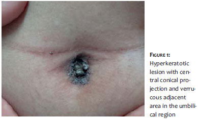
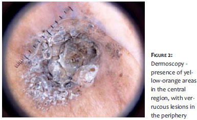
A 27-year-old woman presented a hyperkeratotic lesion with a central conical projection and a verrucous adjacent area, located in the umbilicus for 10 years. The lesion was firm, rough, painless, and not adhered to any deep layers (Figure 1). Dermoscopy evidenced areas of a yellow-orange color in the center, verrucous lesions on the wall of the umbilical scar, and debris. In addition, there was a fine regular pigmented network in the margins of the lesion (Figure 2).
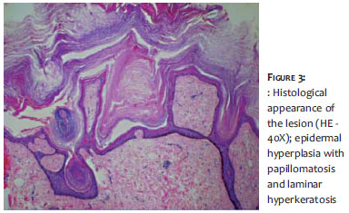
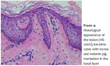
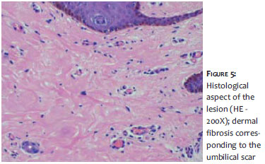
Histologic examination demonstrated laminar hyperkeratosis, acanthosis, and papillomatosis - findings that are consistent with the diagnosis of verrucous epidermal nevus (Figures 3, 4, 5).
The verrucous epidermal nevus appears as papules and/or single or multiple plaques that can be hyperkeratotic or basically verrucous, hyperpigmented, and well-defined, predominantly in the trunk and limbs. Although no specific dermoscopic pattern has been described, the finding of yellow-orange areas suggests the presence of keratin, indicating a keratinocyte proliferation process. The absence of dermoscopic findings typical of other lesions has helped the authors to exclude some differential diagnoses.
In cases where seborrheic keratosis is suspected, come-done-like lesions (yellowish-brown structures), pseudocysts (creamy white structures) and an amorphous yellowish area would be observed. In the case of viral warts, there would be normochromic papules and thrombosed vessels. In angiokeratomas, three patterns are described: dark, wine colored gaps or a whitish veil, peripheral erythema, and hemorrhagic crusts.5
Extremely rare yet important, is the neoplasic transformation of a verrucous nevus into a basal cell carcinoma or a squamous cell carcinoma. Bleeding, ulceration, and thickening can be clinical signs of malignant transformation.2 In the case reported in the present paper, dermoscopic or histological abnormalities suggestive of malignancy were not found.
Based on the case reported, the authors stress the uniqueness of the location and the importance of dermoscopy in the diagnosis and monitoring of this rare malignant transformation.
1. Kluger N. Dermatoses ombilicales et péri-ombilicales. Ann Dermatol Venereol. 2014;141:224-35.
2. Elder DE, Elenitsas R, Johnson BL Jr, Murphy GF, Xu G. Lever's Histopathology of the Skin. 10th ed. Lippincott Williams & Wilkins; 2008.
3. Noronha L, Neto JF, Nones RB, Taniguchi K. Nevo epidérmico: análise clínica e histológica de seis casos. An Bras Dermatol. 1999;74(1):259-62.
4. Kim R, Marmon S, Kaplan J, Kamino H, Pomeranz MK. Verrucous epidermal nevus [Internet]. Dermatol Online J. 2013;19(12):20707.
5. Kim JH, Kim MR, Lee SH, Lee SE. Dermoscopy: a useful tool for the diagnosis of angiokeratoma. Ann Dermatol. 2012; 24(4):468-71.
This study was conducted at the Hospital do Servidor Público Municipal de São Paulo (HSPM-SP) – São Paulo (SP), Brazil.
 All content the journal, except where identified, under the Creative Commons Attribution 4.0 International licence - ISSN-e 1984-8773


 Read in Portuguese
 Portuguese PDF
 Print
Print
 Send this article by email
 How to cite this article
 Submit a comment
 Mendeley
 Pocket