


Andreia Pizarro Leverone1; Bernardo Kawa Kac2; Clarissa Canella3; Claudia Fernanda Dias Souza4; Olga Milena Zarco Suarez4; Fabiana Palmieri Zarur4
Keywords: GLOMUS TUMOR; ULTRASONOGRAPHY, DOPPLER, COLOR; AMBULATORY SURGICAL PROCEDURES.
In dermatology, lesion diagnosis is essentially a clinical matter. However, for the diagnosis of subungual lesions such as glomus tumors, exostosis, mucoid pseudocysts and fibrokeratomas, further assessment through imaging methods is necessary. In addition to identifying alterations, it is possible to assess the precise size and location of these tumors pre-operatively. Ultrasonography is a noninvasive method, which when performed with skill can describe tumors as small as 3 mm.
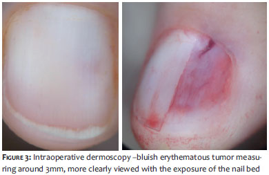
A 38-year-old Caucasian female patient from Nova Iguaçu, RJ - Brazil, who worked as a homemaker, sought care complaining of pain in the left thumb for about three years, accompanied by the sensation of "electric shock" when coming into contact with low temperatures and local trauma. She described progressive worsening, denying comorbidities or family history. On physical examination, erythronychia with undefined limits measuring roughly 3 mm, was observed in the central region of the nail plate, best seen on dermoscopy (Figure 1). The needle puncture test caused local discomfort.
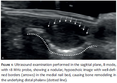
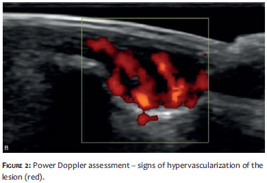
Clinical suspicion of a glomus tumor was raised, with the ultrasound examination revealing a correlation between the pain symptoms and the observed location of the lesion. The analysis showed a nodular, hypoechoic image with well-defined contours (Figure 2) that was hypervascularized under power Doppler examination (Figure 2), occupying the medial ungual bed and causing bone remodeling of the underlying distal phalanx.
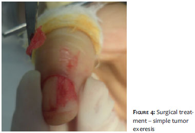
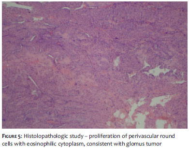
The excision of the lesion was carried out through a longitudinal incision in the nail plate, (Figures 3 and 4) which was replaced and sutured. The histological report revealed proliferation of perivascular round cells with eosinophilic cytoplasm and central vesicular nucleus, and a conclusive diagnosis of glomus tumor. (Figure 5)
Glomus tumors are benign neoplasms of glomus cells derived from neuromyoarterial glomus bodies. In about 75% of cases the lesion is located in the hand, especially in the subungual region where glomus bodies are found in higher concentrations. 1, 2 They occur with any age group and are rare, accounting for only 1-5% of all tumors of the hand. 2 Multiple lesions are infrequent (2-3%), and are more common in children. 3
Most lesions of this type present clinically with the classic triad (paroxysmal pain, hypersensitivity to temperature changes, and local sensitivity). Physical examination reveals bluish erythematous lesions of small dimensions (3-10 mm in diameter). However, due to the fact that they are located beneath the nail plate, it is difficult to assess their exact size and location, sometimes resulting in incorrect diagnoses. 3, 5
Ultrasonography is a useful tool for diagnosis and preoperative localization of the tumor, which facilitates surgery and decreases recurrence rates and is currently the method of choice for the evaluation of lesions that affect the nail bed and plate. Another function of this examination is to dismiss differential diagnoses such as epidermal inclusion and mucous cysts, which are avascular cystic lesions, i.e. devoid of flow under Doppler examination and generally without remodeling of the adjacent bone. 2, 4, 5
1. Song M, Ko HC, Kwon KS, Kim MB. Surgical Treatment of Subungual Glomus Tumor: A Unique and Simple Method. Dermatol Surg 2009;35:786-91.
2. Gencoglan G, Dereli T, Kazandi A. Subungual glomus tumor: surgical and histopathologic evaluation. Cutaneous and Ocular Toxicology, 2011;30(1):72-4.
3. Montandon C et al. Tumores glômicos subungueais: achados de imagem. Radiol Bras. 2009;42(6):371-4.
4. Takemura M, Fujii N, Tanaka T. Subungual glomus tumor diagnosis based on imaging. J Dermatol. 2006;3(6):389-93.
5. Fornage BD. Glomus tumors in the fingers: diagnosis with US. Radiology. 1988;167(1):183-5.
The present study was carried out at the Instituto de Dermatologia Prof. Rubem David Azulay da Santa Casa da Misericórdia do Rio de Janeiro (IDPRDA/SCMRJ) _ Rio de Janeiro (RJ), Brasil.
 All content the journal, except where identified, under the Creative Commons Attribution 4.0 International licence - ISSN-e 1984-8773


 Read in Portuguese
 Portuguese PDF
 Print
Print
 Send this article by email
 How to cite this article
 Submit a comment
 Mendeley
 Pocket