


Natacha Quezada Gaón1; Williams Romero2
Keywords: DERMOSCOPY; EYE; HYPERPIGMENTATION; QUALITY OF LIFE
Periorbital hyperpigmentation is a matter of very common consultation in cosmetic dermatology, one that can have a significant impact on a patient'squality of life.1 The pigmentation of the periorbital region depends on multiple factors: the amount of melanin deposited in the epidermis and dermis, the presence of periorbital blood vessels, reduced thickness of the epidermis (creating a translucent appearance that leaves deep structures visible - the thinnest epidermis of the human body is located in this region), and genetic factors.2-4
The skin of the palpebral region is physiologically thin, and is therefore more sensitive to exposure to irritative, recurrent, and chronic factors (contact dermatitis, blepharitis, etc) that may contribute to the worsening of the picture through post-inflammatory hyperpigmentation. Based on the main causes of pigmentation, periorbital hyperpigmentation has been clinically classified into three types: vascular, pigmented, and mixed.5
The vascular type is genetically determined, with darkening being caused by an extremely thin and translucent skin, favoring the visualization of blood vessels and underlying muscles.4The pigmented type is caused by melanin deposits associated with ethnic factors and exposure to the sun.1 The third type is the mixed type, which results from a combination of factors from the first and second types. It is important to note that all types can be aggravated by post-inflammatory hyperpigmentation, when hemosiderin and melanin deposits can be observed.2-4
The classification described is instrumental for a better therapeutic approach, however recognizing each specific type with the naked eye can be difficult. The dermatoscope is a valuable tool in the dermatological examination; nevertheless it is still seldom used in cosmiatric practice. The authors of the present study propose a very simple exploration of the palpebral skin with the dermatoscope, which facilitates easy identification of the periorbital hyperpigmentation type.
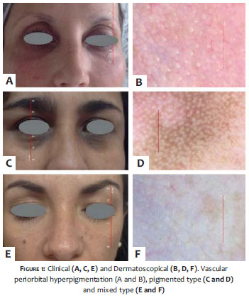
The authors examined 48 patients (40 women and 8 men) between 25- and 53-years-old whose reason for consultation was periorbital hyperpigmentation. They performed clinical examinations with the naked eye under slight local traction, in addition to dermoscopic examination (Handyscope, FotoFinder Systems GmbH, Bad Birnbach, Germany), finding 12 (25%) vascular type patients, 15 (31%) pigmented type patients and 21 (44%) mixed-type patients in the studied series.
In the dermoscopic examination of patients with the vascular type the authors found: diffuse erythema pattern or multiple thin blood vessels or diffuse vascular network.
In the pigmented type, it was possible to observe: a pattern of multiple dots with different sizes and colors, or a diffuse network of pigment. The mixed type was characterized by the combination of the patterns described above. (Figure 1)
In the authors' experience, it was more straightforward to determine and classify the periorbital pigmentation with the assistance of the dermatoscope, especially in cases where the pigment hue was darker, making it difficult to determine the correct pattern with the naked eye. The authors stress that the accurate clinical classification has direct influence on the therapeutic approach. In vascular periorbital hyperpigmentation it is sought to improve the skin's quality and stabilize the walls of the blood vessels, with the literature indicating the use of vitamin C, Phytonadione (vitamin K1), tretinoin etc. for that end.2,4 On the other hand, the pigmented type responds favorably to hydroquinone and other depigmenting elements, in addition to intense pulsed light and lasers.2 In the mixed type, the combination of therapies seems to be the most effective approach.4, 5
Dermoscopy is a simple, minimally invasive and useful tool in the evaluation of periorbital hyperpigmentation.
1. Malakar S, Lahiri K, Banerjee U, Mondal S, Sarangi S. Periorbital melanosis is an extension of pigmentary demarcation line-F on face. Indian J Dermatol Venereol Leprol. 2007;73(5):323-5.
2. Freitag FM, Cestari TF. What causes dark circles under the eyes? J Cosmet Dermatol. 2007;6(3):211-5.
3. Lüdtke C, Souza D, Webwr M, Ascoli A, Swarowski F, Pessin C. Epidemiological profile of patients with periorbital hyperpigmentation, at a dermatology specialist center in southerm Brazil. Surg. Cosmet Dermatol. 2013;5(4)302-8.
4. Roh MR, Chung KY. Infraorbital dark circles: definition, causes, and treatment options. Dermatol Surg. 2009;35(8):1163-71.
5. Souza DM, Ludtke C, Souza ERM, Scandura KMP, Weber MB. Hiperpigmentação periorbital. Surg Cosmet Dermatol. 2011;3(3):233-9.
The present study was carried out at Pontificia Universidad Católica de Chile - Santiago, Chile.
 All content the journal, except where identified, under the Creative Commons Attribution 4.0 International licence - ISSN-e 1984-8773


 Read in Portuguese
 Portuguese PDF
 Print
Print
 Send this article by email
 How to cite this article
 Submit a comment
 Mendeley
 Pocket