


Luiza Eastwood Romagnolli1; Larissa Montanheiro dos Reis2; Fernanda Bebber Douat3; Manuela Ferrasso Zuchi Delfes2; Eveline Roesler Battaglin2; Deborah Skusa de Torre4
Keywords: LUPUS ERYTHEMATOSUS, DISCOID; SKIN TRANSPLANTATION; AMBULATORY SURGICAL PROCEDURES; SKIN.
Lupus erythematosus (LE) is an autoimmune disease of the connective tissue characterized by the presence of localized or disseminated cutaneous-vascular lesions.1 Cutaneous LE presents in a variety of clinical forms, which can be classified into 3 subtypes: acute, subacute, and chronic. Acute cutaneous LE (Acle) arises during systemic disease activity and its most common manifestation is the malar rash. Subacute cutaneous LE (Scle) is characterized by non-infiltrative erythematous plaques, predominantly in areas exposed to the sun. Discoid LE (DLE) is the most common clinical variant of chronic cutaneous LE (Ccle), being characterized by plaques covered with fine scales. Those plaques may be initially hyperpigmented and can develop into cicatricial lesion scars with depigmentation, which are permanent in most cases.2
In the treatment of active DEL, high potency topical corticosteroids, intralesional infiltration with triamcinolone and systemic therapy with the antimalarial chloroquine or hydroxy chloroquine can be used. Other options for resistant cases are systemic corticosteroids, thalidomide, clofazimine, dapsone, acitretin, and immunosuppressants such as methotrexate, azathioprine and cyclophosphamide.1
After adequate control of the disease, DEL can result in unsightly cicatricial lesions, which can be stigmatizing, causing a negative impact on the patient's quality of life. The treatment of achromic DEL cicatricial lesions can be performed through the punch grafting technique, which is traditionally used in cases of localized or segmental refractory vitiligo.
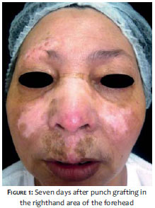
A 47-year-old female patient, bearing DEL for 18 years, sought care presenting achromic and atrophic cicatricial lesions located in the right supraorbital, nasal, and right malar regions, with an absence of signs of disease activity. She had already made prior use of topical corticosteroids, methotrexate, and thalidomide. Initially, punch grafting was performed in the right supraorbital region (test area). The right retroauricular region was chosen as the donor area. Local anesthesia was carried out with 2% lidocaine and 1:100,000 epinephrine, and the collection (four specimens) was performed with a 2.5mm punch. After the incision, the specimens were cut with the assistance of Adson forceps and iris scissors at the dermo-hypodermal junction level, and were subsequently placed on sterile gauze moistened with saline solution. The donor area was sutured with 5.0 nylon monofilament. The receptor area was incised with a 2mm punch, with a distance of about 1cm between each other, with the depigmented fragments being discarded. The implants were placed in the receiving area with the assistance of an Adson forceps, and attached with sterile micropore, which was removed after 7 days. (Figure 1) The patient returned 3 months after the procedure, showing a halo of repigmentation of about 1cm around the implanted grafts.
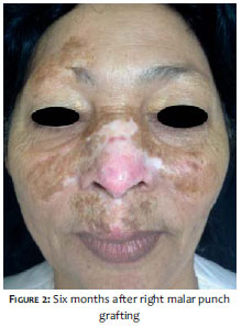
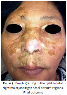
Subsequently, a similar new procedure was performed on the right malar region (2 specimens), according to the technique described above (the right retroauricular region was the donor area again). The patient returned after 6 months with satisfactory repigmentation in the receptor area. The grafting on the nasal dorsum region was then carried out (two specimens) (Figure 2), having as a donor area the left retroauricular region. At the 3-month return the grafting procedure was repeated on the superior nasal dorsum region, according to the techniques described above. It is possible to notice a slight elevation of the grafts in comparison to the surrounding skin, however the patient reported being satisfied with the aesthetic outcome of the treatment nonetheless. (Figure 3)
Residual scar lesions after control of the DEL are disfiguring and cause a worsening in the quality of a patient's life.2
The classical technique for total skin punch grafts harvested from the occipital and temporal regions for the treatment of vitiligo was published in 1959.3 In 1971, Orentreich performed grafts using donor skin from the retroauricular region, with the formation of a halo of pigment diffusion of 1mm.4 Other diseases, such as discoid lupus with achromic cicatricial sequel in the scalp, were treated by Lobuono in 1976 with punch grafts obtained from the occipital region, with the formation of halos of pigment in the receptor area.5 Implants of follicular units have been performed with a spacing of 3-5mm between the insertion points of the follicles. It is possible to notice repigmentation between the 4th and 7th week, with cases of full repigmentation after 6 months.6 In a study published by Fongers et al. in 2009 in which 61 patients with vitiligo vulgaris and 9 patients with segmental vitiligo underwent punch grafting, it was possible to observe that regarding the patients with vitiligo vulgaris, 17 (28%) lesions had excellent repigmentation, 14 (23%) had good, 14 (23%) reasonable, and 16 (26%) poor. Of the patients with segmental vitiligo 7 from the 9 lesions had excellent repigmentation. A cobblestone pattern was observed in 27% of lesions.7
The density of melanocytes varies in an individual, with an average density of 1,560/mm2, and a ratio of 1:13 between melanocytes and malpighian cells. Those figures vary largely depending on the body site. Areas exposed to ultraviolet radiation suffer a 10% reduction in the population of melanocytes every ten years. Therefore, the best donor areas are those that are hidden from sunlight, such as the scalp, the retroauricular region, the sacral region, buttocks, and the dorsa of the feet.3
It is essential that the receiving area be completely inactive in order for the implanted melanocytes not to become targets of immunological aggression. The implant testing area is performed in order to determine the absence - or not - of immune activity that might interfere with repigmentation. In the receiving area to be treated, a 2.5mm implant of normal skin is performed in an orifice of 2mm. The development is observed for two months, during which the implanted skin should not lose its pigmentation.3
Potentially, a 2mm implant might cause a pigmentary halo of 1cm in diameter over the course of 1 year - therefore the implants must be performed with a spacing of approximately 1cm. In the donor area, specimens should be harvested with 0.5mm in excess of the receiving area (Ex: 2.5mm/2mm).3
In line with the positive outcomes described in reports published in the literature,3-7 the present study used an innovative technique aimed at establishing a new treatment option for achromic scars arising from DEL.
Just as Orentreich obtained a halo of pigmentary diffusion, the present study also obtained repigmentation.4 A halo of roughly 1cm emerged around each graft. Thatrepigmentation became more evident between 3 and 6 months after the procedure, falling within the interval of repigmentation mentioned in the literature regarding patients with this disease.6
The treatment of achromic cicatricial lesions arising from DEL is a therapeutic challenge. These unsightly lesions usually have a significant negative impact on the quality of life of affected patients. The punch grafting technique has been described for a long time for the treatment of refractory vitiligo lesions, with good repigmentationrates. Although there are few reports in the literature, this technique can be used in achromic lesions arising from DEL, also with good repigmentation outcomes.
1. Sampaio SAP, Rivitti EA. Afecções do Conectivo. Dermatologia. 3ª Ed. São Paulo: Editora Artes Médicas Ltda; 2007. p.455-86.
2. Ribeiro LH, Nunes MJ, Lomonte ABV, Latorre LC. Atualizações no Tratamento do Lúpus Cutâneo. Rev Bras Reumatol. 2008;48(5):283-290.
3. Machado Filho CDAS, Casabona G, Manzini MC. Técnicas de Repigmentação- Abordagem Cirúrgica. In: Kadunc B, Palermo E, Addor F, Metsavaht L, Rabello L, Mattos R, editores. Tratado de Cirurgia Dermatológica, Cosmiatria e Laser da Sociedade Brasileira de Dermatologia. Rio de Janeiro: Elsevier; 2012. p.387-95.
4. Orentreich N. Hair transplantation: the punch graft technique. Surg Clin North Am. 1971;51(2):511-8.
5. Lobuono P, Shatin H. Transplantation of hair bulbs and melanocytes into leukodermic scars. J Dermatol. Surg. 1976;2(1):53-5.
6. Malakar S, Dhar S. Repigmentation of vitiligo patches by transplantation of hair follicles. Int J Dermatol. 1999;38(3):237-38.
7. Fongers A, Wolkerstorfer A, Nieuweboer-Krobotova L, Krawczyk P, Tóth GG, van der Veen JP. Long-term results of 2-mm punch grafting in patients with vitiligo vulgaris and segmental vitiligo: effect of disease activity. Br J Dermatol. 2009;161(5):1105-11.
The present study was carried out at the Dermatology Service, Hospital Universitário Evangélico de Curitiba - Curitiba (PR), Brazil.
 All content the journal, except where identified, under the Creative Commons Attribution 4.0 International licence - ISSN-e 1984-8773


 Read in Portuguese
 Portuguese PDF
 Print
Print
 Send this article by email
 How to cite this article
 Submit a comment
 Mendeley
 Pocket