


Juliana Augusta Zeglin Nicolau1; Paula Foresti Faria1; Luciana de Oliveira Marques2; Deyse Fabiane Hoepers1; Andressa Dias da Rocha3; Ana Cristina Lira Sobral4
Keywords: ESTROGENS; PROGESTERONE; COLLAGEN; WOUND HEALING.
Cutaneous wounds are the result of multiple factors, such as hypoxia, trauma, or pressure. Their development often follows an already established order: inflammation, proliferation, and remodeling.1 The healing process involves blood cells, extracellular matrix, cell mediators, and parenchymal cells. 2 Several factors, such as malnutrition, various drugs, radiation, and hypoxia may hinder that process.1
The skin is composed primarily of collagen fibers, elastic fibers, and glucosamine protein glucans.3 Particularly in the dermal matrix there are essentially two types of collagen: type I and type III, corresponding to about 80-85% and 15-20% of the total of amount of that protein, respectively. Type I collagen is mainly located in the reticular dermis, the deepest layer of the skin. Type III collagen is present mostly in the papillary dermis, located more superficially. Contrary to what occurs in the untouched dermis, there is a greater proportion of type III collagen in wounds than type I collagen.
Hormones are potentially determining factors in the aging process, and estrogen was shown to be beneficial in accelerating tissue repair related to aging in both genders.4
Some studies show that topical and systemic estrogen therapy stimulates the healing process as it reduces inflammation. In vitro studies also suggest that progesterone can modulate inflammation. Estrogen and progesterone would contribute to macrophage activation in an alternative route, leading to wound healing, angiogenesis, and remodeling.5
In the face of increasing life expectancy, an aging global population, and an increase in cutaneous conditions and healing deficits typical of advanced age, there is a need for research on the effect of progesterone and estrogen as adjuvants in the treatment of skin wounds.5
Therefore, the objective of the present study was to evaluate the effects of topical estrogen and progesterone, isolated and in combination, in healing the wounds of menopausal female rats.
Forty adult female Rattus norvegicus albinus rodentia mammalia, rats from theWistar lineage, originally from the Central Animal Facility of the Faculdade Evangélica do Paraná (FEPAR) in the southern Brazilian State of Paraná, Brazil, were used. All animals were kept in the animal facility of the institution and received water and proper food for the species. The Ethical Principles for handling animal experimentation set by the Ethics Committee on Animal Experimentation (CEEA) were observed.
The project was approved by the Ethics and Research Committee of the institution, registered under the number 4723/11 dated 20th May 2011.
The sample was divided into 4 groups of 10 animals each, according to the proposed treatment, as described below:
Group C (control): received only the vehicle, consisting of non-ionic cream.
Group E (Estradiol): received topical treatment with a cream containing 0.5mg of 17-beta-estradiol in a vehicle consisting of non-ionic cream.
Group P (Progesterone): received topical treatment with ointment containing 15mg of natural progesterone in a vehicle consisting of non-ionic cream.
Group E + P (Estradiol and Progesterone): received topical treatment with an ointment containing 0.5 mg of 17-beta-estradiol, associated with 15mg of natural progesterone in a vehicle consisting of non-ionic cream.
In the first phase of the experiment, all animals were weighed, identified with picric acid and assembled into cages, each with 5 randomly chosen individuals. They were then anesthetized with inhaled isoflurane until deep plane, and underwent bilateral oophorectomy, according to surgical standard technique, for the inductions of the menopausal state.
In the second phase of the experiment, on the 30th day after surgery and under the anesthetic procedure already described, a circular wound of 1cm in diameter was inflicted on the dorsum of each animal, for the removal of the skin and subcutaneous tissue, without damaging the adjacent aponeurosis. From that point on, all animals received the daily topical treatment proposed for each group, always at the same time of day and for 7 continuous days. The animals received a daily dose of morphine (10% solution, 0.1ml/100g of weight), intramuscularly as an analgesic during the healing process.
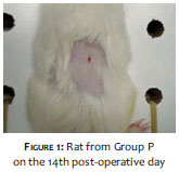
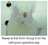
Biopsies were performed on the 3rd, 7th, and 14th days after the beginning of the treatment. Figures 1 and 2 show the appearance of the cutaneous wound on the 14th day of the experiment, in the Group Progesterone and Group Estradiol (Group P and Group E).
In the third phase of the experiment, on the 14th day after the beginning of the treatment, the animals were weighed again and euthanized by anesthetic overdose with inhaled isoflurane.
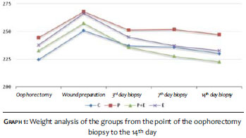
As for the weight analysis, the groups of animals were compared in order to evaluate the hormonal influences at the three separate biopsy points of the experiment (3rd, 7th,, and 14th days).
For the histology and collagen evaluations, the cutaneous scars of the euthanized animals were dissected and fixed in buffered formalin. The preparation of the slides was carried out at the laboratory of histotechnique of the FEPAR through hematoxylin and eosin (HE) staining method (for the histological analysis) and through Sirius Red staining (for the collagen analysis).
Under the first staining technique, the following were analyzed: epithelialization, classification and degree of inflammation, and vascularization. Under the second staining technique, the collagen was quantified and qualified. Microscopic analyses were performed, always by the same pathologist. The total time of study was 44 days.
Statistical tests used for quantitative variables were: ANOVA, and non-parametric tests of Kruskal-Wallis and Friedman. For the analysis of qualitative variables the following were used: the Fisher’s exact test, and the binomial test. Finally, the Shapiro-Wilk test was used for analyzing the normality condition of the variables.
All results received statistical treatment at a 0.05 significance level. The data were analyzed with the software Statistica v. 8.0.
During the study, 3 animals belonging in the control group died: the first died during the preparation of the wound, and the other 2 on the 7th day of the development of the wound. The causes of deaths were linked to the inhalation sedation used during the procedures. The remaining animals were euthanized on the days previously scheduled for the measurement. By the end of the experiment, all wounds had healed.
According to Graph 1, and regarding the weight analysis, the vehicles containing isolated estradiol, estradiol plus progesterone, or isolated progesterone, did not cause changes in the weight of the animals during the study, in the analysis of the 3rd day (p = 0.435), 7th day (p = 0.120) and 14th day (p = 0.130). Due to surgical stress, the occlusive dressings and daily handling, all elements of the 4 groups lost weight significantly (p <0.05) after the oophorectomy and during the healing process, with an absence of statistically significant differences between groups.
In the histological analysis (HE), regarding the development of the re-epithelialization, Group P showed higher and significant re-epithelialization of the wound on the 14th day (p = 0.01).
As for the inflammatory process, there was no significant difference in the three groups at the three time points. The same occurred for the neovascularization, fibrosis, and number of macrophages and fibroblasts in the wounds.
In the intra-group analysis, only progesterone accelerated the re-epithelialization from the 7th to the 14th day (p = 0.016) and increased fibrosis from the 3rd to the 7th day (p = 0.008).
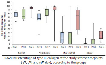
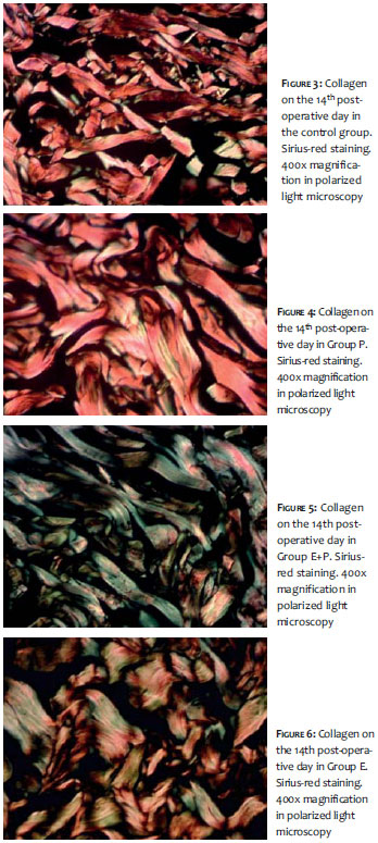
Regarding the analysis of collagen, Group E and Group E+P inhibited the formation of type III collagen in the study’s three time points (p <0.001), as can be seen in Graph 2. The group that obtained the highest percentage of formation of new collagen between the 7th and the 14th day was Group E+P. In Figures 3 to 6, it is possible to note the formation of collagen in the Sirius Red coloration in the different study groups.
According to Brincat, estrogen has many beneficial and protective effects on the skin’s physiology and function, including maintenance of its hydration and thickness, elasticity, and wound healing capacity, in addition to reducing the risk of skin cancer.6
Also, according to Ashcroft, a reduction in estrogen levels leads to impaired quality of skin healing in postmenopausal women and in oophorectomized rats. Topical application of estrogen was also linked to an acceleration of cutaneous healing.7 According to the authors’ study, it was not possible to observe such effects as described in the literature, since only in Group P was there an acceleration of fibrosis and re-epithelialization at certain instances during the study.
Nonetheless, according to Ashcroft, systemic hormone replacement in menopausal females was associated with accelerated wound healing as compared to topical application. On the other hand, estrogen applied topically immediately after the infliction of the wound reduced the incidence of dehiscence and infection.8
In a study of oophorectomized rabbits, chronic therapy with estradiol dipropionate was evaluated, considering the thickness of the skin layers, the percentage of dermal collagen and elastic fibers, and areas of sebaceous glands. Bearing in mind that the thickness of the epidermis varies considerably in different body sites, and that sex steroid hormones are involved in regulating the development and function of the skin, in this study the absence of estradiol decreases the mitotic activity in the basal layer of the epidermis.9
It was observed that estrogens stimulate the synthesis, maturation, and renewal of collagen in rats,10 a fact that is in line with the present study, since an increase of type III collagen in the intra-group analysis was observed. There was an increase in collagen type III from the 7th to the 14th day (p = 0.006) within Group E and within Group E+P (p = 0.001), in the same period.
According to Isaac, there is a higher proportion of type III collagen relative to type I in the wound, meaning that myofibroblasts bind to collagen type III fibers, promoting wound contraction.11 Thus, the higher the number of present type III collagen fibers, the greater the contraction of the wound. Therefore, the present study corroborates the information found in the literature, since Group E and Group E+P inhibited the formation of collagen type III, and only Group P significantly accelerated the wound’s re-epithelialization.
Topical therapy with estradiol and progesterone associated with estradiol inhibited the formation of type III collagen. The isolated progesterone only contributed to the re-epithelialization process of the wounds.
1. Burns JL, Mancoll JS, Philips LG. Impairments to wound healing. Clin Plastic Sur. 2003;30(1):47-56.
2. Santuzzi CH, Buss HF, Pedrosa DF, Freire MOVM, Nogueira BV, Gonçalves WLS. Uso combinado da laserterapia de baixa potência e dainibição da ciclooxigenase-2 na reepitelização de ferida incisional em pele de camundongos: um estudo pré-clínico. An Bras Dermatol. 2011;86(2):278-83.
3. Fang M, Liroff KG, Turner AS, Les CM, Orr BG, Holl MM. Estrogen depletion results in nanoscale morphology changes in dermal collagen. J Invest Dermatol. 2012;132(7):1791-7.
4. Hardman MJ, Ashcroft GS. Estrogen, not intrinsic aging, is the major regulator of delayed human wound healing in the elderly. Genome Biology. 2008; 9(5): 80:1-17.
5. Routley CE, Ashcroft GS. Effect of estrogen and progesterone on macrophage activation during wound healing. Wound Repair Regen. 2009;17(1):42-50.
6. Brincat MP. Hormone replacement therapy and the skin. Maturitas 2000;35(2):107-17.
7. Ashcroft GS, Greenwell-Wild T, Horan MA, Wahl SM, Ferguson MW. Topical estrogen accelerates cutaneous wound healing in aged humans associated with an altered inflammatory response. Am J Pathol 1999;155(4):1137-46.
8. Ashcroft GS, Dodsworth J, van Boxtel E, Tarnuzzer RW, Horan MA, Schultz GS, et al. Estrogen accelerates cutaneous wound healing associated with na increase in TGF- beta 1 levels. Nat Med 3(11):1209-15.
9. Lignières B. Ovariam hormones and cutaneous aging. Rev Fr Gynecol Obstet. 1991; 86(6):451-4.
10. Smith QT, Allison DJ. Changes of collagen content in skin, femur and uterus of 17-beta-estradiol benzoate-treated rats. Endocrinology. 1966;79(3): 486-92
11. Isaac C, Ladeira PRS, Rego FMP, Aldunate JCB, Ferreira MC. Processo de cura das feridas: cicatrização fisiológica. Rev Med (São Paulo). 2010;89(3/4):125-31.
The present study was carried out at the Faculdade Evangélica do Paraná (FEPAR) - Curitiba (PR), Brazil.
 All content the journal, except where identified, under the Creative Commons Attribution 4.0 International licence - ISSN-e 1984-8773


 Read in Portuguese
 Portuguese PDF
 Print
Print
 Send this article by email
 How to cite this article
 Submit a comment
 Mendeley
 Pocket