


Maria Cecilia Closs Ono1; Priscilla Balbinot2; Rosinete Lauren de Souza Lima Morais3; Renato da Silva Freitas4
Keywords: PRIMARY TREATMENT; SURGERY, PLASTIC; TATTOOING.
The practice of tattooing has been performed for thousands of years, and many agents that cause reactions are present in the components of the colors used.1 With the increasing prevalence of tattoos throughout the world, more care is needed with adverse reactions to techniques of body modification.2 The way tattoos are performed has changed over time. In the past, the use of heavy metals was common,3 while more recently, azo dyes are preferred. Green chrome, cobalt-blue, purple manganese, yellow cadmium and red mercury sulfide (cinnabar) have been linked to skin reactions, with the red pigment most commonly causing such reactions.4 Despite the limited use of pigments containing mercury, reactions to red tattoos continue to occur.
Allergy to the red component of tattoos is a well-known phenomenon, and is related to the cinnabar present in the pigment or in other organic compounds. The allergy can manifest in various ways, from simple inflammatory reactions to a generalized allergic response.5-8
Various treatments may be carried out for the condition, such as laser, excisions, and grafting. The objective of the present study is to review the relevant literature, illustrating with a clinical case treated in a public service department.
A literature review related to the management (from diagnosis to treatment) of patients with reactions to red tattoo pigment was carried out. The retrospective study reviewed English and Portuguese language literature on Pubmed and Embase, illustrating with a case report of a patient cared for at the service.
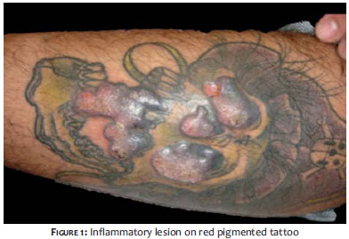
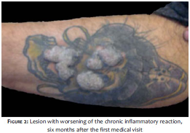
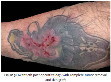
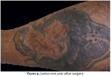
A 27-year-old male patient with a presentation of a chronic pruriginous inflammatory lesion in the lateral portion of the left leg, for a duration of six weeks, over a tattoo performed four months before, sought medical care. On examination, lichenified plaques were observed with bruises over the red pigment area (Figure 1). Biopsy was carried out, showing hyperparakeratosis, chronic lichenoid inflammatory infiltrate with perivascular involvement and deposition of pigment in the dermis consistent with lichenoid dermatitis. The patient underwent treatment with topical and oral corticosteroids without resolution of the clinical picture, and was resistant to any surgical procedure. After six months, the patient returned to the service with a worsening of the inflammatory reaction (Figure 2) and underwent resection of the lesion and a full thickness skin grafting (Figure 3). The result after one year has proved satisfactory, with an absence of inflammatory phenomena. (Figure 4)
Tattoos have been performed for centuries and are still a common practice in many cultures, being associated with the desire for social inclusion and aesthetic improvement.
Historically, tattoos seem to have arisen as blue marks under the skin, seen in Egyptian mummies. The practice was spread by sailors in China, India, the Far East, and also Europe.9
Cutaneous hypersensitivity reactions to tattoo pigments can be classified histologically as granulomatous or lichenoid.10-14 The etiology of these reactions is still uncertain, however the most widely accepted theory is that of a delayed hypersensitivity reaction, related to the pigment itself or the carrier solution. Scleroderma is an uncommon reaction that can occur in tattoos 15 and that complicates a chronic inflammatory reaction to pigments and dyes. 16
Many pigments can induce allergic responses, including mercury sulfide (red) and cadmium sulfide. Histological studies usually reveal dermal inflammation with pruriginous and nodular alterations and epidermal ulcers, and tissue analysis showing the presence of cadmium. 17-22
Other skin diseases related to tattoos, such as pyogenic infection, verruca vulgaris, and zygomycosishave been described. Many skin diseases show a predilection for tattooed skin, and can arise as a primary manifestation or even intensify the occurrence of the Koebner phenomenon, as in lichen planus and psoriasis.23
The transmission of infectious diseases is certainly of greater significance to public health than the reactions to tattoos described above. Infectious diseases can be local or systemic.24 Historically, infections such as erysipelas, cellulitis, and gangrene (requiring amputation) by Staphylococcus sp. and Streptococcus sp. were the most common and alarming infectious complications.25, 26 The reactions are usually diagnosed shortly after the tattoo has been performed or when it is removed with laser. The lesions characteristic of the disease are symmetrical, erythematous, subcutaneous nodules in the legs. Most evidence suggests Type IV hypersensitivity reaction to various antigens.
There are several forms of treatment for reactions to tattoos, including excision with primary closure and laser treatment.1,18, 27, 28 Despite the treatment with intralesional or topical steroids, most reactions persist for months or years. Even the use of systemic corticosteroids might not be sufficient to treat the inflammation in progress. Tattoos can be removed through light-based therapies, including several wavelengths. Those therapies are aimed at reducing the visibility of the reaction to the pigment through the induction of its transepidermal elimination, the removal of the macrophages from the pigment, dispersion of the pigment into smaller particles and alteration of the optics, and refractory properties of the particles. Allergic reactions can manifest in various ways-from simple inflammatory reactions to generalized allergic response. 18, 19
Another treatment option is a CO2 laser-based ablation in order to induce elimination of the pigment through the skin and thus reduce the pigment load and the allergenic stimuli, limiting the reaction of the tattoo. 25, 26, 28-30 For certain anatomical regions and tattoo pigments, erbium or carbon dioxide laser ablation can be the treatment of choice.31 In cases such as a red pigment tattoo on the lips, the treatment with carbon dioxide and erbium can be more problematic due to the lack of dermis. For larger lesions, the treatment of choice is excision, which usually requires grafting.
Despite the popularity of tattoos, adverse inflammatory reactions are common and may present a significant clinical challenge, both for the correct diagnosis and for choosing the effective treatment. Should the topical or systemic treatment fail, total excision may be the only option. The final aesthetic result can be very unsatisfactory depending on the amount of tissue resected.
1. McDaniel DH, Ash K, Lord J, Newman J, Zukowski M. The erbium: YAG laser: a review and preliminary report on resurfacing of the face, neck, and hands. Aesthet Surg J. 1997;17(3):157-64.
2. Corazza M, Zampino MR, Montanari A, Pagnoni A, Virgili A. Lichenoid reaction from a permanent red tattoo: has nickel a possible aetiologic role? Contact Dermatitis. 2002;46(2):114-5.
3. Silberberg I, Leider M. Studies of a red tattoo. Appearances in electron microscope, and analysis by chemical means, laser microprobe and selected-area diffraction of tattooed material. Arch Dermatol. 1970;101(3):299-304.
4. Cruz FAM, Lage D, Frigério RM, Zaniboni MC, Arruda LHF. Reactions to the different pigments in tattoos: a report of two cases. An Bras Dermatol. 2010;85(5):708-11.
5. Dickel H, Gambichler T, Kamphowe J, Altmeyer P, Skrygan M. Standardized tape stripping prior to patch testing induces upregulation of Hsp90, Hsp70, IL-33, TNF-α and IL-8/CXCL8 mRNA: new insights into the involvement of "alarmins". Contact Dermatitis. 2010;63(4):215-22.
6. Engel E, Santarelli F, Vasold R, Maisch T, Ulrich H, Prantl L, et al. Modern tattoos cause high concentrations of hazardous pigments in skin. Contact Dermatitis. 2008;58(4):228-33.
7. Mortimer NJ, Chave T A, Johnston GA. Red tattoo reactions. Clin Exp Dermatol. 2003;28(5):508-10.
8. Yazdian-Tehrani H, Shibu MM, Carver NC. Reaction in a red tattoo in the absence of mercury. Br J Plast Surg. 2001 Sep;54(6):555-6.
9. Lubeck G, Epstein E. Complications of tattooing. Calif Med. 1952;76(2):83-5.
10. Kluger N, Minier-Thoumin C, Plantier F. Keratoacanthoma occurring within the red dye of a tattoo. J Cutan Pathol. 2008;35(5):504-7.
11. Kluger N, Godenèche J, Vermeulen C. Granuloma annulare within the red dye of a tattoo. J Dermatol. 2012;39(2):191-3.
12. Sweeney S a, Hicks LD, Ranallo N, Iv NS, Soldano AC. Perforating Granulomatous Dermatitis Reaction to Exogenous Tattoo Pigment: A Case Report and Review of the Literature. Am J Dermatopathol. 2011 13;35(7):754-6.
13. Taaffe a, Knight a G, Marks R. Lichenoid tattoo hypersensitivity. Br Med J. 197811;1(6113):616-8.
14. Vitiello M, Echeverria B, Romanelli P, Abuchar A, Kerdel F. Multiple eruptive keratoacanthomas arising in a tattoo. J Clin Aesthet Dermatol. 2010;3(7):54-5.
15. Kluger N, Mathelier-Fusade P, Moguelet P. Scleroderma-like reaction restricted to the red parts of a tattoo. Acta Derm Venereol. 2009;89(1):95-6.
16. Wollina U, Gruner M, Schönlebe J. Granulomatous tattoo reaction and erythema nodosum in a young woman: common cause or coincidence?. J Cosmet Dermatol. 2008;7(2):84-8.
17. Duke D, Urioste SS, Dover JS, Anderson RR. A reaction to a red lip cosmetic tattoo. J Am Acad Dermatol. 1998;39(3):488-90.
18. Dave R, Mahaffey PJ. Successful treatment of an allergic reaction in a red tattoo with the Nd-YAG laser. Br J Plast Surg. 2002;55(5):456.
19. Macarthur M, Davies M. Sensitisation to red tattoo pigment. Br J Plast Surg. 2003;56(1):73.
20. Alcantara J. Letters to the editor. J Can Chiropr Assoc. 2013;57(1):97-8.
21. Braithwaite RL, Stephens T, Sterk C, Braithwaite K. Risks associated with tattooing and body piercing. J Public Health Policy. 1999;20(4):459-70.
22. Casparian JM, Krell J. Using a side effect to therapeutic advantage: the darkening of red eyebrow tattoo pigment following Q-switched laser treatment. Dermatol Surg. 2000;26(3):255-8.
23. Morgan CJ, Haworth AE. Allergic contact dermatitis from 1,6-hexamethylene diisocyanate in a domestic setting. Contact Dermatitis. 2003;48(4):224.
24. Preda VA, Maley M, Sullivan JR. Mycobacterium chelonae infection in a tattoo site. Med J Aust. 2009;190(5):278-9.
25. Wolf R, Wolf D. A tattooed butterfly as a vector of atypical Mycobacteria. J. Am. Acad. Dermatol. 2003 May;48(5 Suppl):S73-4.
26. Rodríguez-Blanco I, Fernández LC, Suárez-Peñaranda JM, Pérez del Molino ML, Esteban J, Almagro M. Mycobacterium chelonae infection associated with tattoos. Acta Derm Venereol. 2011;91(1):61-2.
27. Beddoes TP. Case for Diagnosis. Proc R Soc Med. 1913;6(Dermatol Sect):182.
28. McDaniel DH, Lord J, Ash K, Newman J. Combined CO2/erbium:YAG laser resurfacing of peri-oral rhytides and side-by-side comparison with carbon dioxide laser alone. Dermatol Surg. 1999;25(4):285-93.
29. De Argila D, Chaves A, Moreno JC. Erbium:Yag laser therapy of lichenoid red tattoo reaction. J Eur Acad Dermatol Venereol. 2004;18(3):332-3.
30. Newman JB, Lord JL, Ash K, McDaniel DH. Variable pulse erbium:YAG laser skin resurfacing of perioral rhytides and side-by-side comparison with carbon dioxide laser. Lasers Surg Med. 2000;26(2):208-14.
31. Gómez C, Martin V, Sastre R, Costela A, García-Moreno I. In vitro and in vivo laser treatments of tattoos: high efficiency and low fluences. Arch Dermatol. 2010;146(1):39-45.
This study was performed at the Plastic and Reconstructive Surgery Department, Hospital de Clinicas, Universidade Federal do Paraná (PR) - Curitiba (PR), Brazil.
 All content the journal, except where identified, under the Creative Commons Attribution 4.0 International licence - ISSN-e 1984-8773


 Read in Portuguese
 Portuguese PDF
 Print
Print
 Send this article by email
 How to cite this article
 Submit a comment
 Mendeley
 Pocket