


Clarissa Brito Farias; Ana Vitória Lins Paiva Antunes; Aretuza Iolanda Pimentel Torres Portella; Antônio Carlos Evangelista de Araújo Bonfim; Luciana Cavalcanti Trindade
Funding source: None
Conflict of interest: None
Submitted on: 01/13/2026
Final decision: 4/9/2026
How to cite this article: Farias CB, Antunes AVLP, Portella AIPT, Bonfim ACEA, Trindade LC. Odontogenic cutaneous fistula mimicking skin neoplasia: case report. Surg Cosmet Dermatol. 2026;18(2):e20260547.
Odontogenic cutaneous fistulas represent a tract between the oral cavity and the skin. They are manifestations of pulpal necrosis and chronic apical periodontitis, caused by caries, infections, or trauma. Cutaneous manifestations include nodules and cysts and may present with perilesional retraction. They pose a diagnostic challenge, as most patients do not report toothache. We report a case with delayed diagnosis due to lack of clinical suspicion where the patient underwent multiple treatments, including surgery, because of clinical resemblance to cutaneous neoplasia. The lesion resolved completely after dental treatment.
Keywords: Oral Fistula; Fistula; Carcinoma, Basal Cell
Odontogenic cutaneous fistulas (OCFs) represent a communication between the oral cavity and the skin and constitute cutaneous manifestations secondary to pulp necrosis and chronic apical periodontitis.1 The causes include dental caries, pulpal or periapical infection, periodontal diseases, root fractures, and mechanical problems resulting from trauma, with no predilection for sex or age group.2,3
Cutaneous manifestations include lesions such as nodules, cysts, and areas of solution of continuity and may be associated with retraction of the perilesional region. The fistulous tracts most commonly reported are related to involvement of the mandibular dental arch, with drainage to the mandible and chin.4,5
Although well documented, OCFs remain a diagnostic challenge because, in most cases, patients do not report toothache. Definitive diagnosis is usually established through imaging studies, such as panoramic radiography and computed tomography, which find abnormalities of the alveolar process.5
It is estimated that half of all patients with OCFs undergo unnecessary treatments, including multiple dermatologic surgical procedures and prolonged antibiotic therapy, until the correct diagnosis is established.6
In this context, the objective of the present study is to report a case of OCF with delayed diagnosis due to the absence of diagnostic suspicion, underscoring the importance of awareness of this condition and its relationship to oral cavity examination in order to enable appropriate and timely management.
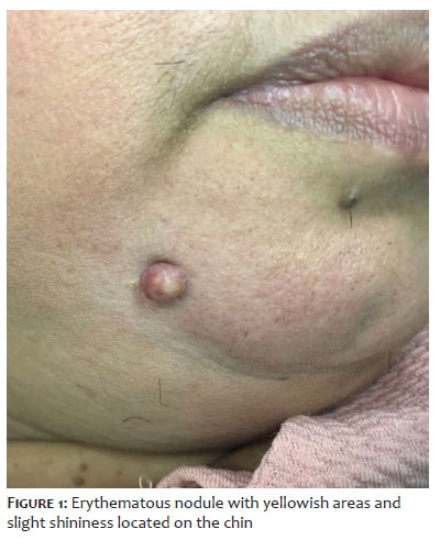
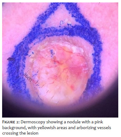
A 43-year-old female patient with a light-brown skin phototype reported the appearance of a painless lesion in the right mandibular region, with progressive growth over the previous 6 months. Physical examination found an erythematous nodule with yellowish areas and slight shininess, of fibroelastic consistency and painless on palpation (Figure 1). Dermoscopy revealed a nodular lesion with a pink background, covered by small adherent yellowish crusts, with homogeneous yellow areas and arborizing telangiectasias crossing the lesion (Figure 2), suggestive of basal cell carcinoma.
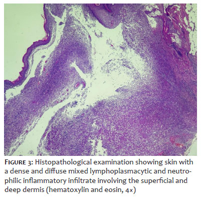
Fusiform resection of the lesion was performed, and histopathological examination revealed skin with a dense and diffuse mixed lymphoplasmacytic and neutrophilic inflammatory infiltrate involving both the superficial and deep dermis, findings that did not support the initial hypothesis of a skin neoplasm (Figure 3).
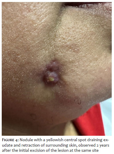
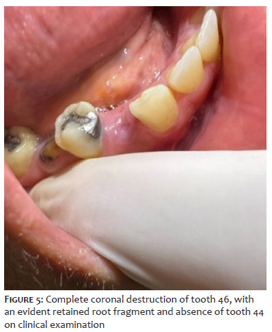
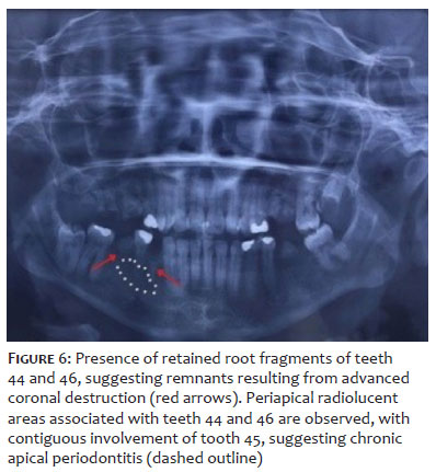
The patient was subsequently lost to follow-up and returned 2 years after the excision, reporting recurrence of the lesion at the same site. On examination, she presented a pink, lobulated nodule with a central yellowish spot draining exudate and retraction of the surrounding skin (Figure 4). At that point, the hypothesis of an OCF was first considered. Examination of the oral cavity revealed right molars and premolars in very poor condition, with the presence of retained root fragments (Figure 5). A panoramic radiograph of the jaws and periapical radiographs of the right mandibular premolars and molars were requested, revealing residual roots of teeth 44 and 46, with a radiolucent area at the root apex of tooth 44, extending into the periapical region of tooth 45 (Figure 6).
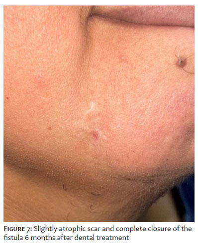
The patient was referred to a dental surgeon, who opted for extraction of the aforementioned root remnants with alveolar curettage and antibiotic therapy with amoxicillin and clavulanate. Tooth 45 was managed conservatively given that pulp vitality was preserved. The patient returned to the outpatient clinic 6 months after completion of dental treatment, showing complete closure of the fistula and a slightly atrophic scar at the site (Figure 7)
OCFs represent a tract between the oral cavity and the skin. They are chronic infections, manifestations of pulp necrosis and chronic apical periodontitis, caused by dental caries, pulpal or periapical infection, periodontal diseases, root fractures, or mechanical problems resulting from trauma. They induce an osteoclastic inflammatory process with abscess formation, which gradually progresses through the alveolar bone, creating a tract toward the area of least resistance.7 Drainage may occur intraorally or extraorally. When not detected early, OCFs may cause aesthetic alterations and psychological distress, compromising patient self-esteem, in addition to complications and secondary infections.
A high degree of diagnostic suspicion is required for OCFs because patients do not always report toothache, and the lesion may be located at a distance from the infectious focus, as in the case of fistulization to the face and neck.8 Lee et al.7 found that, among 33 patients with fistulas of odontogenic origin, 27 were initially misdiagnosed (81.8%). Misdiagnoses included epidermal cyst (24.2%), furuncle (21.2%), subcutaneous mycosis (15.2%), squamous cell carcinoma (9.1%), basal cell carcinoma (6.1%), and foreign body granuloma (6.1%).
Cutaneous manifestations show considerable morphological variability and may present as skin retractions, nodules, abscesses, cysts, ulcers, or draining lesions at different sites, depending on the affected teeth.4 Fistulous tracts are most frequently associated with infections of the mandibular teeth, with preferential drainage to the mandible and chin.5
Diagnosis is based on clinical suspicion and confirmed by panoramic radiography of the face, periapical radiography, and computed tomography.9 Cutaneous ultrasonography is a noninvasive and emerging technique with proven utility in localized lesions,10 representing a safe and accessible tool for the diagnosis of OCFs. The presence of a hypoechoic fistulous tract on color Doppler is a characteristic finding that aids in diagnosis.5
Treatment consists of eliminating the primary cause, resulting in healing and regression of the skin lesion within 1 to 2 weeks. The residual scar may be surgically revised if aesthetically unsatisfactory for the patient.6
Though rare, OCFs should always be considered in the differential diagnosis. Physicians should be familiar with this condition and pay close attention to the oral cavity, particularly when assessing perioral and cervical lesions. When this condition develops and patients are unaware of the underlying dental pathology, they often seek dermatological care before being referred to dental surgeons.
Clarissa Brito Farias
ORCID: 0009-0009-5395-045X
Statistical analysis, Approval of the final version of the manuscript, Conception and design of the study, Preparation and writing of the manuscript, Acquisition, analysis and interpretation of data, Intellectual participation in the propaedeutic and/or therapeutic approach to the cases studied, Critical review of the literature.
Ana Vitória Lins Paiva Antunes
ORCID: 0000-0002-5171-245x
Conception and design of the study, Preparation and writing of the manuscript, Acquisition, analysis and interpretation of data.
Aretuza Iolanda Pimentel Torres Portella
ORCID: 0000-0002-3952-363X
Conception and design of the study, Preparation and writing of the manuscript, Acquisition, analysis and interpretation of data, Intellectual participation in the propaedeutic and/or therapeutic approach to the cases studied.
Antônio Carlos Evangelista de Araújo Bonfim
ORCID: 0000-0001-8739-365X
Approval of the final version of the manuscript, Conception and design of the study, Preparation and writing of the manuscript, Acquisition, analysis and interpretation of data, Effective participation in the conduct of the study, Intellectual participation in the propaedeutic and/or therapeutic approach to the cases studied, Critical review of the literature, Critical revision of the manuscript
Luciana Cavalcanti Trindade
ORCID: 0000-0002-0643-1093
Conception and design of the study, Effective participation in the conduct of the study, Critical review of the literature, Critical revision of the manuscript.
1. Giménez-García R, Martinez-Vera F, Fuentes-Vera L. Cutaneous sinus tracts of odontogenic origin: two case reports. J Am Board Fam Med. 2015;28(6):838-40.
2. Sheehan DJ, Potter BJ, Davis LS. Cutaneous draining sinus tract of odontogenic origin: unusual presentation of a challenging diagnosis. South Med J. 2005;98(2):250-2.
3. Barbosa CAM, Tancredo F, Fonseca CF, Pinho MAB. Diagnosis of cutaneous sinus tract in association with traumatic injuries to the teeth. Braz J Dent Traumatol. 2011;2:75-9.
4. Chhabra A, Chhabra N. Dental infection mimicking dermatological lesion: three case reports of cutaneous fistulae and sinus tracts on face. Indian Dermatol Online J. 2018;9:441-4.
5. Altemir-Vidal A, Iglesias-Sancho M, Quintana-Codina M. Usefulness of high-frequency ultrasonography in the diagnosis of odontogenic cutaneous fistula. An Bras Dermatol. 2021;96:259-60.
6. Cantatore JL, Klein PA, Lieblich LM. Cutaneous dental sinus tract, a common misdiagnosis: a case report and review of the literature. Cutis. 2002;70:264-7.
7. Lee EY, Kang JY, Kim KW, Choi KH, Yoon TY, Lee JY. Clinical characteristics of odontogenic cutaneous fistulas. Ann Dermatol. 2016;28(4):417.
8. Moreira C, Medeiros N, Silva IV, Vaz P, Corrales T, Viegas S. Fístula cutânea odontogénica no pescoço: a importância do exame intraoral. Rev Port Estomatol Med Dent Cir Maxilofac. 2018;59(S1):3.
9. Amorim GM, Paul IR, Molozzi B, Steckert SEV, Biazussi B, Amorim-Filho RM. Fístula cutânea odontogénica. Rev SPDV. 2020;78(3).
10. Wortsman X, Wortsman J. Clinical usefulness of variable-frequency ultrasound in localized lesions of the skin. J Am Acad Dermatol. 2010;62:247-56.
 All content the journal, except where identified, under the Creative Commons Attribution 4.0 International licence - ISSN-e 1984-8773




 Read in Portuguese
 Portuguese PDF
 Print
Print
 Send this article by email
 How to cite this article
 Submit a comment
 Mendeley
 Pocket
 Share on Facebook
 Share on Twitter