


Mariana Sofia Monteiro Silva1; Luiz Fernando Eller Riscaroli1; Eduardo Garbin Rosset2; Ricardo Barbosa Lima1; Luciana Ferreira de Araújo1
Funding source: None
Conflict of interest: None
Submitted on: 12/10/2025
Final decision: 02/16/2026
How to cite this article: Silva MSM, Riscaroli LFE, Rosset EG, Lima RB, Araújo LF, Gomes Neto R. Periocular basal cell carcinoma with orbital invasion: a case report. Surg Cosmet Dermatol. 2026;18(2):e20260542.
Basal cell carcinoma is the most frequent periocular neoplasm. Although mortality is low, orbital invasion represents a severe complication associated with high morbidity. We report the case of a 49-year-old male smoker with an aggressively evolving periocular lesion culminating in amaurosis. Treatment included orbital exenteration and complex reconstruction using local and regional flaps, complicated by partial necrosis and bone exposure, requiring multiple debridements. Histopathological examination revealed a sclerodermiform pattern with squamous differentiation. This case highlights the devastating consequences of locally advanced basal cell carcinoma as well as the importance of early diagnosis and a multidisciplinary approach to prevent deformity and loss of function.
Keywords: Carcinoma, Basal Cell; Orbital Exenteration; Skin Neoplasms
Basal cell carcinoma (BCC) is the most common malignant neoplasm of the periocular region, accounting for approximately 80% to 96% of tumors in this location. Although often considered a tumor with indolent behavior and low metastatic potential, periocular BCC may exhibit locally aggressive behavior. Orbital invasion is an uncommon event, with an estimated incidence ranging from 0.8% to 5.5% in developed countries, reaching alarming rates of up to 17% in developing countries, often due to delays in diagnosis and treatment.1
Clinical progression may be silent, requiring constant vigilance for signs of high-risk tumors. The main factors associated with orbital invasion include multiple recurrences, large tumor size, positive surgical margins, location in the canthal region (medial or lateral), and aggressive histological subtypes, such as morpheaform/sclerodermiform, infiltrative, and basosquamous variants. Perineural invasion, although present in less than 1% of cases, is a marker of poorer prognosis.2
Although the mortality rate associated with periocular BCC remains low, morbidity is high. Tumor progression may result in severe facial disfigurement and vision loss, dramatically affecting patient quality of life.3
The objective of this study is to report a case of periocular BCC with aggressive progression, resulting in visual loss and the need for orbital exenteration, highlighting the serious consequences of delayed specialized care and the importance of early treatment.
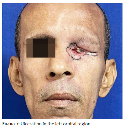
A 49-year-old male patient, smoker, with no comorbidities, was referred to a specialized service with a history of a periocular lesion that had first appeared in 2021. The condition began as a small papule on the lower eyelid and progressed over two years with ulceration, intense pain, and expansive growth, culminating in ipsilateral loss of visual acuity (Figure 1).
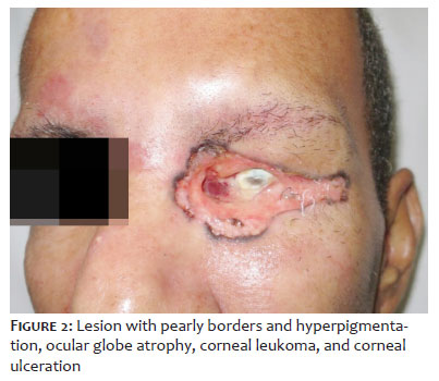
Dermatological examination revealed extensive ulceration in the left orbital region, with pearly borders and large-scale tissue destruction. Ophthalmological examination found ocular globe atrophy and corneal ulceration (Figure 2).
An incisional biopsy was performed, confirming the diagnosis of BCC. Imaging studies found orbital involvement without intracranial invasion. Given the extent of the tumor and the functional loss, orbital exenteration was performed with negative frozen-section margins (Figure 3).
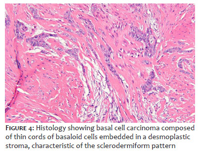
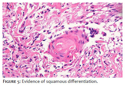
Histopathological examination revealed sclerodermiform BCC with squamous differentiation (Figures 4 and 5). There was no optic nerve invasion.
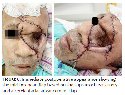
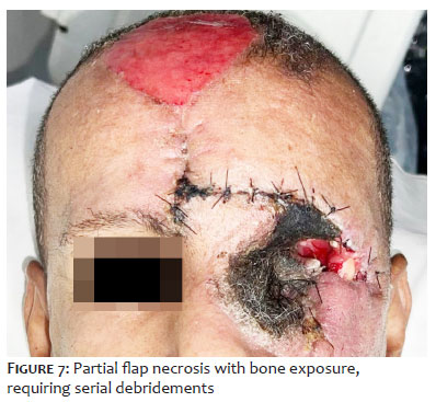
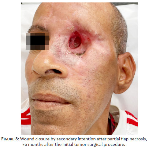
Reconstruction included a mid-forehead flap associated with a cervicofacial advancement flap (Figure 6). The postoperative course was marked by partial necrosis (Figure 7), orbital bone exposure, serial debridements, and subsequent reconstruction using a temporalis muscle flap. Conservative management was adopted, allowing healing by secondary intention (Figure 8). Following these interventions, the patient was lost to clinical follow-up, and resuming it was not possible despite repeated attempts at contact by the medical team
This case illustrates the concept of locally advanced BCC, a challenging condition in which conventional surgery or radiotherapy alone may not be curative or may result in major deformities. The 2023 European consensus-based guideline emphasizes that BCCs should not be underestimated; neglected or recurrent tumors may invade deep tissues, making treatment complex.7
The classification proposed by the European Association of Dermato-Oncology (EADO) and the guidelines of the National Comprehensive Cancer Network (NCCN) (version 2.2024) recommend a multidisciplinary approach for these cases. The first line of treatment for locally advanced BCC remains surgery (with strict margin control) and/or radiotherapy.4
In the present case, radical surgery (orbital exenteration) was required because of orbital invasion and prior visual loss. Histopathological examination revealed aggressive subtypes (sclerodermiform and squamous differentiation), corroborating the rapid and destructive clinical course observed.5,6 The sclerodermiform pattern is known for its poorly defined margins and high recurrence rate, whereas squamous differentiation (basosquamous pattern) confers a more aggressive biological behavior, approaching that of squamous cell carcinoma.8
When surgery and radiotherapy are contraindicated or unsuccessful, current guidelines recommend systemic therapy. Hedgehog pathway inhibitors (such as vismodegib and sonidegib) and immunotherapy with anti-PD-1 checkpoint inhibitors (such as cemiplimab) have emerged as valuable treatment options.4,7 For locally advanced periocular BCCs and metastatic orbital disease, vismodegib (150 mg/day) has demonstrated efficacy and may even be used as neoadjuvant therapy to reduce tumor mass and potentially decrease the rate of orbital exenteration.5
Regarding prognosis, periocular BCC with negative surgical margins presents recurrence rates ranging from 1.5% to 5.6%, depending on the anatomical location. The medial canthus is the site most prone to recurrence and deep invasion because of embryologic cleavage planes that facilitate tumor spread.4 The literature documents that recurrence rates are significantly higher for periorbital malignancies than for BCCs arising at other cutaneous sites.9
This case report highlights the importance of early diagnosis and regular follow-up for periocular lesions. Although BCC is often curable, it may progress to locally advanced forms with orbital invasion, resulting in blindness and mutilation, including the need for orbital exenteration. A multidisciplinary approach involving dermatologists, ophthalmologists, head and neck surgeons, and plastic surgeons is essential for appropriate management, aiming to achieve oncologic cure while minimizing functional and aesthetic sequelae.
Mariana Sofia Monteiro Silva
ORCID: 0009-0007-9928-7337
Approval of the final version of the manuscript, Conception and design of the study, Preparation and writing of the manuscript, Acquisition, analysis and interpretation of data, Critical review of the literature, Critical revision of the manuscript.
Luiz Fernando Eller Riscaroli
ORCID: 0009-0006-6665-0710
Intellectual participation in the propaedeutic and/or therapeutic approach to the cases studied.
Eduardo Garbin Rosset
ORCID: 0009-0002-5743-8743
Intellectual participation in the propaedeutic and/or therapeutic approach to the cases studied
Ricardo Barbosa Lima
ORCID: 0000-0002-4297-3236
Approval of the final version of the manuscript, Effective participation in the conduct of the study, Intellectual participation in the propaedeutic and/or therapeutic approach to the cases studied, Critical review of the literature, Critical revision of the manuscript.
Luciana Ferreira de Araújo
ORCID: 0000-0001-6323-4961
Acquisition, analysis and interpretation of data, Critical revision of the manuscript.
Ruy Gomes Neto
ORCID: 0009-0006-6676-2173
Intellectual participation in the propaedeutic and/or therapeutic approach to the cases studied.
1. Bengoa-González A, Mencía-Gutiérrez E, Garrido M, Salvador E, Lago-Llinás MD. Advanced periocular basal cell carcinoma with orbital invasion: update on management and treatment advances. J Ophthalmol [Internet]. 2024;2024:1-15.
2. Madge SN, Khine AA, Thaller VT, Davis G, Malhotra R, Selva D. Globe-sparing surgery for medial canthal basal cell carcinoma with anterior orbital invasion. Ophthalmology. 2010;117(11):2222–8.
3. Seth R. Periocular basal cell carcinoma: a review of atypical presentations, high-risk features, and management. Ophthalmic Plast Reconstr Surg. 2020;36(4):415–23.
4. Schmults CD. Basal cell skin cancer, version 2.2024, nccn clinical practice guidelines in oncology. J National Compr Cancer Netw [Internet]. 2023;21(11):1181-203.
5. Mathis J, Doerr T, Lin E, Ibrahim SF. Oral hedgehog pathway inhibition as a means for ocular salvage in locally advanced intraorbital basal cell carcinoma. Dermatol Surg [Internet]. 2019;45(1):17-25.
6. Hooper J, Shao K, Feng PW, Falcone M, Feng H. Periocular and ocular surface non-melanoma skin cancer. Clin Dermatol [Internet]. 2023.
7. Peris K. European consensus-based interdisciplinary guideline for diagnosis and treatment of basal cell carcinoma – Update 2023. Eur J Cancer [Internet]. 2023:113254.
8. Crowson AN. Basal cell carcinoma: biology, morphology and clinical implications. Mod Pathol. 2006;19(S2):S127–47.
9. Cook BE Jr, Bartley GB. Treatment options and future prospects for the management of eyelid malignancies: an evidence-based update. Ophthalmology. 2001;108(11):2088–98.
 All content the journal, except where identified, under the Creative Commons Attribution 4.0 International licence - ISSN-e 1984-8773




 Read in Portuguese
 Portuguese PDF
 Print
Print
 Send this article by email
 How to cite this article
 Submit a comment
 Mendeley
 Pocket
 Share on Facebook
 Share on Twitter