


Nivin Mazen Said1; Gisele Alves Morikawa Caldeira1; Airton Kenji Motizuki2; Maraya de Jesus Semblano Bittencourt1; Mariana Bastos Amanajás1; Silvia Ferreira Rodrigues Müller1; Franklin de Souza Rocha1
Funding source: None
Conflict of interest: None
Clinical trial? No
Submitted on: 11/08/2025
Final decision: 05/14/2026
How to cite this article: Said NM, Caldeira GAM, Motizuki AK, Amanajás MB, Bittencourt MJS, Müller SFR, Rocha FS. Atypical location of granular cell tumor in a female patient: a rare case report. Surg Cosmet Dermatol. 2026;18(2):e20260537.
Granular cell tumors are rare neurogenic neoplasms, usually benign, with a nonspecific clinical presentation. We report the case of a 70-year-old woman with an ulcerated nodular lesion on the arm, showing progressive growth, pain, and pruritus. Findings from complementary examinations, including magnetic resonance imaging, histopathological examination, and immunohistochemistry, confirmed, respectively, an expansive subcutaneous mass, a proliferation of polyhedral cells with granular cytoplasm, and positivity for the S100 protein, consistent with a diagnosis of granular cell tumor. The report discusses the importance of early diagnosis, complete surgical excision, and clinical follow-up to prevent recurrences and to monitor the rare risk of malignancy.
Keywords: Granular Cell Tumor; Schwann Cells; Neoplasms by Site
Granular cell tumors (GCTs) are neurogenic neoplasms, typically benign and derived from Schwann cells.1-4 Clinically, they usually present as generally asymptomatic, slow-growing subcutaneous nodules that have nonspecific clinical features, which may hinder their initial diagnosis.1-4 GCTs are found predominantly in adults, especially women and black individuals, and may arise in various anatomical sites, such as the skin, tongue, gastrointestinal tract, peripheral nervous system, and subcutaneous tissue.1-4.
This study consists of a case report of a 70-year-old woman diagnosed with a granular cell tumor in the right arm region. The case is described from diagnosis through treatment of the neoplasm, supported by a literature review conducted using the PubMed, Scientific Electronic Library Online (SciELO), and Latin American and Caribbean Health Science Literature (LILACS) databases. In addition, the study followed the CARE checklist guidelines for case reports and case series. The case report was based on the patient’s medical records and complementary examinations that assisted in diagnosis and clinical management, observing all ethical procedures and after having obtained informed consent from the patient.
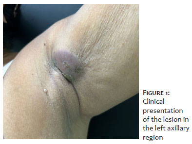
A 70-year-old woman was referred to a dermatologist because of the appearance of a nodular lesion on her left arm approximately 2 years earlier, with progressive growth, pain, and local pruritus, without prior treatment (Figure 1).
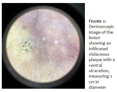
At the initial dermatologic examination, she presented with an infiltrated violaceous plaque with a central ulceration, measuring 2 cm in diameter, and located on the anterolateral aspect of the left arm (Figure 2).
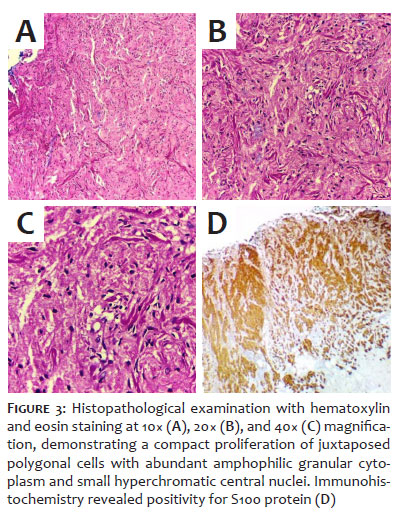
Magnetic resonance imaging (MRI), histopathological examination (HPE), and immunohistochemical analysis (IHC) of the skin lesion were requested. The MRI revealed a heterogeneous expansive mass with lobulated contours permeating the subcutaneous tissue of the left axillary region, measuring approximately 3.8 × 3.3 × 2.5 cm, extending to the adjacent skin and in close contact with the biceps brachii muscle, but without signs of invasion or other abnormalities. Correlation with histopathological findings was considered necessary to establish the diagnosis. The HPE revealed a specimen superficially covered by epidermis without atypia, overlying the mid and deep dermis expanded by a compact proliferation of juxtaposed polygonal cells with abundant amphophilic granular cytoplasm and small hyperchromatic central nuclei (Figure 3A, 3B, and 3C).
The IHC found diffuse positivity for S100 protein and NKI-C3, without evidence of malignancy. Correlation of the IHC with the clinical findings confirmed the diagnosis of granular cell tumor (Figure 3D).
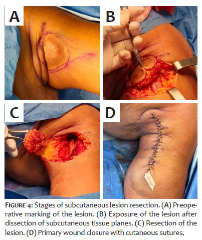
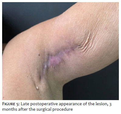
Initial management consisted of requesting preoperative testing for surgical resection and continuing clinical follow-up. The tumor was surgically resected with primary closure (Figure 4). Three months after the procedure, only a residual surgical scar was observed at the lesion site (Figure 5)
Granular cell tumors (GCTs), also known as Abrikossoff’s tumors, are rare neoplasms of controversial origin, predominantly benign, with only a small proportion of malignant cases reported in the literature.1,2 Initially believed to be derived from myoblasts, evidence from immunohistochemical and electron microscopy studies indicates that their true lineage is neurogenic, more specifically originating from Schwann cells, as suggested by the strong expression of the S100 marker.3-5
GCTs may occur in various anatomical sites and are most commonly found in the tongue, skin, and subcutaneous tissues, although they can arise in virtually any part of the body, including the respiratory tract, gastrointestinal tract, breast, and central nervous system. However, skin involvement remains poorly documented in the literature.1,3,4 The clinical presentation is often nonspecific, with slow and painless growth, which may lead to delays in diagnosis.1,4,5 Although infrequent, GCTs have been reported in association with genetic syndromes such as Noonan syndrome and neurofibromatosis. 4-6
They predominantly affect adults between the third and sixth decades of life, with a slight predominance in women and individuals of African descent.2-4 Most tumors are benign, but fewer than 2% do exhibit malignant behavior, with histopathological findings such as necrosis, nuclear pleomorphism, and increased mitotic activity, carrying a risk of metastasis.2
Given their rarity and their potential to mimic other benign or malignant lesions, definitive diagnosis of GCT requires histopathological examination and may be supplemented by immunohistochemistry.2,4 Histopathologically, GCTs are characterized by a nonencapsulated tumor with large polygonal cells, small hyperchromatic central nuclei, and cytoplasm containing abundant eosinophilic granules, extending into the hypodermis.2 Tumor cells frequently display large eosinophilic granules surrounded by a clear halo, known as pustulo-ovoid bodies of Milian.2
The treatment of choice is complete surgical excision with tumor-free margins, and clinical follow-up is essential for the early detection of recurrences, particularly in cases presenting histopathological parameters suggestive of aggressiveness.2
In conclusion, this case documents a rare neoplasm with both a challenging diagnosis and an unusual anatomical site, a presentation that is seldom described in the scientific literature when occurring in the upper limbs. This report also reinforces the importance of early diagnosis of this tumor, given its potential for malignant behavior.
Nivin Mazen Said
ORCID: 0000-0002-0611-5672
Approval of the final version of the manuscript, Conception and design of the study, Preparation and writing of the manuscript, Acquisition, analysis and interpretation of data, Effective participation in the conduct of the study, Intellectual participation in the propaedeutic and/or therapeutic approach to the cases studied, Critical review of the literature, Critical revision of the manuscript.
Gisele Alves Morikawa Caldeira
ORCID:0000-0001-7655-1173
Approval of the final version of the manuscript, Conception and design of the study, Preparation and writing of the manuscript, Acquisition, analysis and interpretation of data, Effective participation in the conduct of the study, Intellectual participation in the propaedeutic and/or therapeutic approach to the cases studied, Critical review of the literature, Critical revision of the manuscript.
Airton Kenji Motizuki
ORCID: 0009-0002-7188-0984
Approval of the final version of the manuscript, Conception and design of the study, Preparation and writing of the manuscript, Acquisition, analysis and interpretation of data, Effective participation in the conduct of the study, Critical review of the literature, Critical revision of the manuscript.
Maraya de Jesus Semblano Bittencourt
ORCID: 0000-0002-7297-0749
Author’s contribution: Approval of the final version of the manuscript, Conception and design of the study, Effective participation in the conduct of the study, Intellectual participation in the propaedeutic and/or therapeutic approach to the cases studied, Critical revision of the manuscript.
Silvia Ferreira Rodrigues Müller
ORCID: 0000-0002-5714-8466
Author’s contribution: Approval of the final version of the manuscript, Conception and design of the study, Effective participation in the conduct of the study, Intellectual participation in the propaedeutic and/or therapeutic approach to the cases studied, Critical revision of the manuscript.
Franklin de Souza Rocha
ORCID:0000-0002-7386-1616
Author’s contribution: Approval of the final version of the manuscript, Conception and design of the study, Effective participation in the conduct of the study, Intellectual participation in the propaedeutic and/or therapeutic approach to the cases studied, Critical revision of the manuscript.
1. Marcoval J, Bauer-Alonso A, Llobera-Ris C, Moreno-Vilchez C, Penín RM, Bermejo J. Granular Cell Tumor: A Clinical Study of 81 Patients. Actas Dermosifiliogr (Engl Ed). 2021;112(5):441-6.
2. Fahim S, Aryanian Z, Ebrahimi Z, Kamyab-Hesari K, Mahmoudi H, Alizadeh N, et al. Cutaneous granular cell tumor: A case series, review, and update. J Family Med Prim Care. 2022;11(11):6955-8.
3. Nasser KR, Cuce LC, Vasconcelos RF, Macedo AC, Rodriguez JGK, Arruda RG, et al. Apresentação atípica de tumor de células granulares. Surg Cosmet Dermatol. 2015;7(1):72-4.
4. Neelon D, Lannan F, Childs J. Tumor de células granulares. Em: StatPearls [Internet]. Treasure Island (FL): StatPearls Publishing; jan. de 2025.
5. Ardeleanu V, Jecan RC, Moroianu M, Teodoreanu RN, Tebeica T, Moroianu LA, et al. Case report: Abrikossoff's tumor of the facial skin. Front Med (Lausanne). 2023;10:1149735.
6. Paul SP, Osipov V. An unusual granular cell tumour of the buttock and a review of granular cell tumours. Case Rep Dermatol Med. 2013;2013:109308.
 All content the journal, except where identified, under the Creative Commons Attribution 4.0 International licence - ISSN-e 1984-8773




 Read in Portuguese
 Portuguese PDF
 Print
Print
 Send this article by email
 How to cite this article
 Submit a comment
 Mendeley
 Pocket
 Share on Facebook
 Share on Twitter