


Olena Baliuk1; Oleh Akimov2; Elena Vazhnichaya1; Vitalii Kostenko2; Ivan Starchenko3
Funding support: None
Conflict of interest: None
Submitted on: 02/20/2024
Approved on: 06/14/2024
How to cite this article: Baliuk O, Akimov O, Vazhnichaya O, Kostenko V, Starchenko I. How to cite this article: Baliuk O, Akimov O, Vazhnichaya O, Kostenko V, Starchenko I. The influence of topical antioxidant use on hair regrowth and skin condition after chemical depilation. Surg Cosmet Dermatol.2024;16:e02024350.
INTRODUCTION: Hair restoration treatments for alopecia using existing medications are not always effective, highlighting the need for new therapeutic options.
OBJECTIVE: This study aimed to evaluate the effects of a topical gel containing ethyl-methyl-hydroxypyridine succinate (EMHPS) on hair regrowth and biochemical and histological skin parameters in animals following chemical depilation.
METHODS: Experiments were conducted on 50 adult male Wistar rats. Alopecia was induced using a commercial depilatory product containing potassium thioglycolate. A 5% EMHPS gel (125 mg/kg) was applied daily to the depilated skin. Trichoscopy, biochemical analysis, and histological examination of skin samples were performed on days 3, 9, and 21 of treatment.
RESULTS: The EMHPS gel demonstrated a tendency to accelerate hair regrowth, reduce lipid peroxidation, normalize antioxidant enzyme activity, and restore hydroxyproline and glycosaminoglycan levels in the treated skin compared to the untreated pathology control.
CONCLUSIONS: The EMHPS gel primarily influences skin biochemical parameters and may be beneficial for treating forms of alopecia associated with oxidative stress.
Keywords: Hair; Hair Removal; Skin.
Healthy hair is often associated with beauty, and hair loss can significantly impact quality of life.1 Alopecia is one of the most common dermatological concerns, with various types requiring pathogenetic treatments aimed at stimulating the anagen phase, delaying catagen, and restoring proper hair thickness.2,3 While oral treatments are available, they carry a high risk of side effects,4 making topical therapy the preferred approach.5
Topical formulations of finasteride and minoxidil are the most widely recognized treatments for alopecia. However, other options include prostaglandins, ketoconazole, vitamins, minerals, herbal preparations, platelet-rich plasma, growth factors, microneedling, laser therapy, and cell-based therapies.6 In cases of scarring alopecia, hair transplantation remains the most effective treatment.7 Despite these options, topical hair restoration treatments are not always effective, highlighting the need for new therapeutic agents or the repositioning of existing drugs.6
From this point of view, antioxidants—particularly ethyl-methyl-hydroxypyridine succinate (EMHPS), also known as Mexidol—are of interest.8 This synthetic heterocyclic antioxidant is considered low-toxic, has a significant safety margin, and is prescribed in some post-Soviet countries for psychoneurological and cardiovascular conditions.9 Computational modeling using the Drug2Ways approach has predicted potential dermatological benefits of EMHPS, suggesting its possible therapeutic application in alopecia.10
Oxidative stress is known to contribute to the development of skin disorders,11 potentially serving as a link between dermatological conditions and hair loss.12 Despite its potent antioxidant properties, topical application of EMHPS in dermatology and cosmetology remains unexplored, and no topical formulations of the drug currently exist. This motivated us to develop an EMHPS gel. The gel formulation was chosen for its ability to deliver bioactive substances promoting tissue regeneration while remaining stable on the treated area and resisting evaporation longer than liquid formulations.13 We decided to investigate the effects of EMHPS gel on hair restoration and skin condition in laboratory rats following chemical depilation, a widely used animal model for studying hair loss in the preclinical testing of drugs and cosmetic products.14 The aim of our study is to evaluate the impact of topical application of the newly developed EMHPS gel on hair regrowth and skin biochemical and histological parameters in animals after chemical depilation.
Gel-forming and auxiliary substances, as well as all reagents for biochemical analysis and dyes for histological examination, were obtained from Merck KGaA (Germany). EMHPS was obtained from SPF Microchem LLC (Ukraine). The gel formulation contained 5.0 g of EMHPS, 0.5 g of sodium metabisulfite, 1.0 g of polyvinyl alcohol, 2.0 g of carbomer 940, 2.8 g of TRIS, and distilled water up to 100.0 g. It was prepared using standard laboratory techniques. First, EMHPS and sodium metabisulfite were dissolved in 2/5 of the required total amount of water. Separately, polyvinyl alcohol was dissolved in another 2/5 of the total water by heating in a water bath. The two solutions were then combined. Finally, carbomer 940 and TRIS, which had been pre-dissolved in the remaining 1/5 of the total water, were gradually added with continuous stirring until a gel was formed.
A total of 50 adult male Wistar rats (122–126 days old, weighing 185–215 g) were housed in groups of five per cage under standard laboratory conditions (standard diet and water ad libitum). They were maintained on a standard laboratory diet with ad libitum access to water in a temperature-controlled room with a 12-hour light-dark cycle. The study protocol was approved by the Committee on Bioethics and Ethical Issues at Poltava State Medical University (No. 220, October 25, 2023).
All rats were pre-selected to be in the telogen phase of the hair growth cycle based on their age.15 They were then randomly assigned to four groups: intact control, pathology control, reference, and experimental groups.
Alopecia was induced by chemically depilating the animals' backs. An 8 cm × 4 cm skin area was treated with a commercial depilatory product containing potassium thioglycolate.16 The product was applied in a thin layer for 10 minutes. Once the hair was dissolved, it was removed, and the skin was thoroughly washed with water and dried with a napkin. Treatment began immediately after the depilated skin area was dried.
The EMHPS gel was applied to the test area at a dose of 125 mg/kg (approximately 0.5 mL per rat). Treatment was administered once daily at the same time each day. After gel application, each animal was isolated for 30 minutes before being returned to its cage. Treatment continued until 24 hours before euthanasia.
As a reference treatment, a 2% minoxidil solution (Industrial Pharmaceutics Cantabria, S.A., Spain) was applied to the depilated skin at a dose of 30 mg/kg (approximately 0.3 mL per rat) once daily.15
Throughout the experiment, animals' behavior was monitored, and their body weight was recorded periodically. On days 3, 9, and 21 post-depilation, animals were euthanized by terminal hemorrhage induced by general anesthesia with sodium thiopental (50 mg/kg, JSC Kyivmedpreparat, Ukraine).17
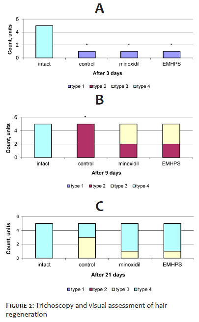
Trichoscopy and photography were performed with a Firefly DE330T digital trichoscope (USA). Hair growth was scored using a four-tier scoring system: type 1 = uneven, weak hair growth with clearly visible skin; type 2 = low hair density with partially visible skin; type 3 = moderate hair density with no visible skin; type 4 = high hair density with full, thick fur.18
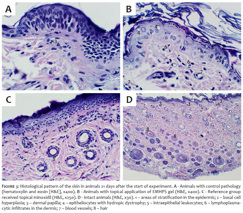
On day 21, skin samples were collected from the test area of euthanized rats and processed for histological analysis. Samples were stained with hematoxylin and eosin (H&E) according to a standard protocol.19 Microscopic examination was performed using an Olympus BX41 microscope (Olympus, Japan).
A 10% tissue homogenate was prepared from the affected skin area using 0.2 M Tris-HCl buffer solution (pH = 7.4). Malondialdehyde (MDA) content was determined based on its reaction with 1-methyl-2-phenyl-indole.20 Superoxide dismutase (SOD) activity was measured by monitoring the kinetics of adrenaline autoxidation.21 Catalase activity was assessed using the molybdate colorimetric method.22 The concentration of free hydroxyproline was determined by a colorimetric assay, which relies on the reaction of pyrrole-2-carboxylic acid, formed during hydroxyproline oxidation, with p-dimethylaminobenzaldehyde in a modified procedure.23 Glycosaminoglycan (GAG) content in skin was analyzed by measuring the concentration of hexuronic acids, which form a colored product in reaction with carbazole, following a modified version of the Dische method.24
All methods were previously validated for 10% tissue homogenate analysis. Optical density measurements were performed using a Ulab 101 spectrophotometer (Ulab, China).
The results of biochemical assays were expressed as mean ± standard error of the mean (M±SE). Data were statistically analyzed using one-way analysis of variance (ANOVA) followed by a post-hoc Tukey test. Data normality was assessed using the Shapiro-Wilk test. The Mann-Whitney U test was applied to evaluate the semi-quantitative assessment of hair regeneration. A p-value < 0.05 was considered statistically significant.
Throughout the observation period, no behavioral abnormalities were detected in the control or experimental groups. Changes in body weight were statistically insignificant compared to baseline values.
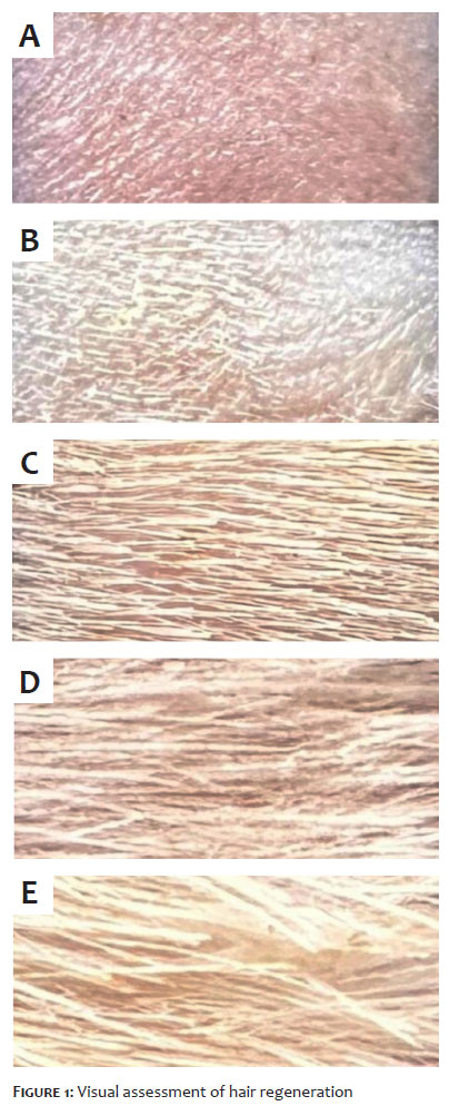
According to visual examination, intact rats exhibited a thick fur coat typical of this species (Figure 1D), corresponding to type 4 (5/5) (Figure 2). Immediately after chemical depilation, all groups displayed a hairless test area with clean skin (Figure 1A). By day 3, hair regrowth had begun, with similar progression across all groups (Figure 1B). At this stage, hair regeneration was classified as type 1 (5/5) and remained significantly different from the intact group (p < 0.005) (Figure 2A).
By day 9, differences between the groups became apparent. In the control pathology group, the test area showed noticeable hair regeneration, classified as type 2 (5/5) (Figure 1C), which remained significantly different from the intact control (p < 0.005) (Figure 2B). In the reference group (minoxidil-treated), there was a trend toward greater hair growth, with type 3 (3/5) (p < 0.1) compared to the control pathology group (Figure 1D, Figure 2B). At the same point, hair regeneration in the EMHPS-treated group was similar to that in the reference group (Figure 1D, Figure 2B), though some hairs in the EMHPS group appeared longer.
By day 21, in the control pathology group, hair coverage had almost returned to normal, with a mix of type 3 (3/5) and type 4 (2/5) pelage (Figure 1D, E). In the EMHPS-treated group, hair regeneration was predominantly type 4 (4/5), though the difference compared to the control pathology group was not statistically significant (Figure 2C). A similar pattern was observed in the reference group, though visually, the fur in this group appeared more uniform than in the EMHPS-treated rats (Figure 2C).
Overall, macroscopic evaluation suggests both the EMHPS gel and the reference treatment (minoxidil) tended to accelerate hair regrowth following chemical depilation.
After 21 days, rats in the control pathology group exhibited basal cell reactivity, indistinct layer boundaries, granular layer hypertrophy, and the presence of single intraepithelial cysts (Figure 3A). Dermal papillae were well or moderately developed, while hair follicles varied in location and diameter.
In the EMHPS-treated group, the epidermis contained numerous epitheliocytes with hydropic dystrophy and intraepithelial leukocytes (Figure 3B). Stratum corneum showed localized hypertrophy, and basal cell reactivity was still present. A few lymphoplasmacytic infiltrates were observed in the dermis, and microvessels displayed signs of reduced blood supply. Papillae formation was moderate, and hair follicles were unevenly distributed, with some sebaceous glands hypertrophied.
In the minoxidil-treated group, a significant number of epitheliocytes with optically empty vacuoles, intraepithelial leukocytes, and areas of basal cell reactivity were noted in the epidermis (Figure 3C). Dermis contained an increased number of cellular elements and focal clusters. Papillae were well or moderately developed, with a substantial number of hair follicles. Their diameters varied slightly, and some sebaceous glands showed signs of hypertrophy.
For comparison, the histological pattern of intact skin is shown in Figure 3D. Epidermis consisted of 2–6 cell layers with distinct boundaries, with single intraepithelial lymphocytes present. Some epitheliocytes showed signs of hydropic dystrophy. Dermis was composed of connective tissue with well-defined collagen fibers and scattered cellular elements. Papillae were moderately developed, and hair follicles were diffusely distributed or grouped in clusters of 3–5, mostly small to medium in diameter. Sebaceous glands were located near hair follicles, sometimes maintaining a visible connection with them.
Overall, hair regeneration after chemical depilation, both in the absence of pharmacological treatment and following topical application of EMHPS or the reference drug (minoxidil), was associated with histological changes in skin. Notably, pharmacotherapy intensified the skin response compared to the control pathology group.
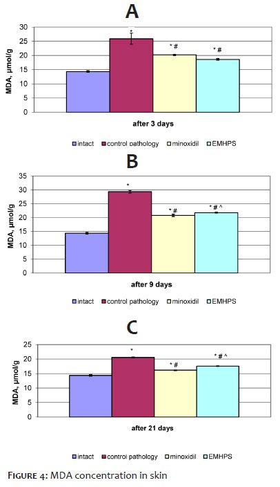
Throughout the observation period, control pathology was associated with increased lipid peroxidation, as evidenced by a significant elevation in MDA concentration (p < 0.001) in the affected skin area compared to intact animals (Figure 4). Minoxidil treatment reduced MDA levels by 1.3-fold (p < 0.001) after 3 days, 1.4-fold (p < 0.001) after 9 days, and 1.2-fold (p < 0.001) after 21 days, compared to untreated depilated skin at the same time points. The EMHPS gel produced a similar effect, reducing MDA concentration by 1.4-fold (p < 0.001) after 3 days, 1.3-fold (p < 0.001) after 9 days, and 1.2-fold (p < 0.001) after 21 days relative to the control pathology group. However, at later observation periods, the EMHPS gel had a slightly weaker effect than minoxidil (p < 0.001).
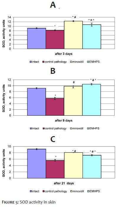
In the control pathology group, SOD activity in the skin decreased 3 days after depilation (p < 0.05) compared to intact rats (Figure 5A). This reduction became even more pronounced after 9 and 21 days (p < 0.001) as the model pathology progressed (Figure 5B, C). Minoxidil treatment increased SOD activity by 1.5- to 1.4-fold (p < 0.001) compared to untreated depilated skin. The EMHPS gel also enhanced SOD activity, with increases of 1.3-fold (p < 0.001) at 3 days, 1.8-fold (p < 0.001) at 9 days, and 1.3-fold (p < 0.001) at 21 days relative to the control pathology group. Notably, after 3 and 21 days, the antioxidant effect of EMHPS was weaker than that of minoxidil (p < 0.002 and p < 0.001, respectively), but after 9 days, it was stronger (p < 0.001).
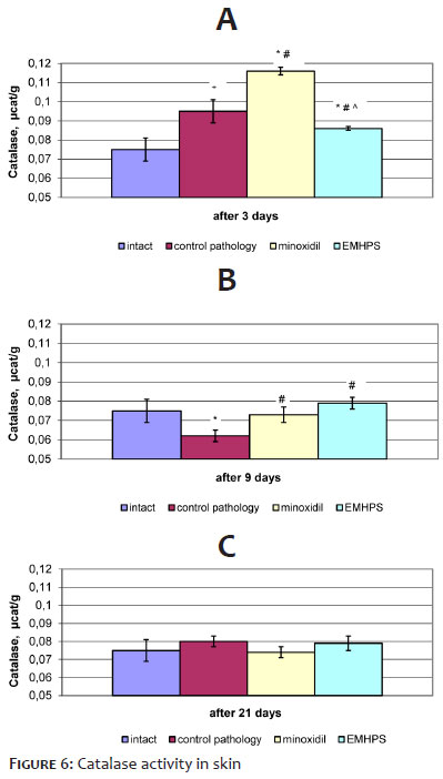
In the control pathology group, catalase activity initially increased after 3 days (p < 0.001), decreased after 9 days (p < 0.002), and remained unchanged after 21 days compared to the intact control (Figure 6). Minoxidil further increased catalase activity by 1.2-fold (p < 0.001) in 3 days, normalized it after 9 days (p < 0.005), and had no significant effect at 21 days compared to the control pathology group. The EMHPS gel initially reduced catalase activity after 3 days (p < 0.05), increased it by 1.3-fold (p < 0.001) after 9 days, and had no significant effect at 21 days. The effect of EMHPS differed from minoxidil only in the early observation period (p < 0.001).
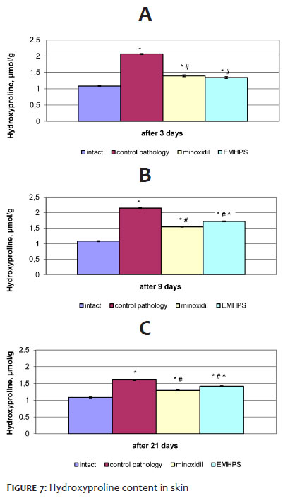
The results of biomarker analysis for connective tissue condition in the affected skin area are shown in Figure 7 and Figure 8. In the control pathology group, free hydroxyproline levels remained elevated throughout the study period. After 3 days, hydroxyproline increased 1.9-fold (p < 0.001), after 9 days, 2.0-fold (p < 0.001), and after 21 days, 1.5-fold (p < 0.001) compared to the intact control (Figure 7). Minoxidil significantly reduced these values, decreasing hydroxyproline concentration by 1.5-fold (p < 0.001) after 3 days, 1.4-fold (p < 0.001) after 9 days, and 1.2-fold (p < 0.001) after 21 days relative to the control pathology group. The EMHPS gel also lowered hydroxyproline levels, reducing them by 1.5-fold (p < 0.001) in 3 days, 1.3-fold (p < 0.001) in 9 days, and 1.1-fold (p < 0.001) in 21 days compared to the control pathology group. In the early observation period, the effects of EMHPS and minoxidil did not differ, but with continued treatment, EMHPS had a weaker effect on hydroxyproline levels than minoxidil (p < 0.001).
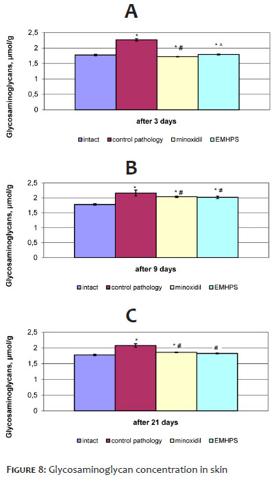
Three days after depilation without pharmacotherapy, GAG concentration in the affected skin increased 1.3-fold (p < 0.001) compared to intact rats (Figure 8A). It remained elevated by 1.2-fold after 9 and 21 days (p < 0.001) (Figure 8B, C). Minoxidil significantly normalized GAG levels after 3 days (p < 0.001) and gradually reduced them over time (p < 0.001) compared to the control pathology group (Figure 8). The EMHPS gel produced a similar effect: after 3 days, it normalized GAG levels to those of the intact control, and after 9 and 21 days, GAG levels decreased similarly to the minoxidil-treated group.
The EMHPS gel effectively inhibited MDA accumulation, modulated SOD and catalase activity, and reduced free hydroxyproline and total GAG concentrations in a manner comparable to minoxidil, though slightly less effective at certain time points.
Visual assessment of hair regeneration after chemical depilation revealed an acceleration of this process in both the EMHPS-treated group and the minoxidil-treated reference group. However, this improvement was observed as a trend rather than a statistically significant effect, likely due to individual variations in hair regrowth among animals, increasing result variability.14 When examining the test area, differences in hair coat uniformity were also noted. The reference group exhibited a more homogeneous hair coat, whereas in the EMHPS-treated group, particularly after 9 days, individual long hairs were observed. This suggests that EMHPS may have a weaker effect on synchronizing follicle development cycles compared to minoxidil, which is known to influence this process.25
Histopathological analysis showed that chemical depilation without treatment led to basal cell reactivity and an increase in intraepithelial leukocytes, which persisted until the end of the experiment. These findings align with previous studies reporting histopathological changes in laboratory mice following exposure to conventional depilatory creams.26 The observed reactive inflammation may be attributed to the alkaline properties of the depilatory agent, which can trigger a stronger reaction in rodent skin due to its thinner structure compared to human skin.14,18
Interestingly, the EMHPS gel enhanced reactive skin changes compared to the control pathology group. At first glance, this might seem contradictory, given that EMHPS inhibits free radical-driven prostaglandin synthesis mediated by cyclooxygenase and lipoxygenase.8 However, in the context of hair regeneration, the interplay between inflammation, damage repair, and regeneration through inflammatory cytokines and Wnt signaling factors may be more relevant.27, 28 This assumption is further supported by the histopathological observations in the minoxidil-treated group, which also exhibited enhanced reactive skin changes compared to control pathology. The only notable difference between the EMHPS gel and minoxidil was that minoxidil produced greater uniformity in hair follicle size and distribution, which could be considered an advantage of minoxidil.
In addition to histopathological changes, untreated depilation induced oxidative stress, as indicated by increased MDA levels, decreased SOD activity, and fluctuations in catalase activity. This suggests that oxidative stress development was linked to cytokine profile modifications caused by depilation,29 and in the early post-depilation phase, potentially to a general adaptation syndrome. Given interspecies differences in skin structure and hair function, chemical depilation over a large body area in animals likely represents a more severe intervention than localized hair removal in humans.14,18
In depilated animals without pharmacological treatment, MDA levels remained elevated, and SOD activity remained suppressed until the end of the experiment. The early-stage suppression of SOD could result from enzyme inhibition by excess reaction products, whereas in the later stages, it may reflect a decrease in superoxide anion radical production. Since catalase works in tandem with SOD, its fluctuations likely correspond to hydrogen peroxide variations in the superoxide dismutase reaction. Both EMHPS and minoxidil reduced MDA levels, increased SOD activity, and modulated catalase activity, demonstrating antioxidant effects. This outcome was expected for EMHPS, given its established antioxidant properties.8 Considering the known role of oxidative stress in hair growth impairment,30 the inhibition of lipid peroxidation suggests a potentially beneficial pharmacodynamic effect of EMHPS on hair regeneration.
The antioxidant activity of minoxidil extended to both inducible antioxidant enzymes (SOD and catalase) and MDA accumulation. Although this property is rarely discussed in literature, it is plausible the effect of minoxidil on oxidative stress biomarkers is related to its ability to chelate intracellular iron.31 Minoxidil exerts multiple pharmacodynamic effects, contributing to hair regrowth through vasodilation, anti-inflammatory action, Wnt/β-catenin pathway activation, and antiandrogenic activity, all of which influence anagen and telogen phase duration.32
When analyzing free hydroxyproline levels, untreated depilation led to a sustained increase throughout the observation period. In this study, a modified hydroxyproline assay (excluding the hydrolysis step) was used, allowing for the interpretation of increased hydroxyproline levels as an indicator of collagen degradation under oxidative stress.33 The EMHPS gel reduced hydroxyproline content, exerting a normalizing effect similar to minoxidil, though less pronounced at 9 and 21 days. For both treatments, this suggests a potential role in regulating the extracellular matrix (ECM) and hair follicle regeneration, possibly by reducing oxidative stress intensity and modulating ROS-related signaling pathways.
The ECM is a complex network composed of collagen, proteoglycans, and GAGs.34 In addition to hydroxyproline, GAG content was analyzed, revealing that untreated depilation led to increased GAG levels, which declined following pharmacological treatment. This suggests that GAG accumulation in depilated skin may be linked to proteoglycan degradation caused by excessive ROS generation. The subsequent GAG reduction under treatment may be at least partially attributable to the antioxidant activity of both pharmacological agents, with potential implications for hair follicle function.35
The initial findings on EMHPS gel use are promising. It stimulates hair regrowth, exhibits antioxidant activity, and positively influences ECM components in a preclinical hair loss model. Further research is necessary to assess its efficacy in more specific experimental models of different alopecia types.
The 5% EMHPS gel, a synthetic antioxidant, promoted hair regrowth, exhibited antioxidant activity, and reduced damage to dermal ECM components caused by chemical depilation in an animal model.
Olena Baliuk
ORCID: 0000-0003-3260-6317
Statistical analysis; manuscript drafting and writing; data acquisition, analysis, and interpretation; active participation in research supervision; intellectual contribution to the propaedeutic and/or therapeutic management of studied cases; critical review of the manuscript.
Oleh Akimov
ORCID: 0000-0002-4958-3695
Statistical analysis; approval of the final version of the manuscript; intellectual contribution to the propaedeutic and/or therapeutic management of studied cases; critical review of the literature; critical review of the manuscript.
Elena Vazhnichaya
ORCID: 0000-0003-2515-7963
Approval of the final version of the manuscript; study design and planning; intellectual contribution to the propaedeutic and/or therapeutic management of studied cases; critical review of the literature; critical review of the manuscript.
Vitalii Kostenko
ORCID: 0000-0002-3965-1826
Author contributions: Statistical analysis; approval of the final version of the manuscript; active participation in research supervision; critical review of the literature; critical review of the manuscript.
Ivan Starchenko
ORCID: 0000-0002-6666-1448
Data acquisition, analysis, and interpretation; active participation in research supervision; intellectual contribution to the propaedeutic and/or therapeutic management of studied cases; critical review of the literature; critical review of the manuscript.
1. Davis DS, Callender VD. Review of quality-of-life studies in women with alopecia. Int J Women Dermatol. 2018;4(1):18-22.
2. Coleman E. Types and treatment of hair loss in men and women. Plast Surg Nurs. 2020;40(1):6-19.
3. Wall D, Meah N, Fagan N, York K, Sinclair R. Advances in hair growth. Fac Rev. 2022;11:1.
4. Vastarella M, Cantelli M, Patrì A, Annunziata MC, Nappa P, Fabbrocini G. Efficacy and safety of oral minoxidil in female androgenetic alopecia. Dermatol Ther. 2020;33(6):e14234.
5. York K, Meah N, Bhoyrul B, Sinclair R. A review of the treatment of male pattern hair loss. Expert Opin Pharmacother. 2020;21(5):603-612.
6. Nestor MS, Ablon G, Gade A, Han H, Fischer DL. Treatment options for androgenetic alopecia: efficacy, side effects, compliance, financial considerations, and ethics. J Cosmet Dermatol. 2021;20(12):3759-3781.
7. Sand JP. Follicular unit transplantation. Facial Plast Surg Clin North Am. 2020;28(2):161- 167.
8. Gupta DS, Parab SB, Kaur G. Promising effects of emoxypine and its succinate derivative in the management of various diseases – with insights on recent patent applications. Curr Res Pharmacol Drug Discov. 2022;3:100121.
9. Burchinskyi SH. Comprehensive correction of anxiety and cognitive disorders in angioneurology: goals, objectives, tools. Int Neurol J. 2022;8(94):.
10. Baliuk OY, Vazhnichaya EM. In silico prediction of possible dermatological effects of a synthetic antioxidant. Actual problems of modern medicine: Bulletin of Ukrainian Medical Stomatological Academy. 2024;24(2):108-12.
11. Nakai K, Tsuruta D. What are reactive oxygen species, free radicals, and oxidative stress in skin diseases? Int J Mol Sci. 2021;22(19):10799.
12. Trüeb RM. Oxidative stress and its impact on skin, scalp and hair. Int J Cosmet Sci. 2021;43 Suppl 1:S9-S13.
13. Fiorillo L, Romano GL. Gels in medicine and surgery: current trends and future perspectives. Gels.2020;6(4):48.
14. Orăsan MS, Coneac A. Evaluation of animal models suitable for hair research and regeneration. In: Experimental animal models of human diseases - an effective therapeutic strategy. InTech; 2018.
15. Orasan MS, Bolfa P, Coneac A, Muresan A, Mihu C. Topical products for human hair regeneration: a comparative study on an animal model. Ann Dermatol. 2016;28(1):65-73.
16. Rowley NL, Ramos-Rivera E, Raiciulescu S, Lee SH, Christy AC. Comparison of two hair removal methods in Sprague-Dawley rats (Rattus norvegicus). J Am Assoc Lab Anim Sci. 2021;60(2):213-220.
17. Parasuraman S, Raveendran R, Kesavan R. Blood sample collection in small laboratory animals. J Pharmacol Pharmacother. 2010;1(2):87-93.
18. Orasan MS, Roman II, Coneac A, Muresan A, Orasan RI. Hair loss and regeneration performed on animal models. Clujul Med. 2016;89(3):327-34.
19. Bancroft JD, Cook HC. Manual of histological techniques and their diagnostic applications. Edinburgh; New York: Churchill Livingstone, 1994. 457p.
20. Gérard-Monnier D, Erdelmeier I, Régnard K, Moze-Henry N, Yadan JC, Chaudière J. Reactions of 1-methyl-2-phenylindole with malondialdehyde and 4-hydroxyalkenals. Analytical applications to a colorimetric assay of lipid peroxidation. Chem Res Toxicol. 1998;11(10):1176-83.
21. Misra HP, Fridovich I. The role of superoxide anion in the autoxidation of epinephrine and a simple assay for superoxide dismutase. J Biol Chem. 1972;247(10):3170-5.
22. Koroliuk MA, Ivanova LI, Maĭorova IG, Tokarev VE. A method of determining catalase activity. Lab Delo. 1988;(1):16-9.
23. Tetianets SS. Method of determining free oxyproline in blood serum. Lab Delo. 1985;(1):61-2.
24. Sharaev PN, Pishkov VN, Solov'eva NI, Shirokova TIu, Solov'eva TV. Method of determining glycosaminoglycans in biological fluids. Lab Delo. 1987;(5):330-2.
25. Rossi A, Cantisani C, Melis L, Iorio A, Scali E, Calvieri S. Minoxidil use in dermatology, side effects and recent patents. Recent Pat Inflamm Allergy Drug Discov. 2012;6(2):130-6.
26. Reichert MN, Koewler NJ, Hargis AM, Felgenhauer JL, Impelluso LC. Effects of depilatory cream formulation and contact time on mouse skin. J Am Assoc Lab Anim Sci. 2023;62(2):153-62.
27. Karin M, Clevers H. Reparative inflammation takes charge of tissue regeneration. Nature. 2016;529(7586):307-15.
28. Cooke JP. Inflammation and its role in regeneration and repair. Circ Res. 2019;124(8):1166-8.
29. Tsai PF, Chou FP, Yu TS, Lee HJ, Chiu CT. Depilatory creams increase the number of hair follicles, and dermal fibroblasts expressing interleukin-6, tumor necrosis factor-α, and tumor necrosis factor-β in mouse skin. Korean J Physiol Pharmacol. 2021;25(6):497-506.
30. Haslam IS, Jadkauskaite L, Szabó IL, Staege S, Hesebeck-Brinckmann J, Jenkins G, et al. Oxidative damage control in a human (mini-) organ: Nrf2 activation protects against oxidative stress-induced hair growth inhibition. J Invest Dermatol. 2017;137(2):295-304.
31. Chung LY, Andrews AM, Schmidt RJ, Turner TD. Effects of minoxidil on cell proliferation and intracellular glutathione status of murine (L929) fibroblasts. In: Harding KG, Leaper DL, Turner TD (eds.). Proceedings of the 1st European Conference on Advances in Wound Management, Cardiff, 4-6 September 1991. Macmillan Magazines, 1992. P. 122–128.
32. Gupta AK, Talukder M, Venkatarama M, Bamimore MA. Minoxidil: a comprehensive review. J Dermatolog Treat. 2022;33(4):1896-906
33. Kruk J, Duchnik E. Oxidative stress and skin diseases: possible role of physical activity. Asian Pac J Cancer Prev. 2014;15(2):561-8.
34. Nikitovic D, Corsini E, Kouretas D, Tsatsakis A, Tzanakakis G. ROS-major mediators of extracellular matrix remodeling during tumor progression. Food Chem Toxicol. 2013;61:178-86.
35. Malgouries S, Thibaut S, Bernard BA. Proteoglycan expression patterns in human hair follicle. Br J Dermatol. 2008;158(2):234-42.
 All content the journal, except where identified, under the Creative Commons Attribution 4.0 International licence - ISSN-e 1984-8773


 Read in Portuguese
 Portuguese PDF
 Print
Print
 Send this article by email
 How to cite this article
 Submit a comment
 Mendeley
 Pocket