


Raman Nitskovich1,2; Izabela Staniszewska1; Irena Walecka1,2
Funding source: None.
Conflict of interest: None.
Submission date: 03/05/2024
Final decision: 08/26/2024
How to cite this article: Nitskovich R, Staniszewska I, Walecka I. Necrotizing soft tissue infection in a patient following cosmetic pedicure: a case report. Surg Cosmet Dermatol. 2025;17:e20250357.
Necrotizing soft tissue infections (NSTIs) are severe infections affecting the skin, subcutaneous tissue, superficial fascia, deep fascia, and musculature, often leading to extensive and permanent tissue loss. Such infections may originate in the skin and subsequently spread to deeper tissues. Cosmetic procedures, such as manicures and pedicures, are becoming increasingly popular. However, they involve disruption of soft tissue integrity, which can increase the risk of localized infections. We present the case of a 41-year-old woman who developed NSTI following a nonsterile pedicure. She was successfully treated with broad-spectrum antibiotics, surgical intervention, and vacuum-assisted closure (VAC) therapy.
Keywords: Soft tissue infections; Fasciitis, Necrotizing; Cosmetics.
Necrotizing soft tissue infections (NSTIs) are severe infections that can rapidly spread across the skin, subcutaneous tissue, superficial fascia, deep fascia, and musculature, often leading to extensive and permanent tissue loss.1 Fortunately, such infections are rare.2 Cosmetic nail procedures have become increasingly popular.3 A cosmetic pedicure involves trimming, filing, and removing thickened skin from the toenails, often followed by nail polish application. The removal of thickened epidermis can disrupt soft tissue integrity, potentially increasing the risk of infection.3,4 We present the case of a 41-year-old woman who developed NSTI following a non-sterile pedicure. She was successfully treated with broad-spectrum antibiotics, surgical intervention, and vacuum-assisted closure (VAC) therapy.
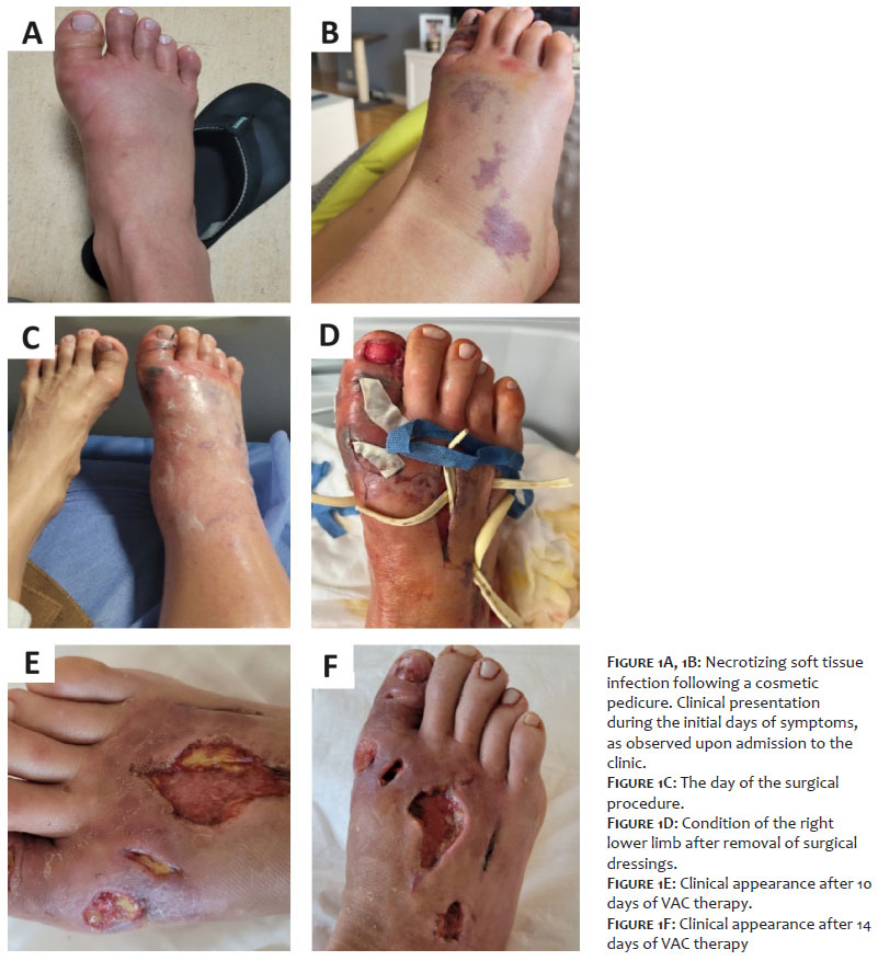
A 41-year-old previously healthy woman, who was not taking any medications, visited a beauty parlor for a pedicure before Easter. The procedure was performed in the beautician's apartment under unsanitary conditions. Several days later, she developed swelling in her right foot along with a low-grade fever (Figure 1A) and severe pain in her right leg.
She was taken to the emergency department, where she was prescribed oral clindamycin (300 mg, three times a day) and a topical antibiotic. However, the swelling remained unchanged. The following day, petechiae, bruising, and worsening swelling appeared on the right foot (Figure 1B), along with the emergence of distinct necrosis on the hallux. A few hours later, blisters and erosions with oozing serous fluid developed.
The patient was referred to the dermatology department due to progressive swelling, now extending from the right foot to the lower leg, along with visible necrosis, blisters, and significant warmth in the affected limb (Figure 1C).
Initial laboratory tests revealed elevated inflammatory markers, with a C-reactive protein (CRP) level of 318 mg/L and procalcitonin at 5.12 ng/mL. The white blood cell count was 15,000/µL, indicating an immune response. Red blood cell parameters showed anemia, with 3.7 × 10⁶/µL erythrocytes, hemoglobin at 9.2 g/dL, and hematocrit at 27%. Additionally, platelet count was reduced to 95,000/µL, consistent with thrombocytopenia. Given the severity of the condition, she was immediately started on broad-spectrum intravenous antibiotic therapy with ceftriaxone and metronidazole. An urgent ultrasound examination of the lower limb veins was performed, and an emergency surgical consultation was scheduled.
The patient underwent urgent surgical intervention, including fasciotomy, decompression, and debridement of necrotic tissue. Filters were placed, and purulent contents were drained. Additionally, the nail plate of the right hallux was removed (Figure 1D).
During the procedure, multiple thrombosed vessels were observed in the subcutaneous tissue, and wound samples were collected for microbiological analysis. The diagnosis of NSTI was confirmed. Intravenous broad-spectrum antibiotic therapy with ceftriaxone and metronidazole was continued.
Microbiological test results identified penicillin-sensitive Streptococcus pyogenes as the causative agent. Based on culture results, antibiotic therapy was switched to intravenous amoxicillin-clavulanic acid. However, the swelling and necrosis continued to progress, leading to a further change in treatment. The antibiotic regimen was escalated to intravenous piperacillin-tazobactam, and the patient underwent a second surgical procedure to remove additional necrotic tissue from the right foot.
During this second intervention, purulent material was drained from the forefoot and the big toe, and new incisions were made to further debride the affected tissues. Additional drains were placed, and VAC therapy was initiated.
VAC therapy proved to be highly effective, leading to a reduction in inflammatory markers and halting the progression of necrosis (Figure 1E). After 14 days of VAC therapy, the patient was in good overall condition (Figure 1F). Due to the significant tissue loss in the subcutaneous and skin layers of the right leg, the patient was referred to a plastic surgery clinic for further reconstructive treatment.
If left untreated, NSTIs are life-threatening, with mortality rates reaching 20–30% even with appropriate treatment.5 Amputation is often required,6 and in nearly all cases, NSTIs result in skin defects, scarring, and limb deformities, significantly reducing patients' quality of life.7
In this case, the infection caused visible, disfiguring lesions on the patient's right lower limb. However, her general condition was stabilized, and she did not develop sepsis or other organ complications. The most crucial aspect of treatment was that amputation of the lower limb was successfully avoided.
Managing NSTIs requires a multimodal approach, where early antibiotic administration is critical for survival. Once necrotic tissue is identified, delaying surgical debridement is futile. Prompt surgical intervention is essential to remove necrotic tissue and prevent further spread of the infection. Additionally, collecting samples for microbiological testing is imperative to guide targeted antimicrobial therapy.3,8
Despite being a well-recognized condition, NSTIs are often misdiagnosed in the early stages. As previously mentioned, immediate antibiotic therapy is crucial for both patient survival and long-term outcomes. Exploratory surgery plays a key role in confirming the diagnosis, allowing for the detection of skin, subcutaneous tissue, fascia, and muscle necrosis, as well as vascular thrombosis, swelling, and exudate.9
This case shows that even minor cosmetic procedures can lead to NSTIs.
Preventing NSTI requires careful selection of sterile environments for cosmetic procedures that breach soft tissue integrity. Proper hygiene and infection control measures significantly reduce the risk of complications. When assessing a patient with suspected necrotizing soft tissue infection, obtaining a detailed history of pain, systemic symptoms, swelling, and local erythema is essential. Immediate initiation of broad-spectrum antibiotic therapy is crucial, along with early surgical intervention when necessary to control the infection and prevent further tissue damage. The primary goal is to preserve the affected limb and minimize complications. If standard treatments fail to promote healing, VAC therapy can be a valuable option. Patients with severe NSTI should be referred for long-term follow-up at a surgical or wound care clinic to ensure appropriate management and recovery.
Availability of data and material: The datasets used and analyzed during this study are available from the corresponding author upon reasonable request.
Conflict of interests: The authors declare no conflict of interests.
Funding: No funding was provided for this research.
Authors contributions
R.N., I.S., and I.W. designed the article, reviewed literature, and wrote the manuscript. R.N. and I.S. had primary responsibility for the final content. All authors read and approved the final manuscript.
Ethics approval and consent to participate
Not applicable.
Consent for publication
Photographs were published with the patient's informed consent.
Raman Nitskovich
ORCID: 0000-0001-5311-3787
Data collection, analysis, and interpretation.
Izabela Staniszewska
ORCID: 0000-0002-1457-0675
Manuscript drafting and writing.
Irena Walecka
ORCID: 0000-0002-3502-3339
Intellectual contribution to the diagnostic and/or therapeutic management of studied cases.
1. Gundersen IM, Bruun T, Almeland SK, Skutlaberg DH, Nedrebø T, Rath E, et al. Necrotising soft tissue infections. Tidsskr Nor Laegeforen. 2024;144(3).
2. Morten H, Martin Bruun M, Lærke Bruun M, Ole H. Incidence, comorbidity and mortality in patients with necrotising soft-tissue infections, 2005–2018: a Danish nationwide register-based cohort study. BMJ Open. 2020;10(10):e041302.
3. Scheers C, Andre J, Richert B. Nail cosmetology. Hand Surg Rehabil. 2024:101657.
4. Yang J, Hall K, Nuriddin A, Woolard D. Risk for hepatitis B and C virus transmission in nail salons and barbershops and state regulatory requirements to prevent such transmission in the United States. J Public Health Manag Pract. 2014;20(6):E20-30.
5. Boyer A, Vargas F, Coste F, Saubusse E, Castaing Y, Gbikpi-Benissan G, et al. Influence of surgical treatment timing on mortality from necrotizing soft tissue infections requiring intensive care management. Intensive Care Med. 2009;35(5):847-53.
6. Peetermans M, Prost N, Eckmann C, Norrby-Teglund A, Skrede S, De Waele JJ. Necrotizing skin and soft-tissue infections in the intensive care unit. Clin Microbiol Infect. 2020;26(1):8-17.
7. Urbina T, Canoui-Poitrine F, Hua C, Layese R, Alves A, Ouedraogo R, et al. Long- term quality of life in necrotizing soft-tissue infection survivors: a monocentric prospective cohort study. Ann Intensive Care. 2021;11(1):102.
8. Stevens DL, Bryant AE. Necrotizing soft-tissue infections. N Engl J Med. 2017;377(23):2253-65.
9. Goh T, Goh LG, Ang CH, Wong CH. Early diagnosis of necrotizing fasciitis. Br J Surg. 2014;101(1):e119-25.
 All content the journal, except where identified, under the Creative Commons Attribution 4.0 International licence - ISSN-e 1984-8773


 Read in Portuguese
 Portuguese PDF
 Print
Print
 Send this article by email
 How to cite this article
 Submit a comment
 Mendeley
 Pocket