


Flavia Alvim Sant’Anna Addor1; Nathalia Terumi Kawakami2; Michelle Sabrina da Silva3
Submission on: 18/10/2023
Approved on: 07/01/2024
Financial support: This study was sponsored by FQM Farmacêutica.
Conflicts of interest: None. Is it a clinical trial? CAAE: 48463321.0.000 Ethics committee number: 5514.
How to cite this article: Addor FAS, Kawakami NT, Silva MS. Efficacy of a dietary supplement on the molecular and clinical parameters of skin aging. Surg Cosmet Dermatol. 2025;17:e20250308.
INTRODUCTION: Oxidative stress, characterized by an imbalance between the production of reactive oxygen species (ROS) and antioxidant defense, seems to be the main process that worsens skin aging, and the mitochondria is an important source of ROS. There is increasing evidence that oral administration of functional nutrients can protect the skin from oxidative damage.
OBJECTIVE: To evaluate the effectiveness of an oral supplement with collagen peptides and other nutrients in reducing the signs of photoaging.
METHODS: In vitro studies evaluated the antioxidant and antiglycant effects under UV light, as well as the synthesis of extracellular dermal matrix components; then, a clinical trial was developed with women between 35 and 60 years old, for 12 weeks. Hydration (corneometry) and skin firmness (cutometry) measurements were performed.
RESULTS: The supplement showed antioxidant and antiglycant properties under the stress generated by UV radiation, as well as the ability to induce synthesis of extracellular dermal matrix elements; There was an improvement in all clinical parameters of aging. Significant improvement in hydration and firmness and elasticity were also observed.
CONCLUSION: Collagen peptides and nutrients association demonstrated an effect in some mechanisms involved in the photoaging process, leading a clinical improvement.
Keywords: Collagen; Exposome; Oxidation; Skin Aging; Glycation End Products, Advanced; Dietary Supplements.
Skin aging consists of a progressive decline in cellular functions, with a consequent reduction in its protective functions against external aggressions. The skin loses its ability to defend itself against physical, chemical, and biological stimuli over the course of a lifetime, and becomes increasingly fragile.1 In the epidermis, the decline in keratinocyte proliferation leads to thinning of the epidermis and stratum corneum, and reduced synthesis of epidermal lipids, with a consequent pH increase on the surface; the barrier function is therefore increasingly compromised, with increased dryness and irritative reactions, accompanied by inflammatory processes. The stratum corneum presents a 30% decrease in its lipid content over the course of life, when compared to the young stratum corneum.2 In the dermis, the process of fibroblast senescence, marked by morphological changes and functional decline, leads to progressively reduced synthesis of collagen, elastin, and proteoglycans, which make up the extracellular matrix; concomitantly, an imbalance occurs between the synthesis of collagen-degrading enzymes – metalloproteinases – and their inhibitors, which may even be reduced.3 These phenomena are concomitantly aggravated by extrinsic factors, solar radiation being the most well-established of them; however, other environmental factors – atmospheric pollution and high temperatures – and lifestyle triggers such as smoking, sleep changes, emotional stress, and nutritional factors have shown evidence of influencing skin aging. They have been considered the main factors for skin aging exposome, due to the current level of evidence that each factor presents;4 however, menopause, chronic diseases, some continuous-use medications can also interfere with aging-related skin damage, contributing to the worsening of its signs.5 Therefore, “intrinsic” and “extrinsic” aging processes overlap, but have a common factor: they are strongly related to the increased generation of free radicals in the skin, the neutralization of which is not complete. Oxidative stress, marked by this imbalance between the production of reactive oxygen species (ROS) and antioxidant defense, appears to be the most harmful process in skin aging.6
Particularly mitochondria are an important source of ROS, probably the most important at an intracellular level.7 Oxidative stress can lead to immune alterations, oncogenesis, and accelerated signs of aging. With regard to aging, ultraviolet A (UVA) radiation is known to generate ROS, leading to damage of keratinocytes, especially fibroblasts, interfering with fibroblast function, and reducing the synthesis of collagen, elastin, and hyaluronic acid, and increasing the expression of protein-degrading enzymes such as metalloproteinases and elastase.8 Glycation is another important mechanism in the aging process; the generation of advanced glycation end products (AGEs), resulting from nonenzymatic reaction between glucose and proteins, leads to changes in the structural properties of collagens 1 and 4, directly resulting in dermal and microvasculature functional impairment, with reduced elasticity, greater fragility to trauma, and damage to the healing process.9 Diabetes mellitus, a more frequent pathology in later life, and even hyperglycemic diets, induce this phenomenon. UVA radiation is capable of potentiating tissue glycation, increasing the expression of elastase and tropoelastin, clinically manifested by elastosis. The oxidation that UVA radiation promotes also increases the generation of AGEs (glycoxidation).10
The production of superoxide by mitochondria can be visualized in fluorescence microscopy using the reagent MitoSOX™ Red, which permeates living cells, selectively targeting mitochondria. This reagent is rapidly oxidized by superoxide, but not by other ROS and reactive nitrogen species (RNS). The oxidized product is highly fluorescent when it binds to nucleic acid, allowing semi-quantification by imaging of this mitochondrial oxidative stress.14
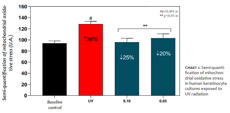
Human keratinocytes were cultured on four plates, after determining the noncytotoxic concentration of the supplement for the experiment. Plate 1 was designated as baseline control (nonirradiated and untreated). Plate 2 was irradiated with 10J/cm2 UVA (365nm) and plates 3 and 4 were treated with different concentrations of the supplement and then irradiated using the same parameters as plate 2 (positive control).
The results found that cultures exposed to UV radiation had mitochondrial oxidative stress increased by 36% (P<0.001) when compared to the baseline control group. The supplement reduced mitochondrial oxidative stress by 25% and 20% (p<0.01) at concentrations of 0.10 and 0.03mg/mL, respectively, when compared to the group exposed to UV radiation, demonstrating its ability to protect cells from damage caused by free radicals. These results are shown in chart 1:
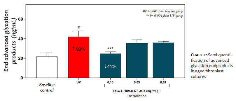
Human fibroblasts were incubated with three concentrations of noncytotoxic AGEs for 48 hours and then exposed to UV radiation. The AGEs were then quantified using the enzyme-linked immunosorbent assay (ELISA) technique. These were cultured on plates after determining the noncytotoxic concentration of the supplement for the experiment. Plate 1 was designated as the baseline control (nonirradiated and untreated) in the three batches. Plate 2 was irradiated with 10J/cm2 UVA (365nm) and plates 3, 4, and 5 were treated with the supplement in three different noncytotoxic concentrations to be irradiated after 48 hours in the same parameters as plate 2 (positive control).
The supplement reduced the generation of AGEs by 41% (p<0.01) at a concentration of 0.10 mg/mL when compared to the group exposed to UV radiation, demonstrating its ability to protect cells from the formation of AGEs triggered by UVA radiation. These results are shown in chart 2:
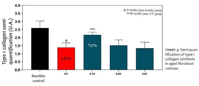
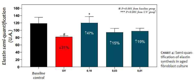
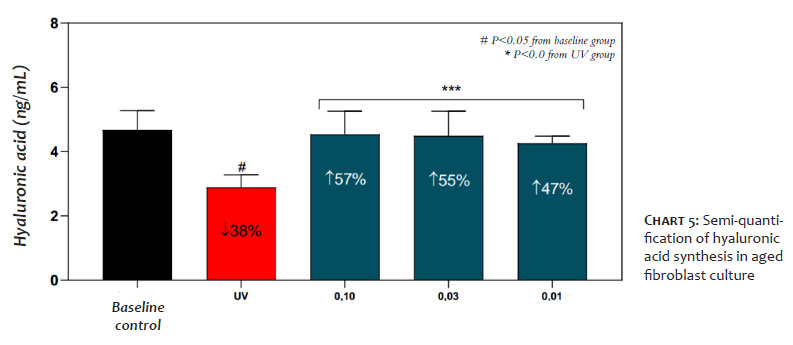
Senescent human fibroblasts were treated with the supplement in three noncytotoxic concentrations for 72 hours. The cells were then fixed in 4% paraformaldehyde for subsequent immunolabeling and semi-quantification of type-1 collagen using an immunofluorescent technique. These cultures were performed on plates after determining the noncytotoxic concentration of the supplement for the experiment. In the three batches, plate 1 was the baseline control (nonirradiated and untreated). Plate 2 was irradiated with 10J/cm2 UVA (365nm) and plates 3, 4, and 5 were treated with the supplement in three different noncytotoxic concentrations to be irradiated after 48 hours in the same parameters as plate 2 (positive control). Each batch was used to assess the effect of the supplement on protecting the synthesis of collagen, elastin and hyaluronic acid.
The results showed that exposing the cultures to UV radiation promoted significant reductions in type-1 collagen, elastin, and hyaluronic acid. (Graphs 3, 4, and 5)
The supplement was able to protect these molecules from UV radiation damage, keeping their concentrations close to the baseline control, which is the nonirradiated culture.
The preservation of the concentration of type-1 collagen, elastin, and hyaluronic acid is 65%, 92%, and 100% respectively, compared to the nonirradiated area, demonstrating their protective effect on fibroblasts.
This is a randomized, double-blind, noncomparative, 12-week study, which included 33 women between 35 and 60 years old with clinical complaints and signs of facial and body aging. Exclusion criteria were any skin anti-aging treatment up to four weeks prior to inclusion in the study, pregnancy or breast-feeding, participants using corticosteroids, immunosuppressants, or with active endocrinopathies, or any other medical condition that could affect the results. All participants were instructed to maintain their normal diet, and not to intentionally expose themselves to the sun.
The study was conducted in accordance with Good Clinical Practice guidelines. This study was performed after ethical approval, issued by the Research Ethics Committee of Universidade São Francisco, SP, Brazil on July 5, 2021, under opinion number 4.826.741 and CAAE: 48463321.0.0000.5514.
All participants were given an oral powder supplement with 11g of protein containing: bioactive collagen peptides (Verisol®), hydrolyzed collagen, vitamin C, lutein, Hibiscus sabdariffa extract and Aristotelia chilensis extract (Delphinol®) in sachets for daily use. They were instructed to consume one sachet a day, diluted in 200mL of water for 12 weeks. All the participants were instructed to record the time they took the supplement and any discomfort or complications in a diary provided by the treatment facility.
The clinical and subjective assessments used parametric scales where 1= worsening and 5= marked improvement; the clinical assessments were conducted by a trained dermatologist.
In the facial region, the following parameters were assessed: fine lines, expression lines, smoothness, elasticity, firmness, and general appearance, using a categorical scale.
In the gluteal and thigh regions, contours, orange peel appearance, elasticity, firmness, and general appearance were assessed using the same scale as the facial region.
All assessments were performed at the beginning of the study, after six weeks and after 12 weeks; at the same time, any condition that could be an adverse reaction related or not to the use of the supplement was assessed.
Instrumental assessments were performed after acclimatization for 20 minutes at a temperature of 20±2ºC and humidity of 50±5%; and the following measurements were taken:
- firmness and elasticity (Cutometer®) in the left periorbital area and right lateral area of the thigh, in the middle third;
- hydration (Corneometer®) on the face (median frontal area).
Of the 33 participants who started the study, three did not attend their visits for reasons unrelated to the study, and were therefore excluded. The study was completed with valid data from 30 participants, and no adverse events related to the use of the supplement were reported.
All participants reported adherence to treatment of 80% or more during the six and 12 weeks of the study.
The statistical analysis of the data was performed using the nonparametric Wilcoxon test for paired samples, based on the scores given at six and 12 weeks versus the baseline; for paired samples that followed a normal distribution, the parametric t-Student statistical test was used.
Of all the parameters assessed for the face and body (buttocks and thighs), some degree of statistically significant improvement was observed;
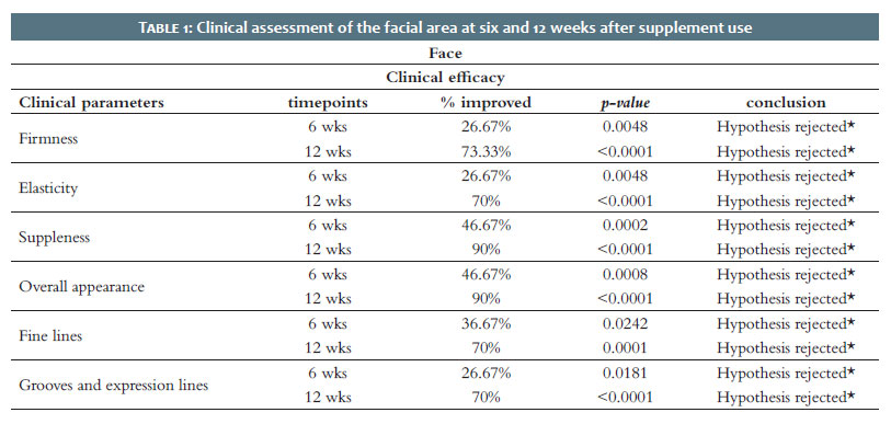
The facial area revealed a significant improvement after six weeks of use in all the items assessed (firmness, elasticity, fine lines, expression lines, glow, and general appearance), as shown in table 1.
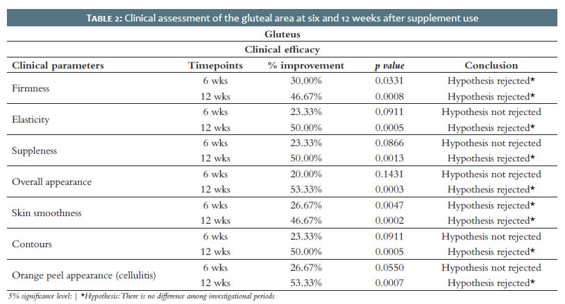
A statistically significant clinical improvement was seen in the gluteal area at six weeks for firmness and skin relief; and in the 12-week assessment, there was a significant improvement for all the items evaluated: firmness, elasticity, skin tone, general appearance, relief, contours, and orange peel appearance, as shown in table 2.
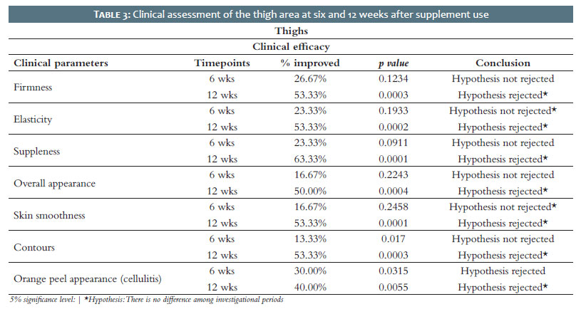
Improvements were clinically observed in the thigh area at six weeks, and were statistically significant for the orange peel aspect; at 12 weeks, all the items assessed, such as firmness, elasticity, skin appearance, general appearance, orange peel aspect, contours, and skin relief, had improved significantly, as shown in table 3.
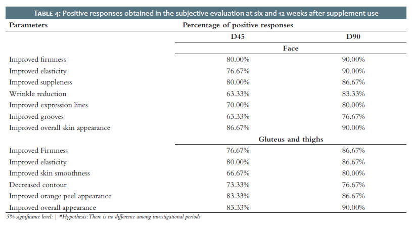
Table 4 shows the percentage of positive responses, grouped together (some degree of improvement) for each item assessed; the responses were grouped into facial and body (buttocks and thighs).
Hydration measured on the face (frontal area) after three months of daily use showed a statistically significant improvement by 16.65% (p<0.05).
The biophysical parameter of firmness, which was measured in two areas: periorbital and thighs, showed an improvement by 33.28% in the periorbital region and 19.8% in the thigh area, both statistically significant (p<0.05).
The biophysical parameter of elasticity was measured in two areas, periorbital and crural, and showed an improvement by 70.3% in the periorbital region and 30.3% in the crural area, both statistically significant (p<0.05).
In the pre-clinical stage, the association of nutrients contained in the supplement demonstrated a broad antioxidant effect, reducing the formation of ROS in keratinocyte culture and exerting a protective effect on the fibroblast against UV radiation, significantly recovering the synthesis of elements fundamental to dermal integrity: type-1 collagen, elastin, and hyaluronic acid.
These effects can be explained by the synergistic antioxidant action of vitamin C (a broad antioxidant found on the epidermal surface which is depleted by sunlight and atmospheric pollution)15 and lutein (a carotenoid which is essential for direct protection against visible light and an antioxidant against the effects of ultraviolet radiation) on different sites.16 As an essential nutrient, its deficiency is relatively common, especially among individuals who do not eat dark leafy vegetables such as watercress, spinach, rocket, etc.17 The polyphenols in Delphinol®, particularly delfinidin, but also other anthocyanidins, have a broad antioxidant and anti-inflammatory action against UV-induced damage.18 In addition to its antioxidant action, delfinidin acts in important stages of the aging inflammation, covering its main mechanisms, such as suppressing epidermal NF-κB and AP-1, and reducing the expression of metalloproteinases.19 Together with these polyphenols, the mechanism of antioxidant action of Hibiscus sabdariffa had already been studied in neoplasms, and was recently evidenced by the isolation of its main compounds with this property: chlorogenic and neochlorogenic acids, cryptochlorogenic acid, rutin, and isoquercitin.20 The compound found in Hibiscus sabdariffa has been shown to be a broad ROS scavenger, and to have cell membrane protective effects against lipid peroxidation and prevent the depletion of endogenous antioxidant systems such as glutathione and superoxide dismutase.21 The anti-glycemic action of Hibiscus sabdariffa has also been documented; its ability to reduce insulin resistance has been demonstrated in patients with type-2 diabetes; at a molecular level, it has been shown to inhibit the expression of receptors for advanced glycation endproducts (RAGEs).22 Studies on the bioavailability of hydrolyzed collagen has shown that after ingestion in the form of peptides, they reach tissues such as skin and cartilage, stimulating the metabolism of dermis cells, improving the synthesis of extracellular matrix proteins, and consequently replacing damaged collagen fibers with new ones, with properties that restore the structure of the dermis.23 Clinical studies with supplements containing collagen peptides have shown improvements in dermal and epidermal parameters related to aging.
One of the largest clinical studies using Verisol® included 114 women aged between 45 and 65 who received 2.5g/day for eight weeks. There was a significant reduction in periocular lines when compared to the control group, and type-1 pro-collagen and elastin synthesis increased significantly in the subgroup subjected to histological assessment.24
The presence of vitamin C as a co-factor in the synthesis of collagen and elastin may also favor the magnitude of these dermal effects.25 The supply of oral amino acids to the skin can also be very beneficial, especially if considered that many populations may be subject to hypoproteinemia – vegetarians, post-bariatric surgery patients, and individuals undergoing crash diets or even with an increased consumption due to heavy physical exercise. A literature review highlighted the characteristics of amino acids and their benefits for skin metabolism:26
- essential amino acids such as histidine, serine, and arginine participate in the production of filaggrin and skin hydration;
- lysine is involved in the maturation of proteins during the hydroxylation reactions of collagen synthesis;
- branched-chain amino acids (isoleucine, leucine, and valine), when supplemented together, have shown benefits in improving wrinkles, healing wounds, and protecting the skin against UVB damage;
- phenylanine and trypsin participate in the production of melanin;
- tryptophan acts as a precursor to melatonin, a hormone that protects the skin against oxidative stress;
- methionine is present in the formation of polysaccharides and glycosaminoglycans;
- threonine plays a role in moisturizing the stratum corneum. The results of the pre-clinical studies showed a clinical correlation, with a significant improvement in signs related to photo-aging, such as fine lines, dynamic lines, and radiance, especially in facial skin, which is more exposed to solar radiation. There was also an improvement in firmness and elasticity, which had already been significantly observed in both clinical and quantitative instrumental assessments.
However, the improvement in dermal parameters such as contours, skin relief, elasticity, and firmness show that the combination of antioxidant nutrients with collagen peptides can positively affect the clinical improvement of signs of skin aging, especially with continued use. After 12 weeks of daily use, a significant improvement was seen in all the parameters evaluated.
Therefore, the results of this study indicate a positive and relevant effect on the entire integument, improving the integrity and functionality of the skin, with progressive improvement with continued use.
The combination of nutrients contained in the supplement studied demonstrated a broad antioxidant effect, preserving keratinocytes and fibroblasts against UV-induced oxidative damage, while maintaining the ability of fibroblasts to synthesize collagen, hyaluronic acid, and elastin.
The benefits of antioxidant action, possibly synergistic with the fibroblast-stimulating effect of collagen peptides on the synthesis of collagen, hyaluronic acid, and elastin, had relevant results in all the signs of aging studied, in all the areas studied.
Daily use of this supplement proved to be safe for continued use, which favors its use both in the prevention of damage caused by extrinsic aging and in the improvement of signs already existing.
Flavia Alvim Sant’Anna Addor
ORCID: 0000-0003-1851-7342
Approval of the final version of the manuscript, study design and planning, preparation and writing of the manuscript, data collection, analysis, and interpretation, effective participation in research guidance, intellectual participation in propaedeutic and/or therapeutic conduct of studied cases, critical literature review, critical review of the manuscript.
Nathalia Terumi Kawakami
ORCID: 0000-0002-2458-7534
Author’s contribution: Statistical analysis, approval of the final version of the manuscript, study design and planning, effective participation in research guidance, critical review of the manuscript.
Michelle Sabrina da Silva
ORCID: 0000-0002-9319-2384
Statistical analysis, approval of the final version of the manuscript, study design and planning, data collection, analysis, and interpretation, effective participation in research guidance, critical literature review, critical review of the manuscript
1. Zargaran D, Zoller F, Zargaran A, Weyrich T, Mosahebi A. Facial skin ageing: key concepts and overview of processes. Int J Cosmet Sci. 2022;44(4):414-420.
2. Ghadially R, Brown BE, Sequeira-Martin SM, Feingold KR, Elias PM. The aged epidermal permeability barrier. Structural, functional, and lipid biochemical abnormalities in humans and a senescent murine model. J Clin Invest. 1995; 95:2281–90.
3. Shin JW, Kwon SH, Choi JY, Na JI, Huh CH, et al. Molecular mechanisms of dermal aging and antiaging approaches. Int J Mol Sci.2019;20(9): 2126.
4. Krutmann J, Bouloc A, Sore G, Bernard BA, Passeron T. The skin aging exposome. J Dermatol Sci. 2017 ;85(3):152-161.
5. Addor FAS. Beyond photoaging: additional factors involved in the process of skin aging. Clin Cosmet Invest Dermatol. 2018;11:437-443.
6. Poljšak B, Dahmane RG, Godić A. Intrinsic skin aging: the role of oxidative stress. Acta Dermatovenerol Alp Pannonica Adriat. 2012;21(2):33-6.
7. Wikramanayake TC, Chéret J, Sevilla A, Birch-Machin M, Paus R. Targeting mitochondria in dermatological therapy: beyond oxidative damage and skin aging. Expert Opin Ther Targets. 2022;26(3):233-259.
8. Imokawa G, Ishida K. Biological mechanisms underlying the ultraviolet radiation-induced formation of skin wrinkling and sagging I: reduced skin elasticity, highly associated with enhanced dermal elastase activity, triggers wrinkling and sagging. Int J Mol Sci. 2015;16(4):7753-75
9. Fournet M, Bonté F, Desmoulière A. Glycation damage: a possible hub for major pathophysiological disorders and aging. Aging and Disease.2018;9(5):897.
10. Pageon H, Zucchi H, Ricois S, Bastien P, Asselineau D. UVA exposure combined with glycation of the dermis are two catalysts for skin aging and promotes a favorable environment to the appearance of elastosis. J Aging Res.2021;2021:6647773.
11. Addor FAS. Antioxidants in dermatology. An Bras Dermatol. 2017;92(3):356-362. 12.Keller KL, Fenske NA. Uses of vitamins A, C, and E and related compounds in dermatology: a review. J Am Acad Dermatol. 1998;39:611-25
12. Geng R, Kang SG, Huang K, Tong T. Boosting the photoaged skin: the potential role of dietary components. Nutrients. 2021;13(5):1691
13. Zielonka J, Hardy M, Kalyanaraman B. HPLC study of oxidation products of hydroethidine in chemical and biological systems: ramifications in superoxide measurements. Free Radic Biol Med. 2009;46(3):329- 38.
14. Dimitrov A, Zanini M, Zucchi H, Boudah S, Lima J, Soeur J, et al. Vitamin C prevents epidermal damage induced by PM-associated pollutants and UVA1 combined exposure. Exp Dermatol. 2021;30(11):1693-1698.
15. Juturu V, Bowman J, Deshpande J. Overall skin tone and skin-lightening-improving effects with oral supplementation of lutein and zeaxanthin isomers: a double-blind, placebo-controlled clinical trial. Clin Cosmet Invest Dermatol. 2016;9:325–332.
16. Balic A, Mokos M. Do we utilize our knowledge of the skin protective effects of carotenoids enough? Antioxidants 2019;8(8):259
17. Watson RR, Schönlau F. Nutraceutical and antioxidant effects of a delphinidin-rich maqui berry extract Delphinol®: a review. Minerva Cardioangiol. 2015;63(2 Suppl 1):1-12.
18. Kang NJ, Lee KW, Kwon JY, Hwang MK, Rogozin EA, Heo YS, et al. Delphinidin attenuates neoplastic transformation in JB6 Cl41 mouse epidermal cells by blocking Raf/mitogen-activated protein kinase kinase/extracellular signal-regulated kinase signaling. Cancer Prev Res (Phila). 2008; 1(7): 522–531.
19. Wang J, Cao X, Jiang H, Qi Y, Chin KL, Yue Y. Antioxidant activity of leaf extracts from different Hibiscus sabdariffa accessions and simultaneous determination five major antioxidant compounds by LC-Q-TOF-MS. Molecules. 2014;19(12):21226- 38.
20. Rocha CI, Bonnlaender B, Sievers H, Pischel I, Heinrich M. Hibiscus sabdariffa L. - a phytochemical and pharmacological review. Food Chem. 2014;165:424-43.
21. Peng CH, Chyau CC, Chan KC, Chan TH, Wang CJ, Huang CN. Hibiscus sabdariffa polyphenolic extract inhibits hyperglycemia, hyperlipidemia, and glycation-oxidative stress while improving insulin resistance. J Agric Food Chem. 2011;59(18):9901-9
22. Murakami H, Shimbo K, Inoue Y, Takino Y, Kobayashi H. Importance of amino acid composition to improve skin collagen protein synthesis rates in UV-irradiated mice. Amino Acids. 2012;42(6):2481-9.
23. Proksch E, Schunck M, Zague V, Segger D, Degwert J, Oesser S. Oral intake of specific bioactive collagen peptides reduces skin wrinkles and increases dermal matrix synthesis. Skin Pharmacol Physiol.2014;27(3): 113–119.
24. Hata R, Senoo H. L-ascorbic acid 2-phosphate stimulates collagen accumulation, cell proliferation, and formation of a three-dimensional tissuelike substance by skin fibroblasts. J Cell Physiol. 1989;138(1):8-16.
25. Solano F. Metabolism and functions of amino acids in the skin. Adv Exp Med Biol. 2020;1265:187-199.
 All content the journal, except where identified, under the Creative Commons Attribution 4.0 International licence - ISSN-e 1984-8773


 Read in Portuguese
 Portuguese PDF
 Print
Print
 Send this article by email
 How to cite this article
 Submit a comment
 Mendeley
 Pocket