


Tércio Elyan Azevedo Martins1,2,3; Andressa Costa de Oliveira2; Beatriz Regazzi de Gusmão4; Alexandre de Almeida Filippo4; Fabiane Mulinari-Brenner5
Submitted on: 06/01/2023
Accepted on: 01/18/2024
Financial support: None.
Conflict of interest: None.
How to cite this article: Martins TEA, Oliveira AC, Gusmão BR, Filippo AA, Brenner FM. Update on hair fiber assessment. Surg Cosmet Dermatol. 2024;16:e20240265.
Damage caused by chemical, physical, and thermal procedures can disrupt the natural morphological characteristics of the hair, causing the strands to become dry, dull, weak, and brittle. The objective of this article was to update dermatologists on the methods available for assessing hair shaft damage. The main laboratory (microscopy, tensile strength, structural assessment, physical and mechanical properties) and outpatient (diagnostic tests and patient history) diagnostic methods for evaluating the quality of the hair shaft are presented.
Keywords: Hair; Dermatology; Keratins.
The study of hair has been one of the most impacted by advances in diagnostic techniques in Dermatology over the last 20 years. A deeper understanding of alterations to the scalp and hair shaft has led to major developments in diagnosis and therapeutic approaches. New entities have been recognized and reclassified with dermoscopy and the new histological cross-sections applied to scalp.1 Hair has multiple functions, mainly physical protection, thermal insulation, camouflage, sebum dispersion, and sensory perception. It affects quality of life, attractiveness, and self-esteem in contemporary human society.2
This review aims to update dermatologists on laboratory and outpatient diagnostic methodologies for the hair shaft. Evaluating the microscopic, dermoscopic, biochemical, and molecular characteristics of hair fibers allows for a better understanding of the biological processes related to the aging process, disorders of hair shaft keratinization, external damage from chemical treatments, and the ability to recover from aesthetic treatments.3
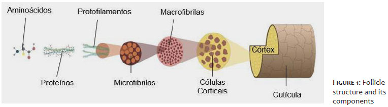
The hair fiber is extremely resistant and has three main morphological constituents: cuticle, cortex, and in some cases, medulla (Figure 1). Overlapping flat, scale-like structures surround the central core of the fiber, such as a thick, chemically resistant protective layer called cuticle. The cuticle prevents the cortex from external environmental damage.4
The protection of hair shaft against environmental and chemical damage begins with the adherence and organization of the cuticular cells. A thick, chemically resistant protective layer is responsible for the shine, resistance, and combability of hair shaft. The human cuticle has around six to eight layers and is made up especially of keratinized cells (80%), structural lipids, and proteins associated with keratin with a high Sulphur content.5 The greater the alignment and integrity of the cuticles, the better the softness, shine, and frizz characteristics. Cuticle cells are classified into the epicuticle, which is more external and hydrophobic, followed by the exocuticle, which has the greatest mass (55%), and the endocuticle, which has the capacity to absorb water.5,6
The cortex accounts for most of the hair fiber’s mass (75%) and plays a significant role in determining the fiber’s intrinsic strength and mechanical resistance. The α-helix proteins in the cortex are held together by chemical bonds such as ionic bonds, hydrogen bonds, Van der Waal bonds, and disulfide bridges. In this region are the melanin granules which provide the color properties of the hair.7 The cuticular membrane complex (CMC), responsible for cohesion between cortical and medullary cells, is rich in 18-methyl eicosanoic acid (18-MEA), providing hair with hydrophobicity and lubrication with reduced friction between the hair fibers.8,9 A reduction in this lipid is related to hair fiber degradation and protein loss.
More internally, and not always present, is the medulla, a cellular vacuole located in the central part of the hair fiber. Several studies correlate the presence of the medulla with an increase in capillary diameter. Structurally, medullary cells are cortical cells together with melanosomes.7,10
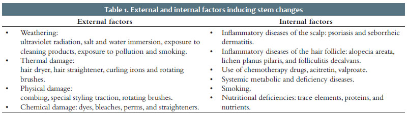
The hair fiber is produced by the hair follicle and any change in the follicle can have repercussions on its structure and quality.11 In routine outpatient evaluation, hair shafts can undergo external and internal changes due to various factors (Table 1).
Hair fibers can be assessed by various clinical and laboratory methods. Clinical methods have a diagnostic purpose in the treatment of hair shaft alterations, allowing for outpatient dermatological evaluation. They allow the evaluation of hair shaft complaints due to internal and external factors. They are performed in the routine dermatological examination, which includes anamnesis, physical examination (pull test), dermoscopy, and the rapid protein loss test (also considered a laboratory test).12
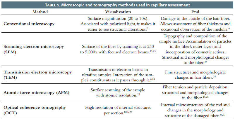
Laboratory methods can identify damage and the response to cosmetic products in the hair fiber and can be conducted on both strands of hair and standardized strands. Cosmetic industry uses these methods as parameters for product development, as they allow physical and chemical characteristics of the hair shaft to be assessed and external damage caused by external agents to be graded.13,14 They include various forms of microscopy, combined with optical coherence tomography, which visually assess the structure of the hair shaft. Spectroscopy techniques, assessment of protein loss, and tryptophan quantification evaluate the stem from a structural point of view. Color and gloss assessment, thermogravimetric analysis, measurement of mechanical strength (traction), combability and elasticity assess the physical properties of the stem.4,14,15
Clinically, the hair shaft can be assessed by exploring the macroscopic properties via clinical examination, from the root to the distal end. The longer the hair, the greater the exposure to external damage. A number of factors are involved in the development of hair shaft alterations. Correct diagnosis can be complex and requires a detailed clinical history, especially focused on routine hygiene care, chemical treatments, use of heat sources for drying, and daily environmental exposures. Epidemiological characteristics, such as age and sex, should be considered in this assessment.
The anamnesis should address the onset, duration, and nature of the complaint; personal and family history, use of inappropriate cosmetics, medications (chemotherapy, retinoids, among others) and protein-restricted diets. During the examination, other hairy areas should be assessed, such as eyebrows and body hair, which can show alterations in systemic diseases and hair shaft abnormalities. Examination of the nails can help diagnose neuroectodermal dystrophies and inflammatory alopecia.16
When excessive hair loss is identified by counting the hairs shed daily, the collected hairs can be analyzed under a microscope and classified according to diameter, indicating possible damage to the fiber.11,17
The tug test is a simple technique used to assess hair fragility. Some hair strands are held, and a traction force is applied to the end of the hair shaft with the other hand. Breakage of the hair shaft during the test indicates fragile hair and abnormalities in the hair shaft.16,17
Growth rates can be measured by scraping a small area of the scalp, which acts as a growth window. Expected growth is around 0.3 cm in a week, or an average of 1.0 cm per month. Growth rates vary between races: African hair has the slowest growth rate (0.9 cm/month), Caucasian hair grows at a rate of around 1.2 cm per month and Asian hair outperforms the other types, with a growth rate of 1.3 cm per month.18
The use of dermoscopy on the scalp has been incorporated into daily practice due to its practicality and potential to minimize the need for invasive examinations. It is useful in various diseases, from pediculosis and hair shaft anomalies to differentiating scarring and nonscarring alopecia. It can be performed using a traditional dermatoscope with polarized light, a videodermatoscope with polarized light or in association with computerized hair analysis.18,19
The traditional 10x dermatoscope is used in routine dermatology; however, the findings of this modality are limited in the study of the hair shaft. Dermoscopic examination of the hair shaft can be performed with or without gel application at magnifications of up to 300x. Trichoscopy without immersion has proven useful for analyzing distal characteristics such as fractures and other disorders of the hair shaft, such as trichorrhexis nodosa.18,19
Alterations to the hair shaft can be evidenced by the trichogram. This technique easily assesses the proportion of vellus and terminal hairs and capillary density, and is useful for monitoring and evaluating patients’ therapeutic response.18,19
It is crucial to detect the first change in the protein in order to avoid more drastic changes in the hair’s properties. Suggesting its protection would also ensure that the hair’s physical properties, such as resistance and elasticity, are preserved against daily harmful exposure.20
A new methodology available is the rapid protein loss test, which uses a chromogenic reagent to quantify total protein. It is based on a conversion of ions that provide the result based on a colorimetric scale according to the amount of protein extracted from the patient’s hair in just 25 minutes.21,22 This test has been adopted as a valuable technique by dermatologists and trichologists in the evaluation of the hair shaft, creating new possibilities for the doctor, allowing a solid and complete diagnosis of the hair shaft,21,22 and directing to more specific and effective treatments.
All these clinical assessments can be complemented by laboratory evaluations, histopathological studies of the hair shafts and scalp biopsies for a general assessment of the patient’s hair system.
Currently, several laboratory techniques can be used to assess the quality and condition of the hair fiber. Microscopy combined with optical coherence tomography allows the structure of the hair shaft to be studied visually. Spectroscopy and protein evaluation methods assess the shaft from a structural point of view. Studies of color, gloss, thermogravimetric analysis and measurement of mechanical strength (tensile strength), combability and elasticity allow a correlation to be established with the physical properties of the stem.5,14,15
Routine external damage to the shaft can affect its mechanical and structural properties by weakening the hair fiber.5 Cumulative exposure to these factors leads to keratin denaturation, degradation of cortex components, rearrangement of disulfide bonds and reduced resistance due to loss of cortical protein, thus resulting in alterations in laboratory tests.14
Microscopic and tomography examinations are subjective but have advantages such as the use of hair strands and standardization of tests. Table 2 shows the characteristics and objectives of the microscopic tests available in laboratories to assess hair fibers and related damage.
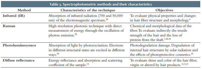
The structure of the hair shaft is mainly made up of proteins, providing resistance to the fiber. In the cortex, they make up 80% of the protein mass of the hair shaft. Spectroscopic and protein loss assessment methods allow structural correlation with the hair shaft.5,15 Currently, several methods have been suggested for quantifying hair protein; however, no method is considered universal. Spectroscopic methods are based on the absorption and emission of electromagnetic radiation on molecules, which occurs due to the movement of electrons between their energy levels.28 Spectrophotometric methods are described in table 3 and their main objective is to assess hair shaft damage due to chemical treatments and physical wear. The technique is used to investigate the mechanism that promotes a reduction in the tensile strength of human hair after the use of chemical treatments that modify the shape of the strand, such as wavy hair that has undergone treatments known as perming; the chronological alterations of the keratin in the fiber of virgin hair with the processes of aging; the influence of chemical treatments that promote reduction, heating and oxidation in the keratin of the hair; and the structure of the virgin white hair shaft (hair graying) that has been subjected to the permanent straightening process.5,7,29 It can be applied in hair research when it is necessary to analyze physical properties, such as moisturizing action, changes in the structure and morphology of the hair fiber promoted by treatments such as bleaching and hair pigmentation.14-16 Another application of the technique is the study of the effects of solar radiation on the degradation of internal hair structures or the efficiency of using photoprotective hair cosmetics.29,30
Exposure of the hair fiber to physical or chemical aggression makes it susceptible to a large number of structural changes, including its protein composition. With the loss of cortical mass, aesthetic changes occur, such as porosity, frizz, and reduced resistance. Protein loss can be assessed by various quantitative tests of amino acids and proteins extracted from the hair.15,32
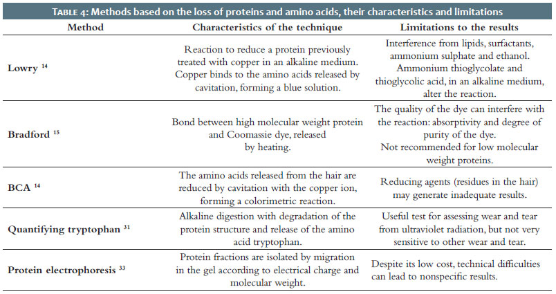
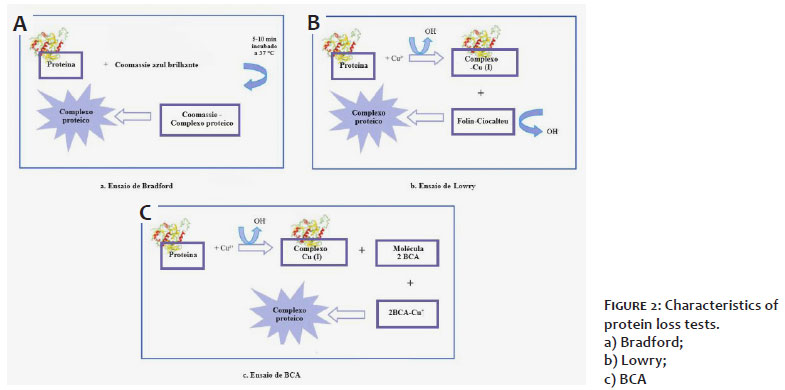
The success or failure of protein quantification depends on the quality of the analytical process used to characterize the samples. Table 4 shows the main methods for quantifying hair fiber proteins and amino acids (Figure 2).
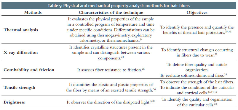
The mechanical strength of the hair fiber can be assessed using methods based on its elastic and plastic properties, using a tension force. The plasticity of the hair fiber allows it to extend by about 2% of its original length. As the load exerted on the fiber continues, the plastic phase begins, and the hair stretches approximately 25 to 30% of its length before breaking.34-36 Table 5 summarizes the methods of physical analysis of the hair fiber.
This article presents the main hair fiber assessment techniques that the cosmetics industry and research centers use, which are still not very accessible or well known in the dermatological field.
Currently, new technologies have been highlighted in the outpatient dermatology field, complementing the assessment and diagnosis of hair health, allowing for a better understanding of disorders and damage to the hair shaft, leading to more specific and effective treatments.
Tércio Elyan Azevedo Martins
ORCID: 0000-0002-7111-1029
Approval of the final version of the manuscript; study design and planning; preparation and writing of the manuscript; critical literature review.
Andressa Costa de Oliveira
ORCID: 0000-0001-8859-5835
Approval of the final version of the manuscript; study design and planning; preparation and writing of the manuscript; critical literature review; critical review of the manuscript.
Beatriz Regazzi de Gusmão
ORCID: 0000-0001-6390-9447
Drafting and editing the manuscript; data collection, analysis and interpretation.
Alexandre de Almeida Filippo
ORCID: 0000-0001-9550-5156
Drafting and editing the manuscript; data collection, analysis and interpretation.
Fabiane Mulinari Brenner
ORCID: 0000-0001-7970-522X
Approval of the final version of the manuscript; drafting and editing of the manuscript; effective participation in research orientation; critical literature review; critical review of the manuscript.
1. Schneider MR, Schmidt-Ullrich R, Paus R. The hair follicle is a dynamic miniorgan. Curr Biol. 2009;19(3):R132-R142.
2. Hoover E, Alhajj M, Flores JM. Physiology, Hair. Treasure Island: StatPearls; 2022.
3. Richena M. Efeitos da irradiação na morfologia e estrutura do cabelo. [Tese]. Campinas: Universidade Estadual de Campinas; 2015.
4. Cruz CF, Costa C, Gomes AC, Matamá T, Cavaco-Paulo A. Human hair and the impact of cosmetic procedures: a review on cleansing and shape-modulating cosmetics. Cosmet. 2016;3(26):103390.
5. Velasco MVR, Dias TCS, Freitas AZ, Júnior NDV, Pinto CASO, Kaneko TM, et al. Hair fiber characteristics and methods to evaluate hair physical and mechanical properties. Braz J Pharm Sci. 2009;45(1):153-62.
6. Lee SY, Choi AR, Baek JH, Kim HO, Shin MK, Koh JS. Twelve-point scale grading system of scanning electron microscopic examination to investigate subtle changes in damaged hair surface. Skin Res and Technol. 2016;22(4):406- 11.
7. Wagner R, Joekes I. Hair medulla morphology and mechanical properties. J Cosmet Sci. 2007;58(4):359-68.
8. Bhushan B. Nanoscale characterization of human hair and hair conditioners. Prog Mat Sci. 2008;53(4):585–710.
9. Wei G, Bhushan B, Torgerson PM. Nanomechanical characterization of human hair using nanoindentation and SEM. Ultramicroscopy. 2005;105(1-4):248-66.
10. Sakai M, Kikuchi K, Fujii M. Quaternary and secondary structural imaging of a human hair by a VSFG-detected IR super-resolution microscope. Chem Phys. 2013;419:261-5.
11. Koch SL, Tridico SR, Bernard BA, Shriver MD, Jablonski NG. The biology of human hair: a multidisciplinary review. Am J Hum Biol. 2020;32(2):e23316.
12. Camargo FB, Minami MM, Rossan MR, Magalhães WV, Ferreira VTP, Campos PM BGM. Prevention of chemically induced hair damage by means of treatment based on proteins and polysaccharides. J Cosmet Dermatol. 2022;21(2):827-35.
13. Santos JD, Edwards H, Oliveira LD. Raman spectroscopy and electronic microscopy structural studies of Caucasian and Afro human hair. Heliyon. 2019; 5(5):e01582.
14. Gama RM, Baby AR, Velasco MVR. In vitro methodologies to evaluate the effects of hair care products on hair fiber. Cosmetics. 2017;4(2):1-10.
15. Sá-Dias TC, Baby AR, Kaneko TM, Velasco MVR. Protective effect of conditioning agents on Afro-ethnic hair chemically treated with thioglycolate- based straightening emulsion. J Cosmet Dermatol. 2008;7(2);120–6.
16. Mubki T, Rudnicka L, Olszewska M, Shapiro J. Evaluation and diagnosis of the hair loss patient: Part I. History and clinical examination. J Am Acad Dermatol. 2014;71(3):415.e1-415.e15.
17. Cheng AS, Bayliss SJ. The genetics of hair shaft disorders. J Am Acad Dermatol. 2008;59(1):1–22.
18. Burgess C, Roberts W, Downie J, Kera M, Kogan S, Belpulsi D. A closer look at a multi-targeted approach to hair loss in African American women. J Drugs Dermatol. 2020;19(1):95-8.
19. Kibar M. Trichoscopy and Trichogram. In: Kutlubay Z, Serdaroglu S, editors. Hair and Scalp Disorders. InTech; 2017. p.81-101.
20. Tubia C, Fernández-Botello A, Dupont J, Gómez E, Desroches J, Attia J, et al. A new Ex Vivo model to evaluate the hair protective effect of a biomimetic Exopolysaccharide against water pollution. Cosmet. 2020;7(4):78.
21. Martins TEA, Werner B, Filippo A, Regazzi B, Mulinari-Brenner FM. Proceedings of the 25th Congress on Dermatology and Aesthetic & Plastic; 2023 Jan 26-28; Paris, France. Rapid protein loss test as a guide to hair damage.
22. Mulinari-Brenner F, Regazzi B, Filippo A, Martins T, editors. Proceedings of the 25th Congress on Dermatology and Aesthetic & Plastic; 2023; Paris, France. Analysis of protein loss as a marker of broad hair shaft damage: evaluation by Lowry and rapid (RPLT) tests.
23. Duarte LC, Juchen PL, Pulz GM, Brum TM, Chodur N, Liccardo A, et al. Aplicações de microscopia eletrônica de varredura (MEV) e sistema de energia dispersiva (EDS) no estudo de gemas: exemplos brasileiros. Pesquisas Geociências. 2003;30(2):3–15.
24. Sasaki K, Murata H, Kuroda K, Saka H. Conventional transmission electron microscope observation of electric and magnetic fields. In: Maaz K, editor. The Transmission Electron Microscope. Croatia: InTech Open; 2012. p.1–27.
25. Latorre C, Bhushan B. Investigation of scale effects and directionality dependence on friction and adhesion of human hair using AFM and macroscale friction test apparatus. Ultramicroscopy. 2006;106(8):720–34.
26. Freitas AZ, Amaral MM, Raele MP. Optical coherence tomography: development and applications. In: Duarte FJ, editor. Laser Pulse Phenomena and Applications. Croatia: InTech Open; 2010. p. 409–32.
27. Velasco MVR, Abreu SRP, Freitas AZ, Bedin V, Baby AR, Gama RM. Optical coherence tomography to evaluate the effects of oxidative hair dye on the fiber. Skin Res Technol. 2016;22(1):430–6.
28. Rodrigues AG, Galzerani JC. Espectroscopias de infravermelho, raman e de fotoluminescência: potencialidades e complementaridades. Rev Bras Ensino Fis. 2012;34(1):1–9.
29. Kuzuhara A. Internal structural changes in keratin fibres resulting from combined hair waving and stress relaxation treatments: A Raman spectroscopic investigation. Int J Cosmet Sci. 2016;(38):201–209.
30. Miyamae Y, Yamakawa Y, Ozaki Y. Evaluation of physical properties of human hair by diffuse reflectance near-infrared spectroscopy. Appl. Spectrosc. 2007, 61, 212–7.
31. Dario MF, Freire TB, Pinto CASO, Prado MAS, Baby AR, Velasco MVR. Tryptophan and kynurenine determination in human hair by liquid chromatography. J Chrom B. 2017;1066(2):59–62.
32. França-Stefoni AS, Dari MF, Sá-Dias TC, Bedin V, Almeida AJ, Baby AR, et al. Protein loss in human hair from combination straightening and coloring treatments. J Cosmet Dermatol. 2015;14(1):204–8.
33. Westermeier R, Gronau S, Becket P, et al. Electrophoresis in Practice: A guide to methods and mpplications of DNA and protein separations. Germany: Wiley; 2005. p.427
34. Lima CRRC, Almeida MM, Velasco MVR, Matos JR. Thermoanalytical characterization study of hair from different ethnicities. J Therm Anal Calorim. 2016;123(1):2321–8.
35. Dario MF, Pahl R, Castro JR, Lima FS, Kaneko TM, Pinto CASO, et al. Efficacy of Punica granatum L. hydroalcoholic extract on properties of dyed hair exposed to UVA radiation. J Photochem Photobiol B Biol. 2013;120(1):142–7.
36. Velasco MVR, Balogh TS, Kagiyama JW, Dario MF, Gama RM, Bedin V, et al. Influence of brazilian vegetable oils on mechanical resistance of hair fiber. Biomed Biopharm Res. 2015; 12(1): 99–106.
37. Robbins CR, Crawford RJ. Cuticle damage and the tensile properties of human hair. J Soc Cosmet Chem. 1991: 42(1):49–58.
 All content the journal, except where identified, under the Creative Commons Attribution 4.0 International licence - ISSN-e 1984-8773


 Read in Portuguese
 Portuguese PDF
 Print
Print
 Send this article by email
 How to cite this article
 Submit a comment
 Mendeley
 Pocket