


Gabriela Alfier1; Srecko Budi2; Zrinka Sudar-Magas3; Milan Miocinovic4; Saida Rezakovic5
Submitted on: 13/05/2023
Approved on: 24/10/2023
Financial support: None.
Conflict of interest: None.
How to cite this article: Alfier G, Budi S, Sudar-Magas Z, Miocinovic M, Rezakovic S. Perianal extramammary Paget’s disease: a rare case report and review of the literature. Surg Cosmet Dermatol. 2024;16:e20240262.
Extramammary Paget’s disease (EMPD) is a rare malignancy and is quite difficult to diagnose. We report a case of a 51-year-old female patient with perianal EMPD. Because experience with this disease is still very limited, we searched international databases for similar publications. After consultation with an oncologist, the treatment was wide excision without adjuvant therapy. This treatment option was curative.
Keywords: Paget Disease, Extramammary; Surgery, Plastic; Anal Gland Neoplasms.
EMPD is an uncommon intraepithelial malignancy reported in the worldwide medical literature.1 It is considered a form of adenocarcinoma that can invade the dermis and spread via the lymphatic vessels.2 EMPD was originally described by Darier and Couillaud in 1893. The first cases were reported in the perianal region (Crocke 1889), vulva (Dubreuilh 1901), and axilla (Satani 1920).3 Regardless of the similar clinical and histopathologic appearances of Paget’s disease of the nipple and the anus, mammary Paget’s disease is undoubtedly connected with underlying ductal carcinoma, while perianal Paget’s disease may or may not be associated with an underlying malignancy (33%-86% of cases).4 The most common cancers related to EMPD are colorectal and tubo-ovarian.5 In the literature, a recent large series reported that perianal EMPD was associated with an adnexal adenocarcinoma in 7% of cases and an internal malignancy in 14%.6 However, in most cases EMPD arises primarily as an intraepidermal neoplasm,7 characteristically in skin areas rich in apocrine glands, such as the genital region. Because of its rareness and multicentric nature, diagnosis and treatment represent a major challenge. In this context, the aim of this report is to present the case of a 51-year-old woman admitted to our outpatient clinic and summarize the latest evidence about EMPD.
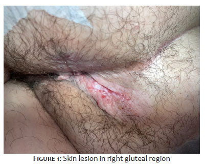
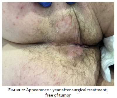
A 51-year-old obese woman presented to our outpatient service with pruritus ani as her sole symptom. She was not taking any medications and had no other comorbidities. There was no associated pain or any other gastrointestinal complaints. Clinical examination revealed a white, moist, raised lesion at the 2 o’clock position of the right gluteus, measuring 3.5×2×0.7cm. Rectal examination and proctosigmoidoscopy were negative. There were no palpably enlarged lymph nodes. The patient had been undiagnosed for 1 year. Prior to surgery, dermatoscopy was made by dermatologist in order to marke the border of tumor, which showed pink structureless area with dotted and short linear vessels. After consultation with a dermatologist and oncologist, a wide excision was performed, encompassing the perianal skin together with the anal mucosa up to the level of the dentate line, preserving the internal sphincter. The surgical defect was successfully closed. Histological examination described squamous epithelium containing nests of atypical mucus-producing Paget cells, with large, pale cytoplasm and hyperchromatic nuclei in hyperplastic epidermis, confirming the diagnosis of EMPD. Resection margins were clear, and there was no evidence of other underlying carcinoma, tumor, or distant metastases. The patient was discharged on the same day. A radiation oncologist confirmed there was no need for adjuvant therapy, only regular follow-up every 3 months during the first year. At the time of writing, the patient has remained disease-free for 1 year after surgical treatment. (Figure 1 and 2).
We searched major international electronic bibliographic databases for articles related to our topic. The search terms were extramammary Paget’s disease and perianal extramammary Paget’s disease, excluding other possible locations of extramammary Paget’s disease, such as the perineum, vulva, scrotum, and penis. The search was limited to studies published in the English language. In brief, perianal EMPD affects patients between the ages of 50 and 80 years, especially white (Caucasian), peaking at age 65, but the true incidence is difficult to measured due to its rarity.4,8 Overall, women are more commonly affected (1.4:1 female-to-male ratio). Its pathogenesis is unclear, but most cases are thought to arise as a primary intraepidermal neoplasm of glandular origin.7,9,10 Clinically, lesions usually present as well-defined, elevated erythematous or white nonhealing plaques, averaging 6-12 cm in diameter, with accompanying irritation and ache.1,11,12 They may also present as annular or hypopigmented plaques with scales, excoriations, and/or erosions.13 Typical presenting symptoms are anal rash and irritation. Anal pain, bleeding, mucoid discharge, lumps, and difficulty defecating may also occur.14 The most commonly affected sites are vulvar, perineal, perianal, scrotal, and penile skin; rare sites include the thighs, buttocks, axilla, eyelids, and external ear canal. EMPD has also been reported in ovarian teratomas and bronchial epithelium.15,16,17 The term ectopic EMPD refers to cases affecting areas in which apocrine glands are not usually found, such as the lateral aspect of the back or lower portion of the chest. The differential diagnosis includes many conditions, such as psoriasis, contact dermatitis, fungal infection, lichen sclerosus, histiocytosis, Pagetoid basal cell carcinoma, mycosis fungoides, and hemorrhoids.18,19 Once a diagnosis of EMPD has been confirmed, the next step is to rule out metastases. In a study by Williams et al., among 7 patients who presented with EMPD, in no one was the correct diagnosis made clinically.20 When there is a diagnosed underlying malignancy, up to 50% of EMPD cutaneous lesions have already metastasized. In these cases, an average survival is limited to 3 years.19 Due to that, suitable diagnostic procedures to exclude other underlying carcinomas include: pelvic ultrasound scan, hysteroscopy, laparoscopy, and/or an MRI scan of the pelvis; colonoscopy, sigmoidoscopy, and/or barium enema; cystoscopy and intravenous pyelogram (IVP); mammogram and chest imaging.21 An ideal modality treatment should offer both minimal tissue destruction and low recurrence rates. This modality must also overcome inconvenient features of EMPD, namely multicentricity and irregular histological margins that extend well beyond the clinically visible lesions. Wide local excision with 2-cm margins or Mohs’ micrographic surgery is the favored approach for noninvasive, locally confined disease.22 Intraoperative frozen sections can be misleading.23 Furthermore, wide local excision requires four-quadrant biopsies, including grossly normal skin. A study performed at Roswell Park Cancer Institute between 1970 and 1998 concluded that surgery offers a moderate chance of cure in advanced cases; long-term multimodal approaches are still needed.13 Nevertheless, surgical biopsy is crucial to confirm the right diagnosis.21 Surgical removal of tumors is considered curative if radical resection with histologically clear margins can be achieved. However, many patients present with advanced primary tumors, so curative surgery is not feasible. Since radical excision usually results in significant tissue loss, the defect frequently needs to be covered with local flaps or skin grafts. When the disease is associated with an underlying anorectal carcinoma, the procedure of choice is abdominoperineal resection with wide excision of the cutaneous lesion.24 Other treatment methods include radiotherapy, laser therapy, and topical and systemic chemotherapy; photodynamic therapy is most promising among recent options. Besa et al. and Burrows et al. found that radiotherapy might be an treatment modality appropriate for patients with non-invasive EMPD who are not surgical candidates.26,27 On the other hand, Thirlby showed that radiotherapy alone is not adequate treatment.23,24,25 In a few cases, only combined chemoradiotherapy was associated with full response on long-term follow-up. Zampogna et al. reported two cases treated with imiquimod cream, which can be used only in a setting of limited primary cutaneous EMPD.11 Finally, the physician must maintain a high index of suspicion, especially in cases with characteristic lesions unresponsive to conventional dermatologic therapy. In view of the above, treatment of EMPD remains a challenge shared by surgeons, pathologists, and dermatologists. Due to its malignant potential, many studies suggest that EMPD should be grouped with other cutaneous carcinomas. Close follow-up for at least 8 years is mandatory for all patients presenting with this rare disease. Bech et al. suggested a follow-up program which includes at least a complete physical examination, proctosigmoidoscopy, and random biopsy of the perianal region once a year. Colonoscopy should be performed at 2-to-3-yearly intervals.5
This case report with review of the literature draws attention to a rare condition that should be always kept in the differential diagnosis of perianal disorders. It must be noted that there is usually a delay in establishing the right diagnosis. Treatment and prognosis depend primarily on the presence and type of underlying carcinoma. Therefore, it is crucial to have a group of specialists involved in management, including s dermatologist, surgeon, pathologist, and oncologist. We concluded that, in this case, wide excision was curative. In other, more advanced cases, adjuvant chemo- or radiotherapy would probably be recommended. Adequate evaluation and long-term follow-up are crucial in all patients with EMPD to identify recurrence and potential development of other malignancies.
The datasets generated during and/or analyzed during the current study are available from the corresponding author on reasonable request.
All aspects of the work covered in this manuscript have been conducted with the ethical approval of all relevant bodies and such approvals are acknowledged within the manuscript.
Gabriela Alfier
ORCID: 0000-0001-8225-6429
Preparation and writing of the manuscript; collecting, analyzing, and interpreting data; effective participation in research guidance
Srecko Budi
ORCID: 0009-0009-7452-621X
Approval of the final version of the manuscript; study design and planning; collecting, analyzing, and interpreting data; effective participation in research guidance; critical review of the literature; critical review of the manuscript
Zrinka Sudar-Magas
ORCID: 0000-0003-0380-2239
Approval of the final version of the manuscript; collecting, analyzing, and interpreting data
Milan Miocinovic
ORCID: 0009-0003-0213-9970
Approval of the final version of the manuscript; intellectual participation in propaedeutic and/or therapeutic conduct of studied cases
Saida Rezakovic
ORCID: 0000-0002-5065-637X
Participation would be approval of the final version of the manuscript, obtaining, analyzing and interpreting data
1. Breen JL, Smith CI, Gregori CA. Extramammary Paget's disease. Clinical Obstetrics and Gynecology. 1978;21(4):1107-15.
2. Darier J, Coulillaud P. Sur un cas de maladie de Paget de la region perinea-anale et scrotale. Ann Dermatol Syphiligr. 1893;4:25-33.
3. Heymann WR. Extramammary Paget’s disease. Clinics in Dermatology. 1993;11(1):83-7.
4. Kyriazanos ID, Stamos PN, Miliadis L, Noussis G, Stoidis CN. Extra-mammary Paget’s disease of the perianal region: a review of the literature emphasizing the operative management technique. Surgical Oncology. 2011;20(2):e61-71
5. Beck DE, Fazio VW. Perianal Paget’s disease. Diseases of the Colon & Rectum. 1987;30(4):263–6.
6. Marchesa P, Fazio VW, Oliart S, Goldblum JR, Lavery JR, Milsom JW. Long- term outcome of patients with perianal Paget’s disease. Annals of Surgical Oncology. 1997;4(6):475–80.
7. Lloyd J, Flanagan AM. Mammary and extramammary Paget’s disease. Journal of Clinical Pathology. 2000;53(10):742–9.
8. Lam C, Funaro D. Extramammary Paget’s disease: summary of current knowledge. Dermatologic Clin. 2010;28(4):807–26.
9. Shepherd V, Davidson EJ, Davies-Humphreys J. Extramammary Paget’s disease. BJOG. 2005;112(3):273–9.
10. Kanitakis J. Mammary and extramammary Paget’s disease. J Eur Acad Dermatol Venereol. 2007;21(5):581-90.
11. Zampogna JC, Flowers FP, Roth WI, Hassenein AM. Treatment of primary limited cutaneous extramammary Paget’s disease with topical imiquimod monotherapy: two case reports. J Am Acad Dermatol. 2002;47(4):S229–S235.
12. Tulchinsky H, Zmora O, Brazowski E, Goldman G, Rabau M. Extramammary Paget’s disease of the perianal region. Colorectal Dis. 2004;6(3):206–9.
13. Zollo JD, Zeitouni NC. The Roswell Park Cancer Institute experience with Extramammary Paget’s disease. Br J Dermatol. 2000;142(1):59–65.
14. Lock MR, Katz DR, Parks A, Thomson JP. Perianal Paget’s disease. Postgrad Med J. 1977;53(626):768–72.
15. Heymann WR. Extramammary Paget’s disease. Clinics in Dermatology. 1993..;11(1):83–7.
16. oshiaki Saida, Iwata M. “Ectopic” Extramammary Paget’s disease affecting the lower anterior aspect of the chest. Journal of The American Academy of Dermatology. 1987;17(5):910–3.
17. Higashiyama M, Doi O, Kodama K, Tateishi R, Kurokawa E. Extramammary Paget’s disease of the bronchial epithelium. Archives of pathology & laboratory medicine. 1991;115(2):185–8.
18. Arminski TC, Pollard RJ. Paget’s disease of the anus secondary to a malignant papillary adenoma of the rectum. Diseases of the Colon & Rectum. 1973;16(1):46.
19. Balducci L, Crawford ED, Smith GF, Lambuth B, McGehee, Hardy C. Extramammary Paget’s Disease: An Annotated Review. Cancer Investigation. 1988;6(3):293–303.
20. Williams SL, Rogers LW, Quan SH. Perianal Paget’s disease: report of seven cases. Diseases of the Colon & Rectum. 1976;19(1):30–40.
21. Al Hallak MN, Zouain N. Extramammary Perianal Paget’s disease. Case Reports in Gastroenterology. 2009;3(3):332–7.
22. Coldiron BM, Goldsmith BA, Robinson JK. Surgical treatment of extramammary Paget’s disease. A report of six cases and reexamination of Mohs micrographic surgery compared to with conventional surgical excision. Cancer 1991;67:993-8
23. Thirlby RC, Hammer CJ, Galagan K, Travaglini J, Picozzi VJ. Perianal Paget’s disease. Diseases of the Colon & Rectum. 1990;33(2):150–2.
24. Berardi RS, Lee S, Chen HP. Perianal extramammary Paget’s disease. Surgery, gynecology & obstetrics. 1988;167(4):359–66.
25. Jensen SL, Sjølin KE, Shokouh-Amiri MH, Harling H. Paget’s disease of the anal margin. Br J Surg 1988;75:1089-92
26. Besa P, Rich TA, Delclos L, Edwards CL, Ota DM, Wharton JT. Extramammary Paget’s disease of the perineal skin: role of radiotherapy. International Journal of Radiation Oncology Biology Physics. 1992;24(1):73–8.
27. Burrows N, Jones DH, Hudson PM, Pye RJ. Treatment of extramammary Paget’s disease by radiotherapy. British Journal of Dermatology. 2010;132(6):970–2.
 All content the journal, except where identified, under the Creative Commons Attribution 4.0 International licence - ISSN-e 1984-8773


 Read in Portuguese
 Portuguese PDF
 Print
Print
 Send this article by email
 How to cite this article
 Submit a comment
 Mendeley
 Pocket